
Tumor boundary reconstruction using hyperspectral imaging

a hyperspectral imaging and tumor technology, applied in the field of tumor boundary reconstruction using hyperspectral imaging, can solve the problems of increasing the effectiveness of the procedure, challenging the removal of the complete tumor, and no reliable way to provide real-time feedback to the surgeon

Active Publication Date: 2020-04-30
KONINKLJIJKE PHILIPS NV
View PDF0 Cites 10 Cited by
  • Summary
  • Abstract
  • Description
  • Claims
  • Application Information

AI Technical Summary

Benefits of technology

The patent describes a computer method for analyzing tissue samples to determine which part of the tissue needs to be removed in a surgical procedure. The method uses two independent techniques to determine the boundary of the tumor or diseased tissue. By comparing the similarity of the boundaries, the method calculates the probability that the boundary is correct, which helps the physician decide which part of the tissue needs removal. The method can be used for both tumors and diseased tissue. The classification system uses a spectral camera to capture spectral images during medical interventions and a combination device to generate a combined tumor boundary or diseased tissue boundary with an indication of reliability. The training data used for the classification system is relatively small, but the method can still recognize a relatively large number of objects in the tissue images. The spectral analysis can be performed for each pixel or clusters of neighboring pixels to reduce computation time. The technical effects of this invention are an improved accuracy in tumor and disease boundary detection, assistance in decision making for surgical procedures, and reliable indication of the extent of disease in tissue samples.

Problems solved by technology

Removal of the complete tumor is challenging, since the procedure relies on palpation.
Currently there is no reliable way to provide real-time feedback to the surgeon, and tissue must be sent to the pathology department for analysis.
This is a time costly process and technology that can support the surgeon in realtime will increase the effectiveness of the procedure.
Compared to regular RGB data it is challenging to process the HSI data due to the size of the data: hundreds of color bands in a multi-megapixel image results in large files with varying amounts of redundant information.
Larger numbers of channels have been considered in the same network architecture, but did not increase performance significantly.
That might be caused by the limited data set, or by the fact that the network is very shallow.
The imaging system uses a line-by-line scanning method, which makes real time applications difficult.

Method used


Image

  • Tumor boundary reconstruction using hyperspectral imaging
  • Tumor boundary reconstruction using hyperspectral imaging
  • Tumor boundary reconstruction using hyperspectral imaging

Examples


Embodiment Construction

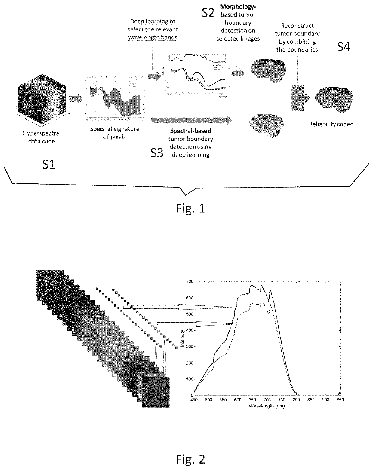
[0077]FIG. 1 shows a block diagram of a computer implemented method of determining a boundary of a tumor according to an exemplary embodiment of the present invention. Starting with receiving (S1) a hyperspectral data cube of a tissue sample, which includes the spectral signature of each pixel of the image, a two-fold approach is performed: On the one hand, relevant wavelength bands are selected, preferably by deep learning or a neural network. The image in the respective wavelength band is analysed in a morphological approach (S2) thereby generating a morphological tumor boundary. On the other hand, the spectral information of each pixel or a group of neighbouring pixels is analysed in a spectral approach (S3). Preferably, an AI module or deep learning, as described herein in detail, is used to classify the pixel or the group of neighbouring pixels in tumorous or healthy tissue, thereby generating a spectral tumor boundary. By combining (S4) the morphological tumor boundary and the...


PUM


Abstract

In a computer implemented method of determining a boundary of a tumor region or other diseased tissue, hyper- or multispectral image data of a tissue sample including a tumor region or other diseased tissue is taken. The analysis includes a morphological analysis and a spectral analysis of the hyper- or multispectral image data resulting in a morphological tumor boundary and a spectral tumor boundary or a morphological diseased tissue boundary and a spectral diseased tissue boundary. These two boundaries are combined resulting in a combined tumor boundary or combined diseased tissue boundary, wherein an indication of reliability of the combined tumor boundary or combined diseased tissue boundary is given.

Description

FIELD OF THE INVENTION[0001]The present invention relates to a computer implemented method of determining a boundary of a tumor region or other diseased tissue, a computer program product comprising instructions which when executed on a processor cause the processor to carry out a method of determining a boundary of a tumor region or other diseased tissue, and an apparatus for determining a boundary of a tumor region or other diseased tissue.BACKGROUND OF THE INVENTION[0002]For the physician, knowing the diseased tissue boundary is important since the treatment depends heavily on it. Knowing where to cut and what critical tissues can be spared is very important for a good surgical outcome and for low comorbidity.[0003]Surgical removal of breast tumors (e.g. lumpectomy) is one of the most frequently performed surgical resections with significant rate increase, due to the leading incidence and prevalence of the breast neoplasms. The clinical goal of the breast surgery is to remove the...

Claims


Application Information

Patent Timeline
Patent Type & Authority Applications(United States)
IPC IPC(8): G06T7/00G06T7/11
CPCG06T7/11G06T2207/30096G06T2210/12G06T2207/10036G06T2207/20084G06T7/0012G06T2207/20081G06T7/12G06T7/174
Inventor HENDRIKS, BERNARDUS HENDRIKUS WILHELMUSLIU, HONGSHAN, CAIFENG
Owner KONINKLJIJKE PHILIPS NV
Learn More