In vitro biliary excretion assay

a biliary excretion and in vitro technology, applied in the field of in vitro biliary excretion assay, can solve the problems of difficult acquisition of primary hepatocytes, high cost, and inability to direct measurement of biliary accumulation in the two-culture assay forma

Inactive Publication Date: 2020-05-07
HUREL CORP
View PDF0 Cites 0 Cited by
  • Summary
  • Abstract
  • Description
  • Claims
  • Application Information

AI Technical Summary

Benefits of technology

[0039]In some embodiments of the methods the activity of at least one hepatocyte transport protein is inhibited in the hepatocytes of the cell culture.

Problems solved by technology

Therapeutic chemical entities are often undesirably removed from an animal's circulatory system by first-pass metabolism in the liver.
A first drawback is the simple reality that creating a single biliary excretion data point using this assay format requires two cultures of primary hepatocytes.
Primary hepatocytes are difficult to procure and therefore are very expensive.
A second drawback is that biliary accumulation of a chemical entity is necessarily calculated in a two-culture assay format by measuring two values that are not biliary accumulation, namely total cellular accumulation (cytoplasm and bile) and cytoplasmic accumulation, and then calculating the difference between these values.
A direct measurement of biliary accumulation is not possible in the two-culture assay format.
However, the previously taught assay is indirect.
This assay format has several drawbacks including low accuracy.

Method used


Image

  • In vitro biliary excretion assay
  • In vitro biliary excretion assay
  • In vitro biliary excretion assay

Examples


example 1

e-Stromal Cell Cocultures

[0131]Cryopreserved human hepatocytes were removed from liquid nitrogen and thawed. After thawing, cells were resuspended in medium and cell number and cell viability was determined using trypan blue exclusion. Stromal cells were passed in a CO2 incubator until used for experimental plating. On plating day cells were detached from the plate, washed, and resuspended in medium. Cell number and viability were determined using trypan blue exclusion.

[0132]Hepatocytes and stromal cells were seeded into collagen-coated 96-well plates at a density of 30,000 hepatocytes per well. The stromal cells were growth arrested prior to seeding. Cultures were maintained for 7 days before the start of any biliary excretion assays. Before the experiments were started, the cells were stained with CDFDA on day 7 to ensure canalicular formation.

example 2

of Test Compound into Bile Canaliculi in Hepatocyte-Stromal Cell Cocultures

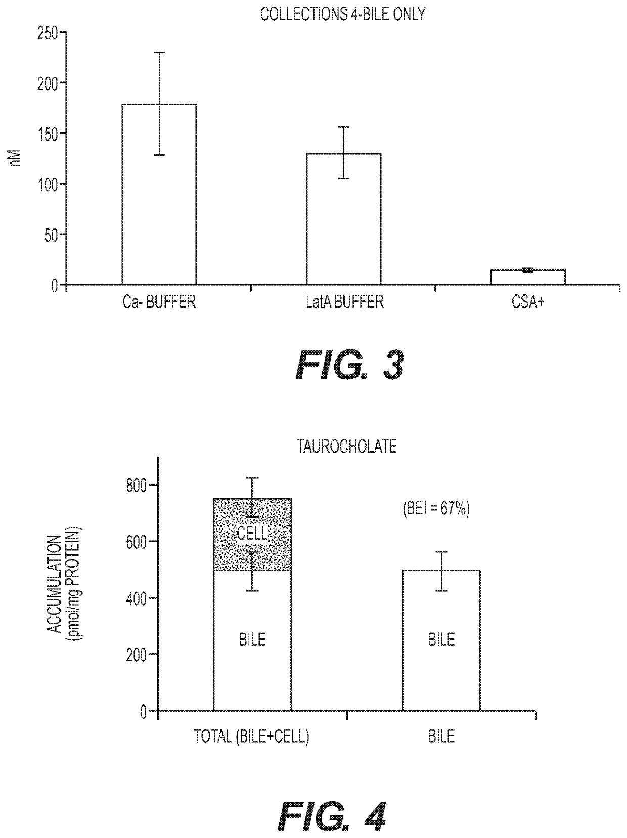
[0133]In this experiment the presence of bile canaliculi in hepatocyte-stromal cell cocultures prepared according to Example 1 was assessed. Cocultures were incubated with 5 uM 5-(and-6)-Carboxy-2′, 7′-dichloro-fluorecein diacetate (CDFDA) for 20 min. CDFDA that is taken up by hepatocytes is hydrolyzed to fluorescent 5-(and-6)-Carboxy-2′, 7′-dichloro-fluorecein (CDA) by the hepatocyte and then secreted in this fluorescent form into the canaliculus via the Mrp2 transporter. The cocultures comprised several canaliculi that are readily apparent under the microscope (data not shown). Cyclosporin A (CSA) is a known inhibitor of the Mrp2 transporter. In a subsequent experiment a coculture was incubed with CDFDA in the presence of 50 uM CSA for 20 min. No fluorescent canaliculi were visible following incubation with CDFDA in the presence of CSA (data now shown). This result demonstrates that bile canaliculi are pres...

example 3

n of Canaliculi

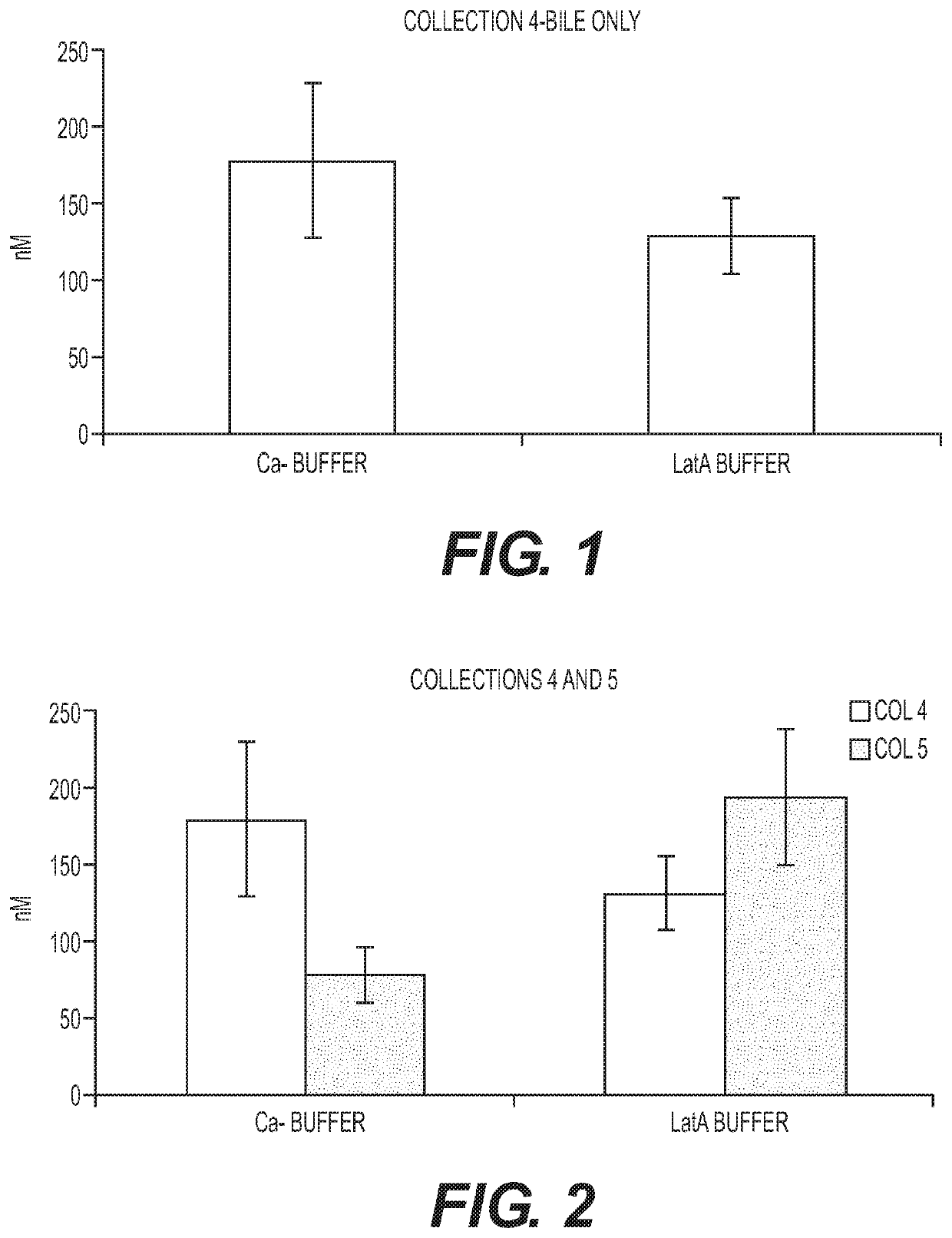
[0134]Assessing excretion of a chemical entity into the bile in the hepatocyte-stromal cell coculture requires a method of distinguising between the chemical entity present in canaliculi of the coculture and chemical entity that may be present in the cytoplasm of cells in the coculture. Two methods of canalicular disruption were tested. In the first method, the cultures were exposed to a calcium free (Ca−) free buffer. The lack of calcium inhibits the ability of the cell to form tight junctions which allows bile present in the canaliculi to escape. Cells were first washed two times in HBSS buffer with Ca+. Cells were then incubated with 5 uM CDFDA in HBSS buffer without Ca− for 20 min. Cells were then washed two times in HBSS buffer without Ca−. Because accumulated CDF escaped the canaliculi after exposure to Ca− buffer no CDF staining was apparent (data not shown).

[0135]In the second method, the cultures were exposed to buffer containing latranculin A (LatA). The Lat...


PUM

PropertyMeasurementUnit
flow rateaaaaaaaaaa
chemicalaaaaaaaaaa
movementaaaaaaaaaa

Abstract

An in vitro methods of characterizing biliary excretion of a chemical entity using a single hepatocyte culture. Comprising providing cell culture comprising hepatocytes forming at least one bile canaliculus; contacting the cell culture with a first chemical entity for a time sufficient to allow uptake of the chemical entity by hepatocytes in the culture; disrupting the at least one bile canaliculus without lysing the hepatocytes and detecting the amount (if any) of the first chemical entity and / or a metabolite thereof released by the at least one bile canaliculus; and lysing the hepatocytes and detecting the amount of the first chemical entity and / or a metabolite thereof released by the hepatocytes.

Description

INTRODUCTION[0001]Therapeutic chemical entities are often undesirably removed from an animal's circulatory system by first-pass metabolism in the liver. If a chemical entity is taken up by hepatocytes and excreted in bile via the bile canaliculi the chemical entity will never reach its therapeutic target. Transport proteins endogenous to hepatocytes are responsible for moving substrates across the sinusoidal membrane of the hepatocytes and then into bile canaliculi. Bile canaliculi are structures within liver tissue that receive excreted components from the hepatocytes and transport the bile to a common bile duct for removal from the animal. Biliary excretion of substrates is thus a complex process involving translocation across the sinusoidal membrane, movement through the cytoplasm, and transport across the canalicular membrane.[0002]Understanding that hepatobiliary excretion of parent drugs or their metabolites often play a significant role in the overall clearance of a drug has ...

Claims


Application Information

Patent Timeline
Patent Type & Authority Applications(United States)
IPC IPC(8): G01N33/50
CPCG01N33/5067G01N33/5091
Inventor NOVIK, ERICCHO, CHEULPAREKH, AMITFREEDMAN, ROBERTYARMUSH, MARTIN L.PLUDWINSKI, ERIC
Owner HUREL CORP
Why Eureka
Try Eureka