System and method for three-dimensional label-free optical imaging of a biological cell sample in an environmental chamber

a biological cell and environmental chamber technology, applied in material analysis, phase-affecting property measurement, instruments, etc., can solve the problems of significant imaging artifacts, lack of sensitivity of conventional intensity-based light microscopy approaches, and insufficient quantitative results

Active Publication Date: 2020-06-11
RAMOT AT TEL AVIV UNIV LTD
View PDF0 Cites 8 Cited by
  • Summary
  • Abstract
  • Description
  • Claims
  • Application Information

AI Technical Summary

Problems solved by technology

Biological cells are three-dimensional (3-D) entities and imaging them in three dimensions while they are in vitro, without using staining to enhance contrast, is a great challenge since they are mostly transparent under regular light microscopy.
However, cells in vitro (in a dish or in a tube) are mostly-transparent 3-D objects with absorbance and reflection characteristics that are very similar to their surroundings, and thus conventional in...

Method used


Image

  • System and method for three-dimensional label-free optical imaging of a biological cell sample in an environmental chamber
  • System and method for three-dimensional label-free optical imaging of a biological cell sample in an environmental chamber
  • System and method for three-dimensional label-free optical imaging of a biological cell sample in an environmental chamber

Examples


Embodiment Construction

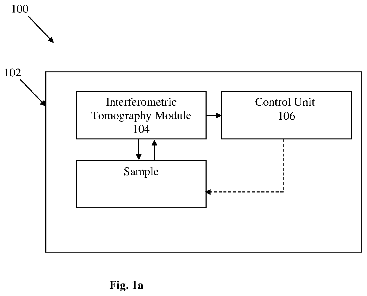
[0052]Reference is made to FIG. 1a illustrating a system 100 for non-invasive imaging of a sample comprising at least one biological cell without labeling according to some embodiments of the present invention. The system 100 comprises inter alia an environmental chamber 102 for accommodating the sample. The environmental chamber 102 comprises an interferometric tomography module 104 configured and operable for generating an illumination beam towards the sample and generating multiple interferometric projections of the cell at various angles. The interferometric tomography module 104 is placed within the environmental chamber 102 such that the sample should not be taken out of the environmental chamber to be inspected. The system 100 also comprises a control unit 106 configured and operable to perform the steps of (1) at least one of rotation of the illumination beam (i.e. sequentially actuating at least one optical element for scanning the sample by illumination at different incide...


PUM


Abstract

The present invention relates to a novel label-free tomographic interferometry technique implemented inside an environmental chamber (e.g. imaging incubator) that provides rapid capturing of the three-dimensional (3-D) refractive index distribution of biological cells.

Description

TECHNOLOGICAL FIELD[0001]The present invention relates to a system and method for three-dimensional label-free optical imaging of a biological cells sample accommodated in an environmental chamber.BACKGROUND ART[0002]References considered to be relevant as background to the presently disclosed subject matter are listed below:[0003]1. Choi, W. et al. Tomographic phase microscopy. Nat. Methods 4, 717-719 (2007).[0004]2. Isikman, S. O. et al. Lens-free optical tomographic microscope with a large imaging volume on a chip. Proc. Natl. Acad. Sci. U.S.A. 108, 7296-7301 (2011).[0005]3. Kim, K. et al. High-resolution three-dimensional imaging of red blood cells parasitized by plasmodium falciparum and in-situ hemozoin crystals using optical diffraction tomography. J. Biomed. Opt. 19, 011005 (2014).[0006]4. Sung, Y. et al. Optical diffraction tomography for high resolution live cell imaging. Opt. Express 17, 266-277 (2009).[0007]5. Hsu, W. C., Su, J. W., Tseng, T. Y. & Sung, K. B. Tomographic...

Claims


Application Information

Patent Timeline
IPC IPC(8): G01N21/45G03H1/00G01N15/14
CPCG01N15/1475G01N21/453G03H1/0005G03H2001/005G01N2015/1445G01N21/45G01N2015/1454G01N2015/1006G01N15/1434G01N15/1484Y02A50/30
Inventor SHAKED, NATAN TZVI
Owner RAMOT AT TEL AVIV UNIV LTD
Why Eureka
Try Eureka