Image processing method and device

a processing method and image technology, applied in the field of image processing, can solve the problems of poor user interaction effect, inability to operate professionals, and inability to support the head of a scanning gun to roam freely in the oral cavity

Active Publication Date: 2020-06-11
BEIJING KEEYOO TECH CO LTD
View PDF0 Cites 5 Cited by
  • Summary
  • Abstract
  • Description
  • Claims
  • Application Information

AI Technical Summary

Benefits of technology

[0005]Embodiments of the present invention provides an image processing method and an image processing device, which are used for solving the problems of poor user interaction effect and difficulty in supporting self-service three-dimensional true color modeling of users in oral cavity image presentation in the prior art.

Problems solved by technology

However, the scanning gun head is not supported to roam freely in the oral cavity, and is highly professional, has poor user interaction effect, and requires professionals to be operated.
However, endoscopic images can only show a very small part at a time.
Although the images of teeth can also be seen, it is difficult to confirm which tooth this is and which specific position of the oral cavity the currently seen details are in.
In addition, there is a lack of three-dimensional image information, which makes it impossible to mold the full dentition, to generate the current digital model of the full dentition in real time, and thus to support dental applications such as three-dimensional printing of the dental shell type dentifrice.

Method used


Image

  • Image processing method and device
  • Image processing method and device
  • Image processing method and device

Examples


embodiment 1

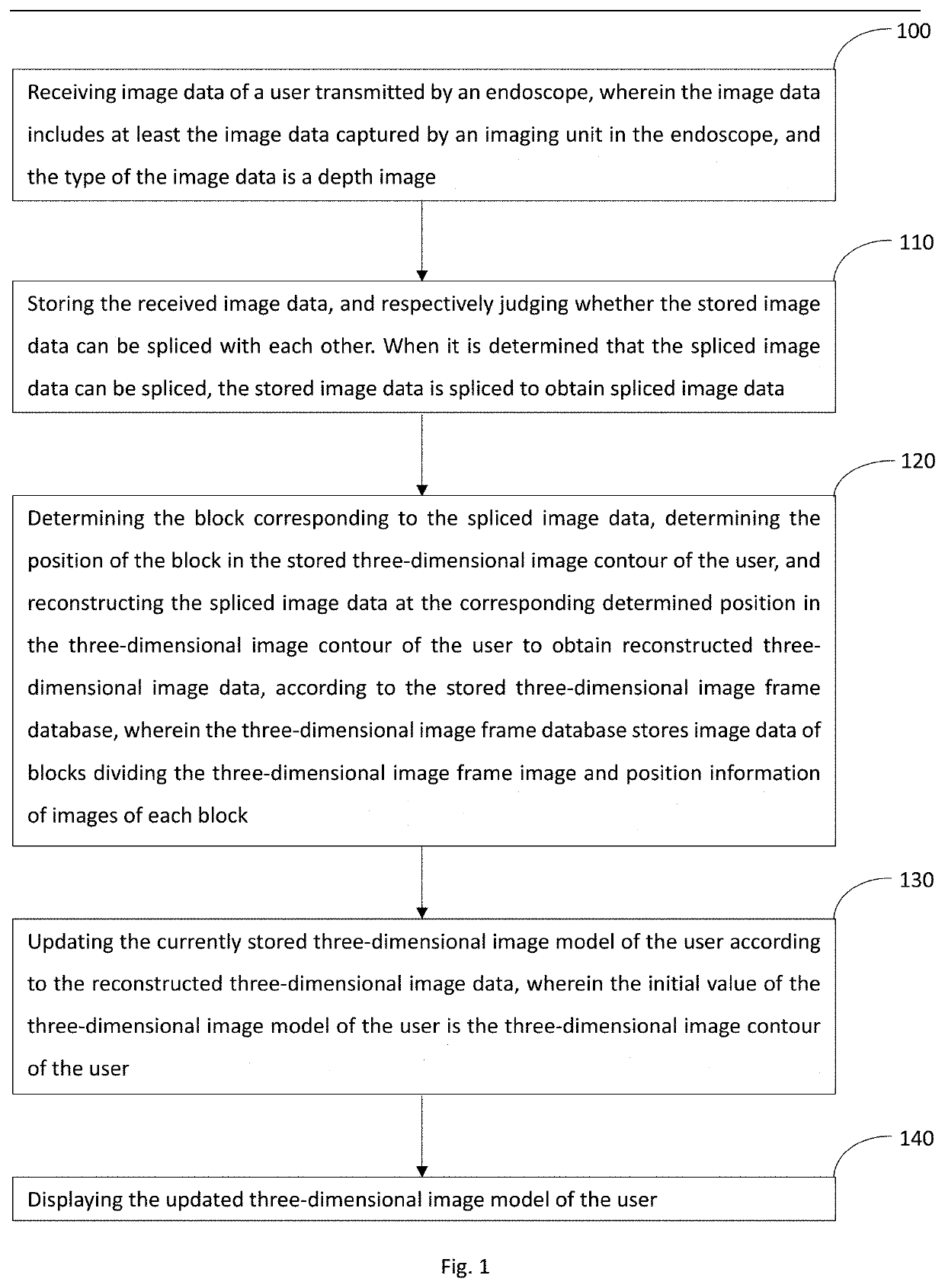
[0078]Referring to FIG. 1, in an embodiment of the present invention, the specific flow of the image processing method is as follows:

[0079]Step 100: receiving image data of a user transmitted by an endoscope, wherein the image data includes at least the image data captured by an imaging unit in the endoscope, and the type of the image data is a depth image.

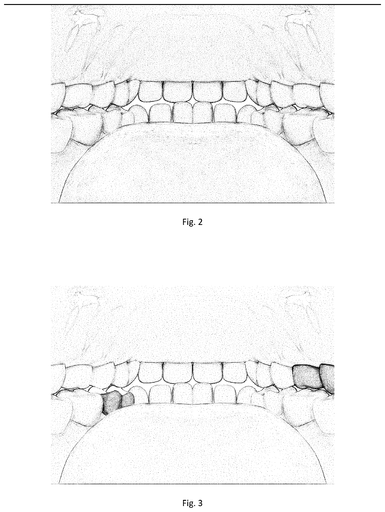
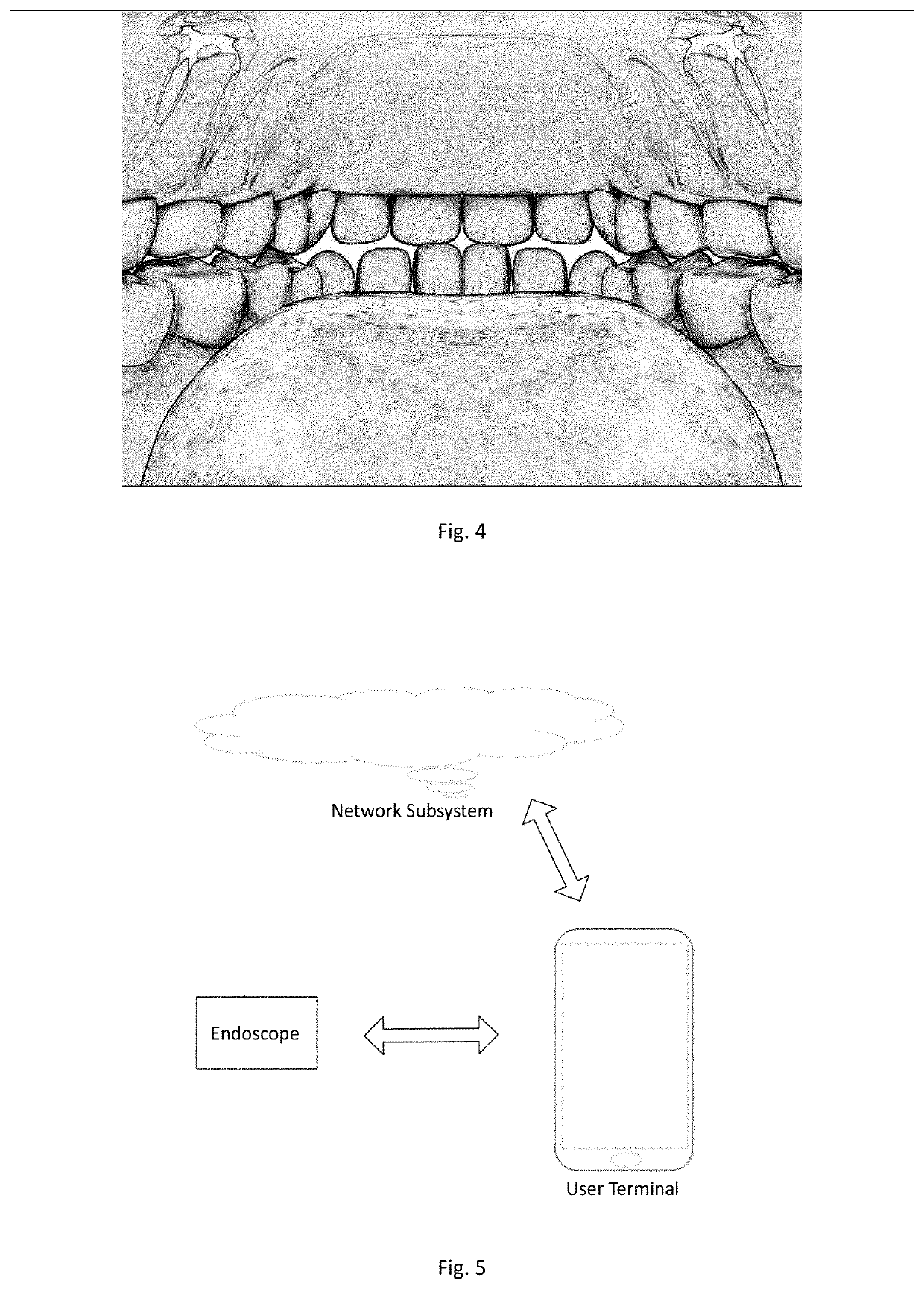
[0080]In practice, users often have a need to view images of the oral cavity. For example, when having a toothache or a tooth is broken, an image in the oral cavity can be obtained by scanning the oral cavity through an oral endoscope. However, in the prior art, after scanning the oral cavity, only a partial image can be obtained, and the whole three-dimensional image cannot be presented, so that the user cannot view the whole three-dimensional image of the oral cavity, and cannot determine the specific position of the broken tooth or the part with problems in the oral cavity. At present, there is also a technology that can presen...

embodiment 2

[0154]In step 120 of Embodiment 1, according to the stored three-dimensional image frame database, the block corresponding to the spliced image data is determined, and the position of the block in the stored three-dimensional image contour of the user is determined. The specific implementation method is described below.

[0155]Specifically, it determines the block in the user's three-dimensional image frame database corresponding to the spliced image data according to at least image characteristic information of the block in the three-dimensional image frame database, and it determines the position in the three-dimensional image contour of the user corresponding to the spliced image data according to the block in the user's three-dimensional image frame database corresponding to the spliced image data.

[0156]Specifically, it can be divided into the following ways:

[0157]The first method:

[0158]1) respectively matching the spliced image data with the images of the blocks in the three-dime...

embodiment 3

[0198]Based on the above-mentioned embodiments, the three-dimensional image frame database and the three-dimensional image contour will be described in detail below.

[0199]1) Three-Dimensional Image Frame Database.

[0200]In the embodiment of the invention, the three-dimensional image frame database is constructed based on various conditions of the human oral cavity, and the three-dimensional image frame database stores general frame data of a three-dimensional image model of the human oral cavity, and the frame data covers image characteristic information of all surface areas of the human oral cavity under various conditions, such as information of shape characteristics, color characteristics, texture characteristics and the like. These situations include normal healthy oral cavity scenes, dirty oral cavity scenes, pathological oral cavity scenes, abnormal oral cavity scenes, traumatic oral cavity scenes, mixed tooth scenes in which deciduous teeth grow towards permanent teeth of adul...


PUM


Abstract

An image processing method and an image processing device includes: receiving image data of a user transmitted by an endoscope, storing and splicing to obtain spliced image data; determining the block corresponding to the spliced image data, determine the position of the block in the stored three-dimensional image contour of the user, reconstructing the spliced image data at the corresponding determined position in the three-dimensional image contour of the user to obtain reconstructed three-dimensional image data and update the currently stored three-dimensional image model of the user, according to the stored three-dimensional image frame database; and displaying the updated three-dimensional image model of the user. The three-dimensional image of the oral cavity can be reconstructed without continuous and orderly scanning of the oral cavity, the dynamic display can be performed, the user interaction effect can be improved, and the self-service three-dimensional true color model selection can be supported.

Description

CROSS-REFERENCE TO RELATED APPLICATION[0001]This application claims priority to Chinese Patent Application No. 201710744863.3, filed on Aug. 25, 2017, entitled: “image processing method and device” the entire contents of which are herein is incorporated by reference.TECHNICAL FIELD[0002]The invention relates to the technical field of image processing, in particular to an image processing method and device.DESCRIPTION OF THE RELATED ART[0003]The traditional oral endoscope is a kind of equipment used for dental optical impression-taking. When in use, the optical gun head used for impression-taking needs to be moved orderly in the upper dentition and lower dentition of a user. However, the scanning gun head is not supported to roam freely in the oral cavity, and is highly professional, has poor user interaction effect, and requires professionals to be operated.[0004]At present, there is also a user self-service oral endoscope, which the user holds, puts a camera part into the oral cavi...

Claims


Application Information

Patent Timeline
Patent Type & Authority Applications(United States)
IPC IPC(8): G06T19/00G06K9/62G06T7/73G06V10/44
CPCG06T7/74G06T2207/30036G06T2207/10068G06T19/00G06K9/6201G06T17/00G06T7/337G06T2200/08G06T2210/41G06V20/64G06V10/44G06V2201/03G06F18/22
Inventor OUYANG, CONGXING
Owner BEIJING KEEYOO TECH CO LTD
Why Eureka
Try Eureka