Diagnosis and treatment of endometriosis by screening for l-form bacteria

a technology of endometriosis and l-form bacteria, which is applied in the direction of antibacterial agents, instruments, drug compositions, etc., can solve the problems that endometriosis may often go undetected, and achieve the effects of reducing patient/subject anxiety, saving time, and saving costs

Inactive Publication Date: 2020-07-30
SOFT CELL BIOLOGICAL RES LLC
View PDF0 Cites 1 Cited by
  • Summary
  • Abstract
  • Description
  • Claims
  • Application Information

AI Technical Summary

Benefits of technology

[0004]In some aspects, the disclosure relates to methods for culturing and identifying L-form bacteria within a subject's sample (e.g., blood, saliva, etc.) in order to diagnose the subject as having endometriosis and/or an ovarian fibroid tumor. Such methods beneficially enable the detection of such conditions that w...

Problems solved by technology

Endometriosis may often go undetected until prolonge...

Method used


Image

  • Diagnosis and treatment of endometriosis by screening for l-form bacteria
  • Diagnosis and treatment of endometriosis by screening for l-form bacteria
  • Diagnosis and treatment of endometriosis by screening for l-form bacteria

Examples


example 1

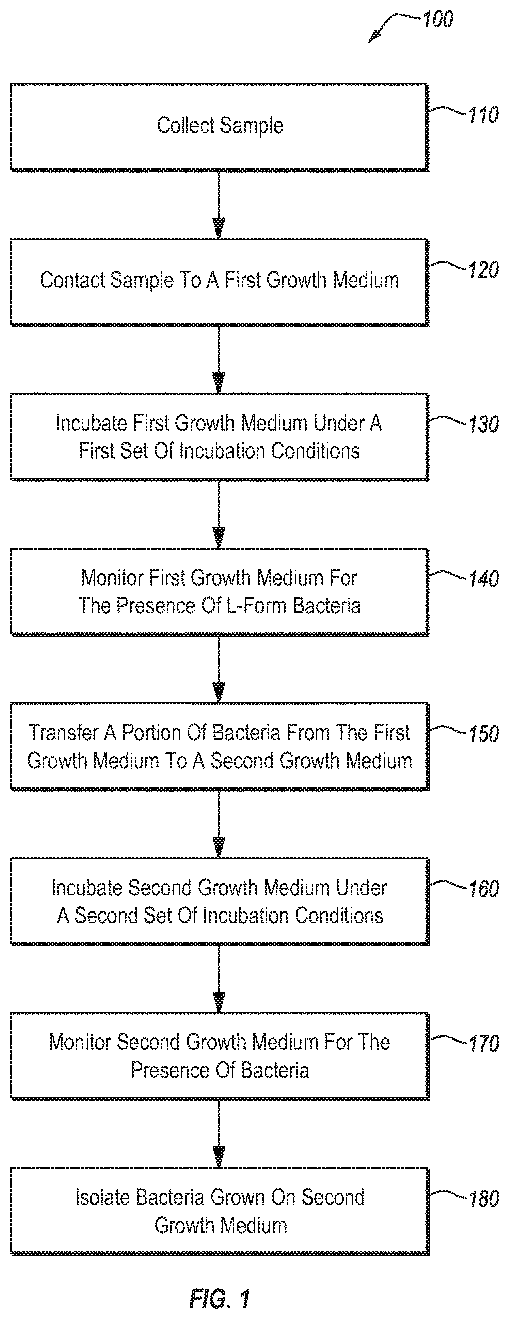
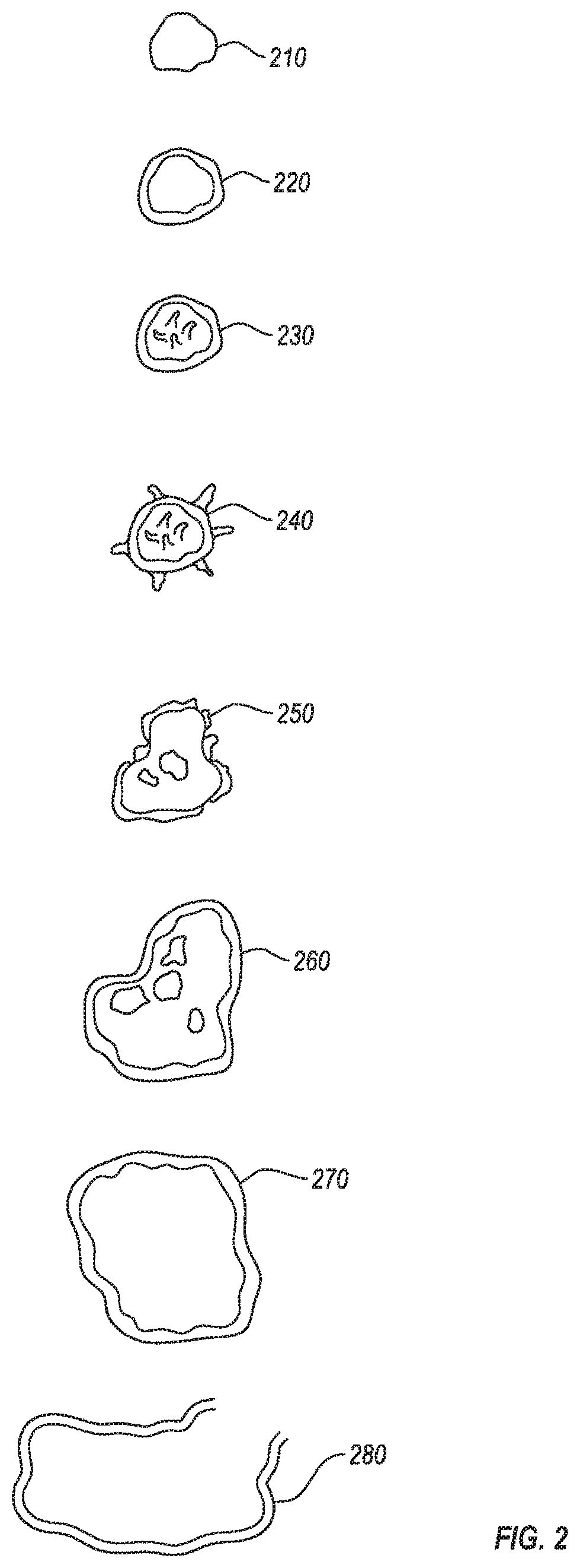
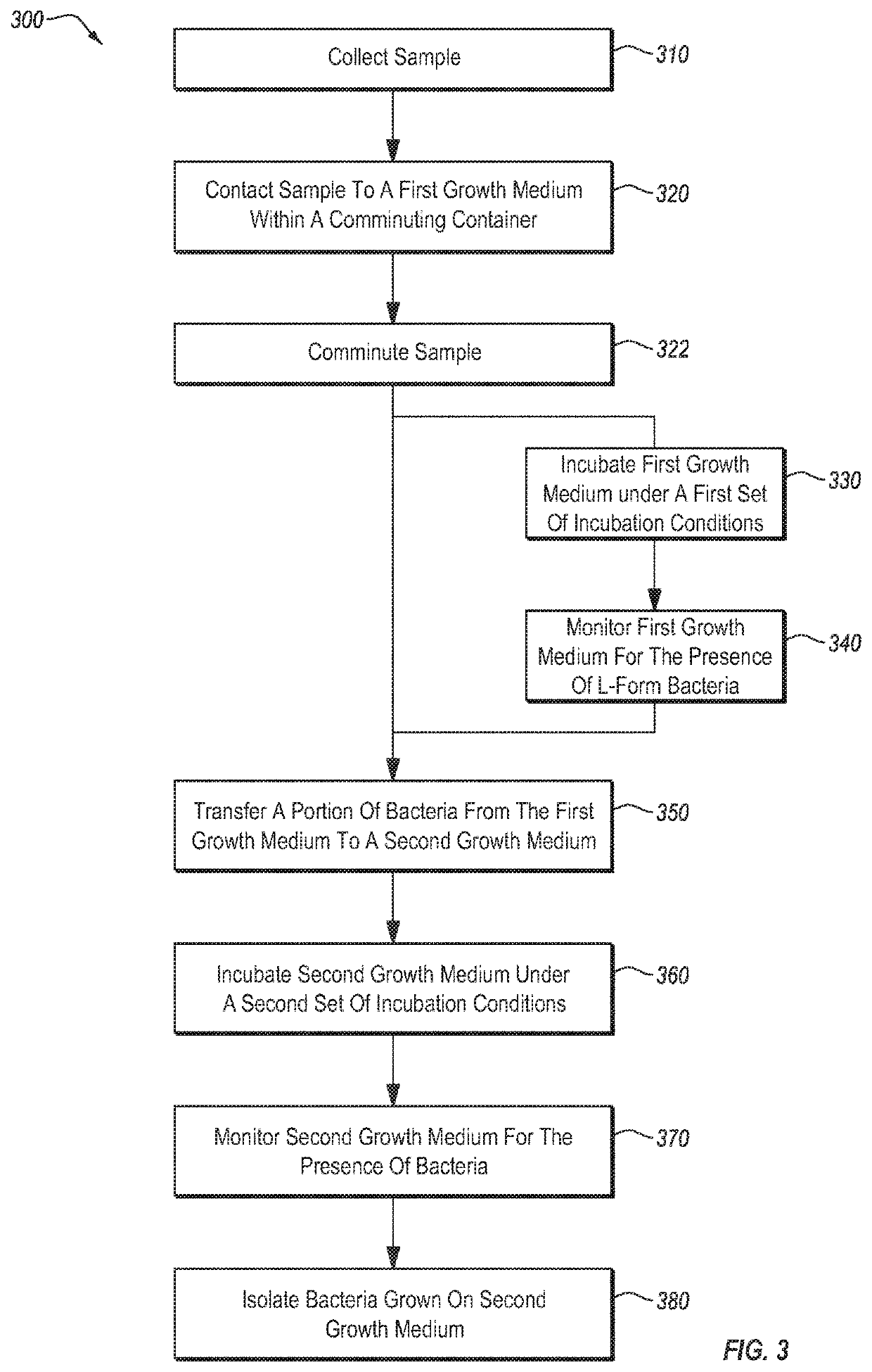
[0126]Over 500 separate blood samples were collected (about 120 of these were from individuals who gave more than one sample). More than 30 separate synovial fluid samples and 1 lymphatic fluid sample were also collected. For each sample, about 0.5 ml or less of the sample (about 2 drops) was added to a tube containing 10-15 ml of bovine serum and a tube containing 10-15 ml of BHI broth. The inoculated tubes were incubated at 27° C. Development of L-form culture was monitored by preparing wet mount live slides daily. Samples were monitored for a period of up to 30 days. Samples that showed indications of L-form bacterial growth were typically incubated for at least 48 hours, and typically began to show signs of progressive growth within 48-72 hours. L-form bacteria were not observed to progress to a CWS form while within the broth.

[0127]For samples in which L-form bacterial growth was detected, the broth was used to inoculate a variety of agarose plates (mannitol salt, BHI, tryptic ...

example 2

[0129]A comparative study was conducted to compare a standard culturing process to the process of Example 1. Each sample was divided into two portions. The first portion was used to directly inoculate two nutrient agars, which were then incubated and monitored for growth. The second portion was used as inoculant in the L-form growth protocol of Example 1. Results of the comparative study are shown in Table 1 (samples which showed no growth in either protocol are omitted).

TABLE 1Bacteria cultured Bacteria cultured Sample via directvia process ofTypeinoculationExample 1BloodNo growthAcintobacter genomospecies 15tuMicrococcus luteus AUnknown RodBloodBacillus pumilus / Bacillus pumilus / safensisStaph. capitis ss Bacillus pumilus / safensisBloodNo growthBacillus plakortidisBrachybacterium sacelii (26C)Unknown BacteriaBloodNo growthBacillus pumilus / safensisBloodNo growthBacillus pumilus / safensisBloodNo growthBacillus lichenformisBloodNo growthBacillus pumilus / safensisBloodNo growthBacillus pum...

example 3

[0131]Of the subjects tested using the culturing process described in Example 1, 10 subjects were found to harbor the Microbacterium maritypicum strain DSM 12512. Of the 10 subjects with this Microbacterium strain, 8 were female, and 7 of the 8 had indicated in a health questionnaire that they suffered from endometriosis or fibroid tumors of the ovaries.


PUM

PropertyMeasurementUnit
Fractionaaaaaaaaaa
Fractionaaaaaaaaaa
Fractionaaaaaaaaaa

Abstract

In some aspects, the disclosure relates to methods for screening clinical samples for the presence of L-form bacteria associated with endometriosis and/or ovarian fibroid tumors. The disclosure is based, in part, on screening methods used to diagnose a subject as likely having or likely not having endometriosis and/or an ovarian fibroid tumor. In some embodiments, the disclosure relates to methods of treating endometriosis by administering an antibiotic to a subject that has been determined to be infected by one or more strains of L-form bacteria.

Description

RELATED APPLICATIONS[0001]This application is a national stage filing under 35 U.S.C. § 371 of international PCT application, PCT / US2018 / 044885, filed Aug. 1, 2018, which claims priority under 35 U.S.C. § 119(e) to U.S. provisional patent application, U.S. Ser. No. 62 / 539,688, filed Aug. 1, 2017, the entire contents of each of which are incorporated herein by reference.BACKGROUND[0002]Endometriosis is a condition in which endometrium tissue abnormally grows at locations outside the uterus. Because such tissue growth often takes place on the ovaries, fallopian tubes, and surrounding tissues, the condition can cause infertility and chronic pelvic pain. Endometrial tissue will bleed as part of the menstrual cycle, and areas of endometriosis can become inflamed and scarred as a result. It is estimated that endometriosis affects 6 to 10% of women. Among women with endometriosis, about 40% will have infertility issues.[0003]Diagnosis of endometriosis can be challenging. Although a physici...

Claims


Application Information

Patent Timeline
IPC IPC(8): A61K31/65C12Q1/04A61K31/407A61K31/426A61K9/00A61K31/424A61P31/04
CPCA61K31/65A61K31/426C12Q1/04A61K31/424A61P31/04A61K9/0053A61K9/0019A61K31/407A61K45/06A61K31/43C12Q1/02A61P15/00G01N33/56911G01N33/57449G01N2800/364A61K2300/00
Inventor HUNT, JOHN BRENT
Owner SOFT CELL BIOLOGICAL RES LLC
Why Eureka
Try Eureka