Diagnostic Test for Hepatocellular Carcinoma

a hepatocellular carcinoma and diagnostic test technology, applied in the field of biomarkers and assays, can solve the problems of high cost, inability to detect tumors at an early stage, and insufficient diagnostic sensitivity and specificity of afp and dcp to detect h

Active Publication Date: 2020-10-08
GLYCOBOND
View PDF0 Cites 0 Cited by
  • Summary
  • Abstract
  • Description
  • Claims
  • Application Information

AI Technical Summary

Benefits of technology

[0018]The invention also relates to antibodies capable of specific binding to an epitope overlapping at least in part amino acid residues 183-189 of human AGP, and to IgG antibodies capable of specific binding to an epitope overlapping at least in part amino acid residues 183-201 of human AGP.

Problems solved by technology

However the diagnostic power of AFP to detect HCC is limited.
However, both AFP and DCP show insufficient diagnostic sensitivity and specificity to be used as screening markers and their use as diagnostic markers in surveillance is not recommended in international guidelines5.
Ultrasound has better diagnostic sensitivity and specificity (60-80%) but suffers from high costs and inability to detect tumors at an early stage6.
However studies using lectins such as the fucose-binding lectin from Aleuria aurantia (AAL), that has a broad specificity towards fucose, often shows problems in differentiating between patients with HCC and patients with liver cirrhosis14, 15.

Method used


Image

  • Diagnostic Test for Hepatocellular Carcinoma
  • Diagnostic Test for Hepatocellular Carcinoma
  • Diagnostic Test for Hepatocellular Carcinoma

Examples


example 1

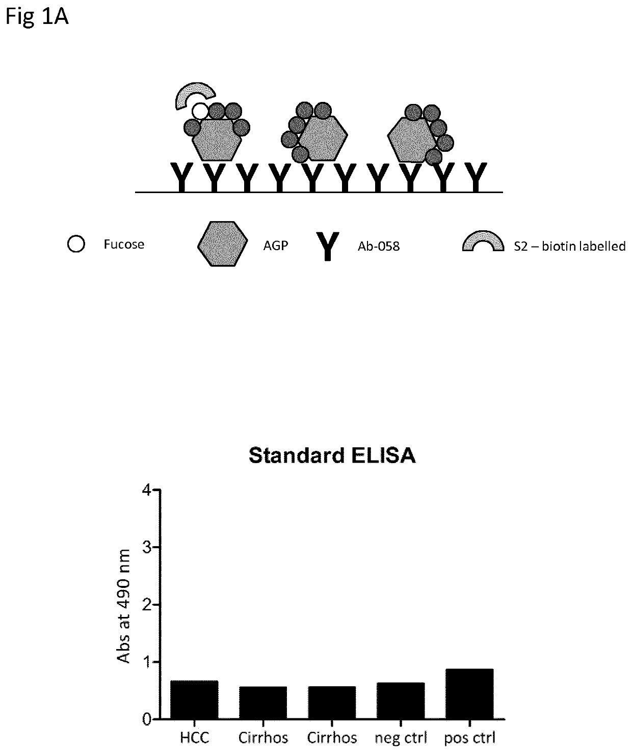
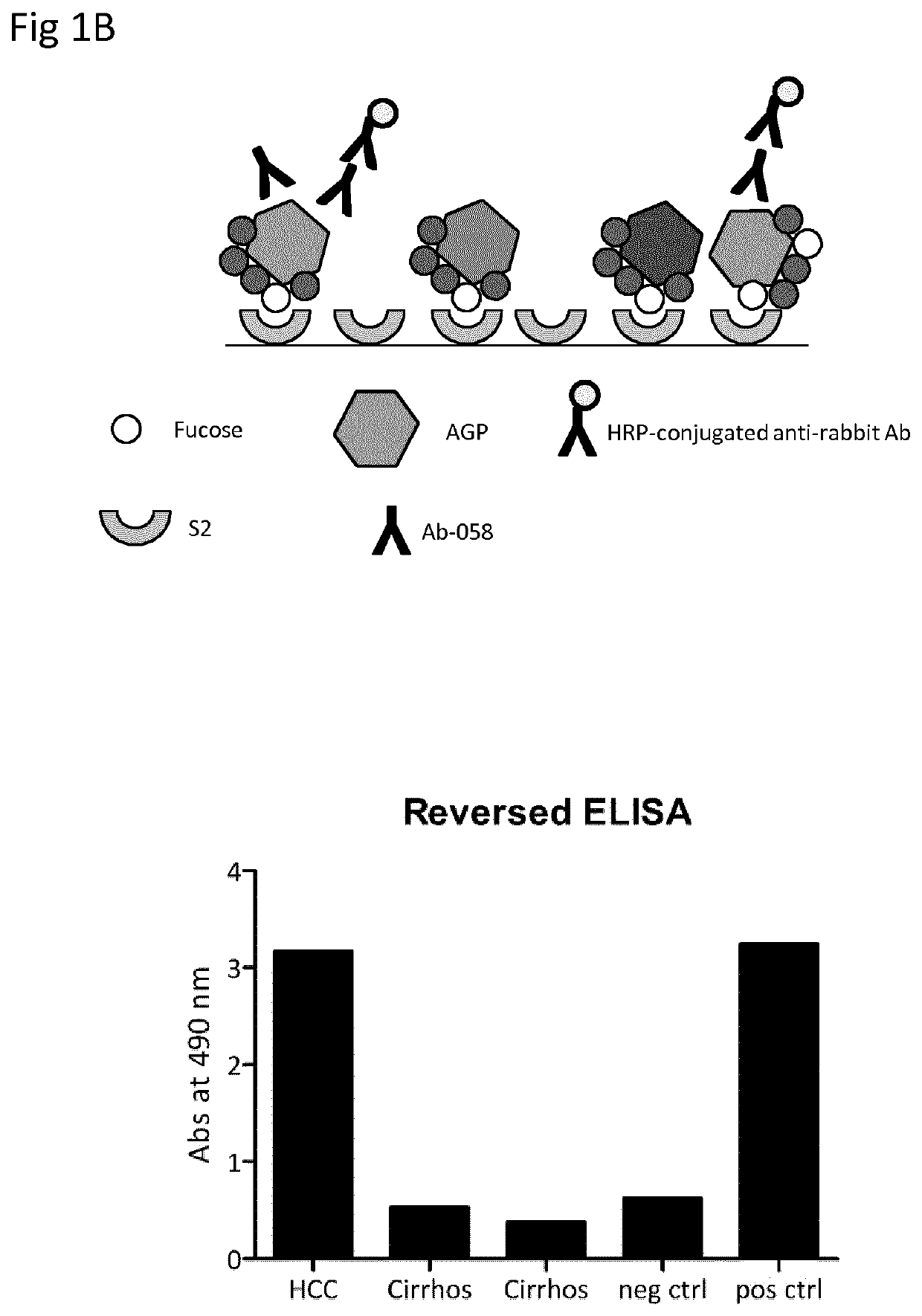
, Comparison of Standard ELISA and Reversed ELISA

[0084]Patient Samples

[0085]Plasma samples from one patient with HCC och two patients with cirrhosis were obtained from Karolinska University Hospital at Huddinge, Sweden. Normal control was a plasma pool from healthy blood donors. Positive control was dsAGP at a concentration of 2 μg / ml.

[0086]Production of Antibodies

[0087]Production of the polyclonal anti-human α1-acid glycoprotein antibody 058 was done by Agrisera antibodies (Vännäs, Sweden). Shortly, a synthetic peptide corresponding to the C-terminal amino acids 183 to 201 of human α1-acid glycoprotein was synthesized by Agrisera. The peptide was conjugated to KLH via its terminal cysteine using a maleimide crosslinker and rabbits were immunized four times with the KLH-conjugated peptide. Anti-AGP antibodies were purified from sera using the synthetic peptide coupled to 2 mL of UltraLink Iodoacteyl resin (Pierce, Rockford, Ill., USA).

[0088]Standard Lectin ELISA

[0089]Microtiter plat...
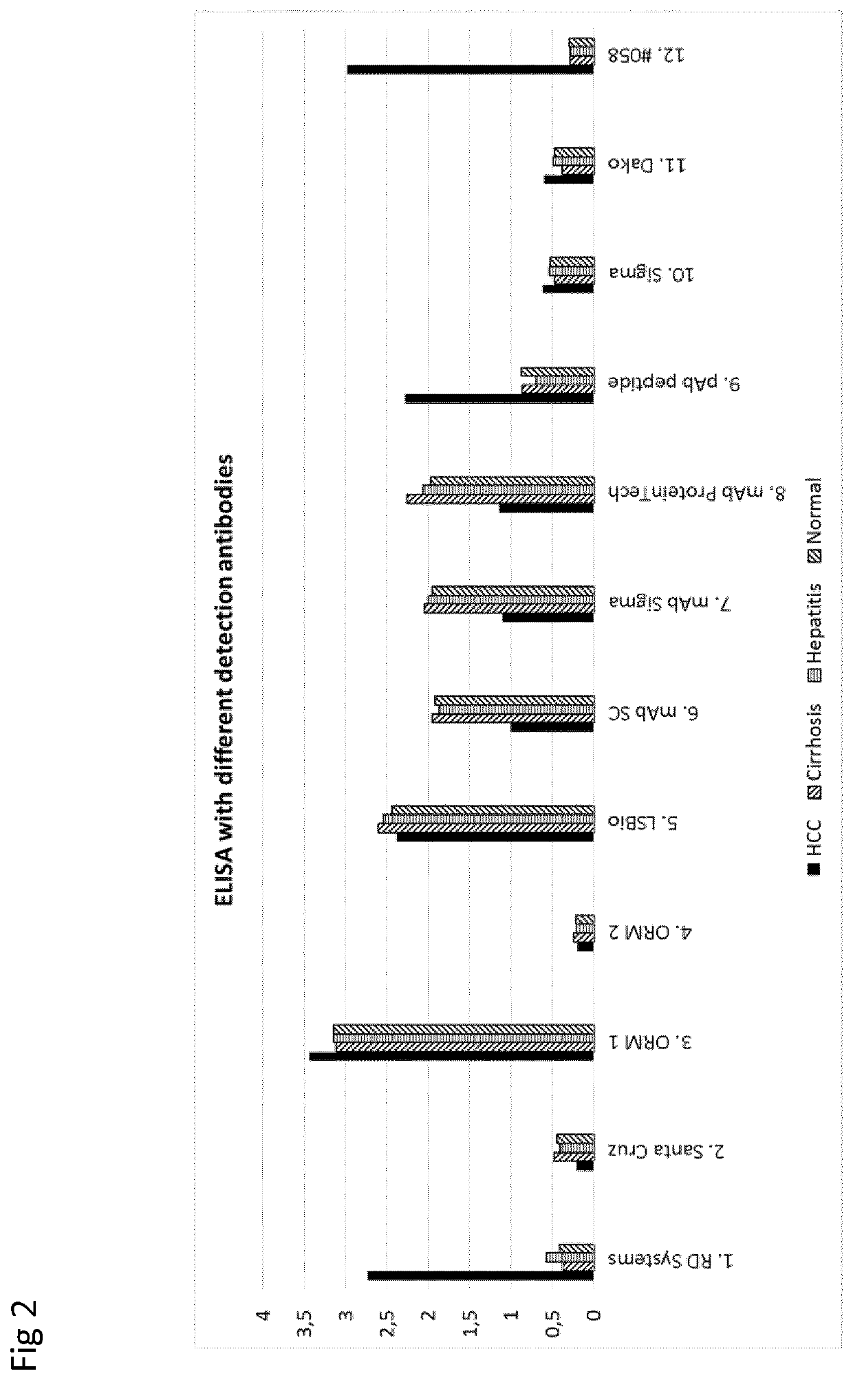
example 2

, Comparison of Detection Antibodies

[0094]Patient Samples

[0095]The analyses were performed on plasma samples from one patient with HCC, one patient with cirrhosis, one patient with hepatitis and a plasma pool from healthy blood donors (Normal).

[0096]Assay

[0097]The reversed S2-ELISA (as exemplified above) was used to compare commercial anti-AGP antibodies with Ab-058. The 058 antibody (sample 12 in FIG. 2) was compared to a number of commercial polyclonal or monoclonal anti-AGP antibody according to the list below.

[0098]Detection Antibodies

[0099]The following antibodies were used for detection.[0100]1. Human α1-acid Glycoprotein antibody (R&D Systems, cat no AF3694)[0101]Source: Polyclonal Goat IgG[0102]Immunogen: Human plasma derived α1-acid glycoprotein[0103]2. Human α1-acid Glycoprotein antibody (AGP-1 / 2, Santa Cruz Biotechnology, cat no sc-51018)[0104]Source: Polyclonal Goat IgG[0105]Immunogen: peptide mapping within an internal region of AGP-1 of human origin[0106]3. ORM 1 Polyc...

example 3

, Validation of Reversed S2-ELISA Using Purified AGP Samples

[0139]Purification of AGP

[0140]AGP was isolated from plasma samples using a two-step ion exchange chromatography method according to Asao et al.20. One mL plasma samples were applied to a HiTrap Desalting column (GE Healthcare, Uppsala, Sweden) equilibrated with 20 mM citrate-phosphate buffer, pH 4. The desalted peak was applied to a HiTrap DEAE column (GE Healthcare) equilibrated with 20 mM citrate-phosphate buffer, pH 4. Fractions containing AGP were eluted with 20 mM citrate-phosphate buffer, pH 7, containing 200 mM NaCl, pooled and applied on two joined HiTrap Desalting columns equilibrated with 20 mM citrate-phosphate buffer, pH 4. The desalted peak was applied to a HiTrap SP column (GE Healthcare) equilibrated with 20 mM citrate-phosphate buffer, pH 4, and AGP was eluted with 20 mM citrate-phosphate buffer, pH 4.8. The eluted fractions were dialyzed against water and lyophilized.

[0141]Production of Polyclonal Anti-Hum...


PUM

PropertyMeasurementUnit
pHaaaaaaaaaa
concentrationaaaaaaaaaa
pHaaaaaaaaaa

Abstract

The present invention relates to a method for detecting fucosylated alpha1-acid glycoprotein (AGP) in a sample, comprising the steps providing a monovalent fucose-binding peptide having at least 80% identity, such as 85, 90, 95, 99 or 100% identity, to a peptide having an amino acid according to SEQ ID NO: 1, immobilised on a solid phase; bringing the sample into contact with the immobilised fucose-binding peptide; and detecting any fucosylated AGP bound to said fucose-binding peptide. The invention further relates to a method for assessing a risk that a human individual suffers from hepatocellular carcinoma, and to a kit of parts and antibodies useful in the methods according to the invention, and to a peptide useful as an immunizing antigen in production of such antibodies.

Description

FIELD OF THE INVENTION[0001]The present invention relates to the field of biomarkers and assays for use in detection of biomarkers, and to antibodies useful in such assays. In particular, the invention relates to assays for detecting biomarkers indicative of primary liver cancer (hepatocellular carcinoma, HCC).BACKGROUND[0002]Primary liver cancer (hepatocellular carcinoma, HCC) is one of the most prevalent human cancers and the third deadliest. Most cases of HCC develop on the background of liver cirrhosis and the major etiology for developing cirrhosis and HCC is chronic hepatitis B virus (HBV) or hepatitis C virus (HCV) infection. HBV and HCV infections are associated with >80% of all HCC cases worldwide1-3. Non-viral background for development of HCC includes liver cirrhosis caused by alcoholic disease, diabetes and metabolic and autoimmune diseases.[0003]If HCC tumors are detected at an early stage, curative treatment using surgical resection, transplantation or radio-frequen...

Claims


Application Information

Patent Timeline
Patent Type & Authority Applications(United States)
IPC IPC(8): G01N33/574G01N33/543
CPCG01N33/57488G01N2333/42G01N33/54306G01N2400/02G01N33/57438G01N33/68
Inventor PÅHLSSON, PETER
Owner GLYCOBOND
Why Eureka
Try Eureka