
Three-dimensional medical image analysis method and system for identification of vertebral fractures

a three-dimensional medical image and imaging technology, applied in image enhancement, tomography, instruments, etc., can solve the problems of difficult clinical assessment of vertebral fractures, low screening and early diagnosis of patients with vertebral fractures by bone care givers in clinical centers

Active Publication Date: 2020-11-19
UCB PHARMA SRL
View PDF0 Cites 11 Cited by
  • Summary
  • Abstract
  • Description
  • Claims
  • Application Information

AI Technical Summary

Benefits of technology

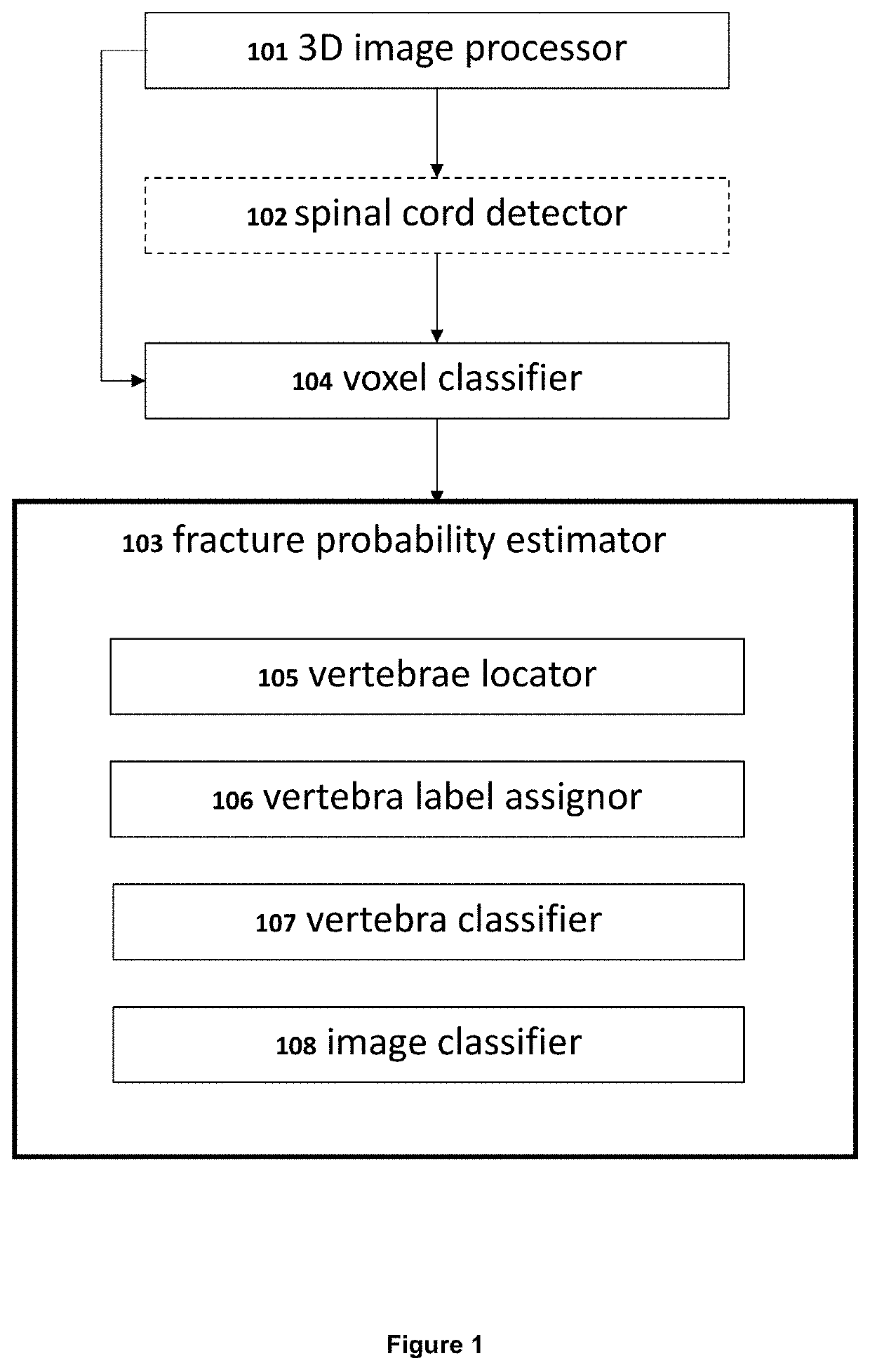
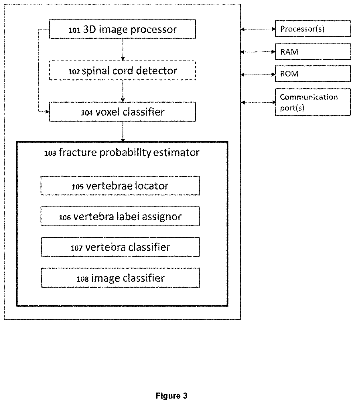
The present patent is about a system and method for analyzing 3D images of a person's spinal cord to predict if they have a vertebral fracture. The system includes a processor that receives and processes image data, creating sets of voxels that contain equal amounts of data at different dimensions. The system then uses a computational model to assign each voxel a probability of containing a fracture. The method involves identifying specific voxels that are classified as containing a fracture and predicting the presence of a vertebral fracture based on this identification. Overall, this system and method can help healthcare professionals quickly and accurately detect spinal fractures, potentially improving patient outcomes.

Problems solved by technology

Clinical assessment of vertebral fractures is difficult because many patients are unaware that they have suffered a vertebral fracture.
Despite efforts from the International Osteoporosis Foundation to raise awareness of vertebral fractures and provide training on vertebra fracture detection, bone care givers in clinical centers have little means for screening and early-stage diagnosis of patients with vertebral fractures.
As clinical imaging data volumes keep growing steadily, developing a three-dimensional image processing system and a method is a technical problem of great clinical importance to reduce inter-observer variability and to allow for screening for vertebral fractures.

Method used


Image

  • Three-dimensional medical image analysis method and system for identification of vertebral fractures
  • Three-dimensional medical image analysis method and system for identification of vertebral fractures
  • Three-dimensional medical image analysis method and system for identification of vertebral fractures

Examples


example 1

ce of the System for Identification of Vertebral Fractures on a Set of CT Images

[0123]120 anonymized image series of abdomen and thorax CT examinations from the imaging database of one university hospital were used for the method described herein. These images were acquired in February and March 2017 on four different scanners (Siemens, Philips and two types of GE scanners) including patients with various indications above seventy years of age (average age was 81 years, range: 70-101, 64% female patients). As a result, this dataset contains a heterogeneous range of protocols, reconstruction formats and patients representing a sample from clinical practice for this patient cohort. The vertebrae distribution contains a total of 1219 vertebrae of which 228 (18.7%) are fractured. The dataset has been curated by one radiologist (S.R.) providing Genant grades (normal, mild, moderate, severe) for every vertebra in the field-of-view.

[0124]Table 1 summarizes that the patient-level prediction...


PUM


Abstract

A machine-based learning method estimates a probability of bone fractures in a 3D image, more specifically vertebral fractures. The method and system utilizing such method utilize a data-driven computational model to learn 3D image features for classifying vertebra fractures. A three-dimensional medical image analysis system for predicting a presence of a vertebral fracture in a subject includes a 3D image processor for receiving and processing 3D image data of a 3D image of the subject, producing two or more sets of 3D voxels. Each of the sets of 3D voxels corresponds to an entirety of the 3D image and each of the sets of 3D voxels consists of equal 3D voxels of different dimensions. The system also includes a voxel classifier for assigning the 3D voxels one or more class probabilities each of the 3D voxels contains a fracture using a computational model, and a fracture probability estimator for estimating a probability of the presence of a vertebral fracture in the subject.

Description

[0001]The present disclosure relates to medical image analysis and provides a system and a method for identification of bone fractures in 3D images, more specifically vertebral fractures.BACKGROUND[0002]Osteoporosis is a disease affecting bones where increased bone weakness increases the risk of bone fractures. Common osteoporotic fractures occur in the vertebrae in the spine, the bones of the forearm, and the hip. Every 3 seconds an osteoporotic fracture occurs, with vertebral fractures being the most common (Johnell et al, 2006). Vertebral fractures are predictive of subsequent fractures, e.g. patients with moderate or severe vertebral fractures have 4.8 to 6.7 times higher risk of subsequent hip fracture (Buckens et al., 2014). Roux et al demonstrated that 1 out of 4 patients with incident mild vertebral fractures will most probably have a fracture again within subsequent 2 years (Roux et al., 2007).[0003]Clinical assessment of vertebral fractures is difficult because many patien...

Claims


Application Information

Patent Timeline
Patent Type & Authority Applications(United States)
IPC IPC(8): G06T7/00G06T7/11G06T15/08G06K9/62
CPCG06T2207/30012G06T2207/20084G06T7/0012G06T15/08G06T7/11G06T2207/20081G06K9/6277A61B6/032G16H50/30A61B6/035A61B6/5217G16H30/40G16H50/50A61B6/505G06T2207/10081G16H50/00A61B2576/00G16H50/20G16H50/70G06F18/2415
Inventor NICOLAES, JOERI
Owner UCB PHARMA SRL
Learn More