Automated cardiac function assessment by echocardiography

an automatic assessment and cardiac function technology, applied in the field of automatic cardiac function assessment by echocardiography, can solve the problem of prohibitive cost of imaging each individual with cardiac risk factors over time, and achieve the effect of high accuracy

Pending Publication Date: 2021-01-07
RGT UNIV OF CALIFORNIA
View PDF1 Cites 9 Cited by
  • Summary
  • Abstract
  • Description
  • Claims
  • Application Information

AI Technical Summary

Benefits of technology

[0006]Embodiments can allow high accuracy (>98%) of image identification using the CNN. This lays the groundwork for using automated interpretation to support the use of handheld cardiac ultrasound in primary care practices and may enable large-scale analysis of collected echocardiograms already archived within the healthcare system. Example embodiments comprise a fully automated cardiac echocardiogram interpretation system that can be deployed on the web.

Problems solved by technology

However, the cost of imaging each individual with cardiac risk factors over time is currently prohibitive to incorporating this approach into the standard cardiovascular health practice.

Method used


Image

  • Automated cardiac function assessment by echocardiography
  • Automated cardiac function assessment by echocardiography
  • Automated cardiac function assessment by echocardiography

Examples


example improvements

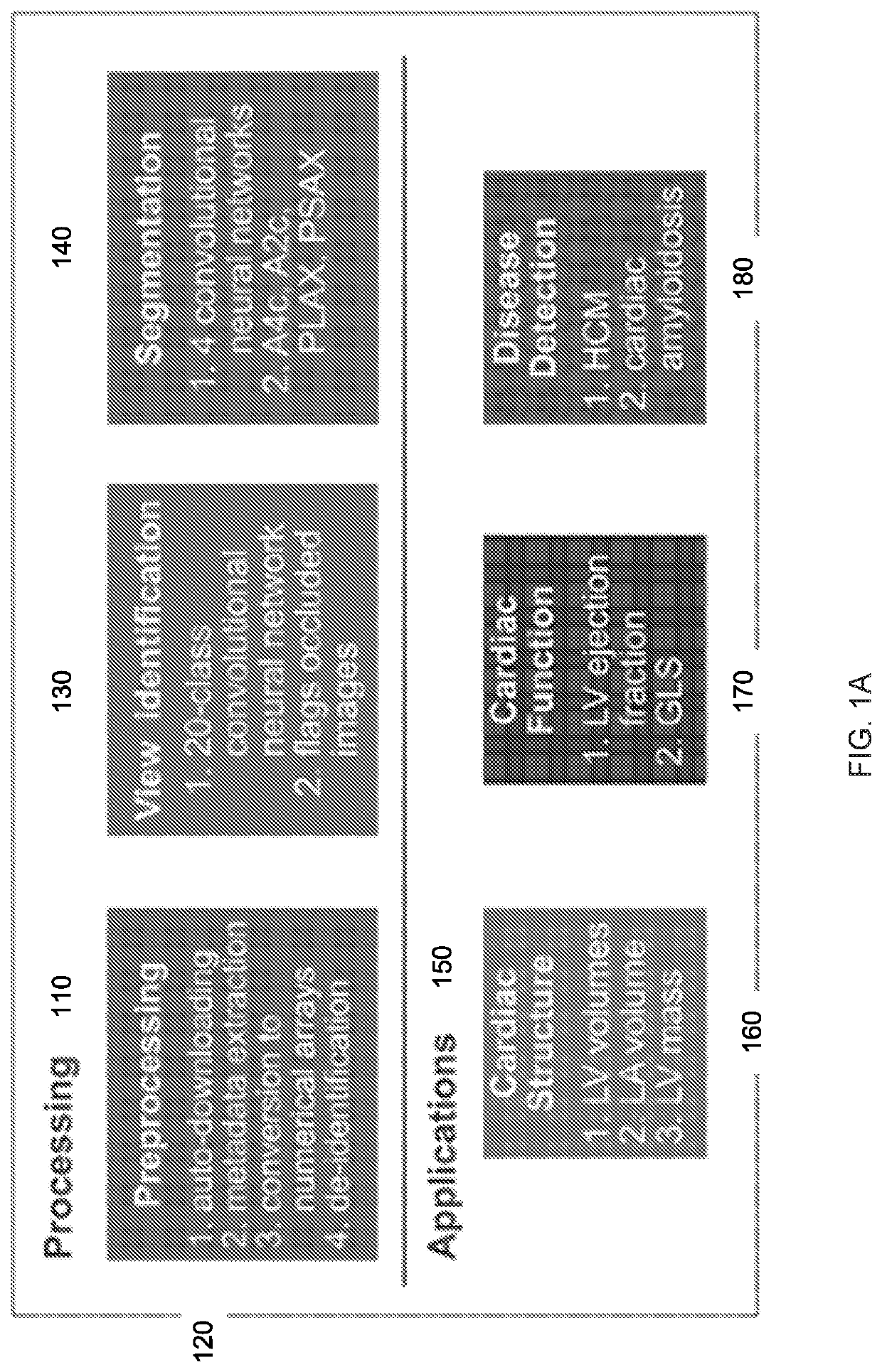
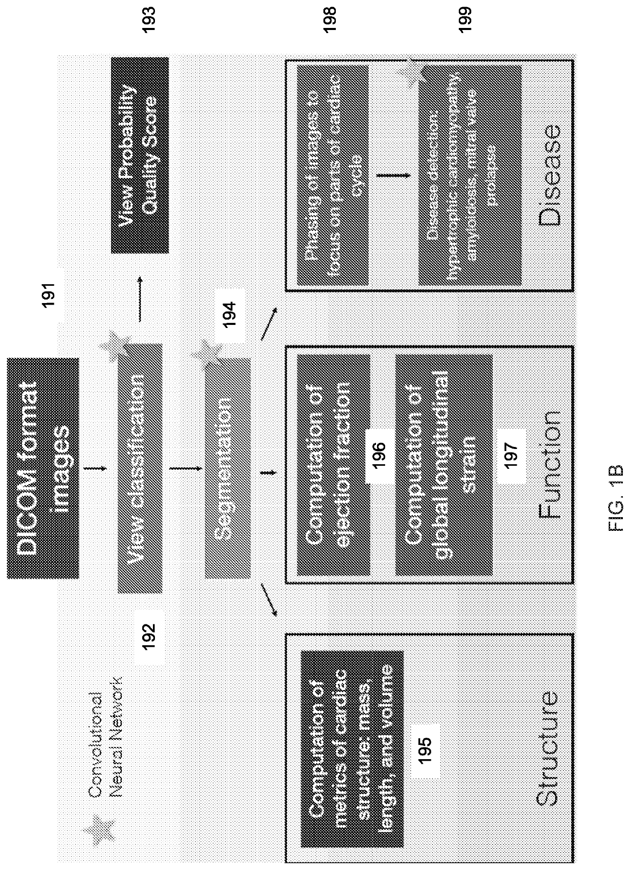
[0101 provided by this work are the application of CNNs to segment echo images, the development of an empirically validated automated quality score (e.g., VPQS) for studies, the automation of common 2D measurements, the validation of automated values against measurements from thousands of studies, and the creation of a complete pipeline that can be deployed on the web. More training data may be used for improved performance, although it is remarkable to note how few images (<200) were used to train each of our segmentation models.

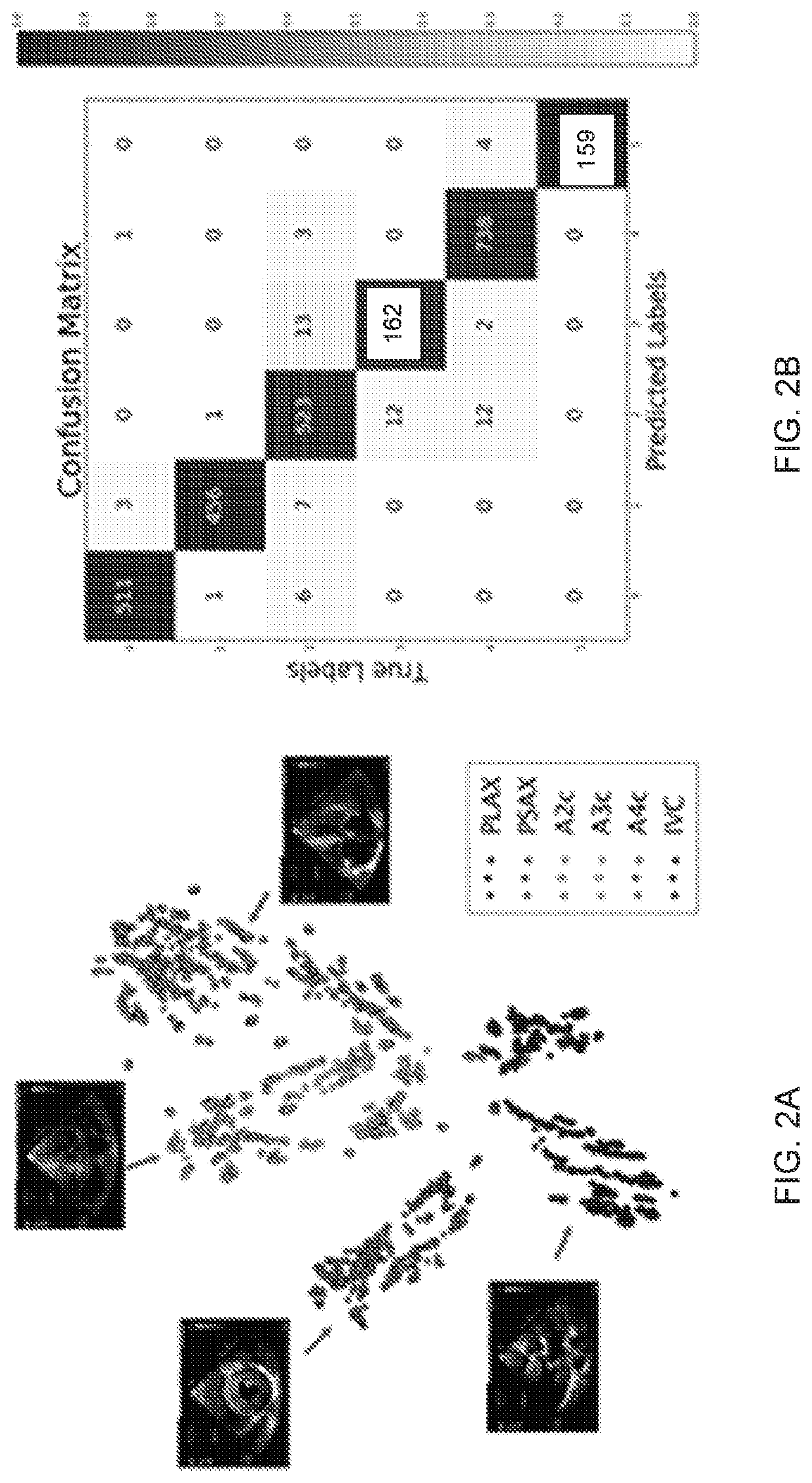
[0102]Embodiments can benefit from averaging across multiple measurements, and we demonstrate the utility of multiple studies in improving concordance between manual and automated measurements. Our results also show benefits from building more redundancy into their acquisition of echo images for using an automated computer vision pipeline for study interpretation. In particular, there is typically only 1 PSAX video available to compute left ventricular mass...


PUM


Abstract

A computer vision pipeline is provided for fully automated interpretation of cardiac function, using a combination of machine learning strategies to enable building a scalable analysis pipeline for echocardiogram interpretation. Videos from patients with heart failure can be analyzed and processed as follows: 1) preprocessing of echo studies; 2) convolutional neural networks (CNN) processing for view identification; 3) segmentation of chambers and delineation of cardiac boundaries using CNNs; 4); particle tracking to compute longitudinal strain; and 5) targeted disease detection.

Description

CROSS-REFERENCES TO RELATED APPLICATIONS[0001]The present application claims priority from and is a PCT application of U.S. Provisional Application No. 62 / 642,930, entitled “Automated Cardiac Function Assessment By Echocardiography” filed Mar. 14, 2018, the entire contents of which are herein incorporated by reference for all purposes.STATEMENT AS TO RIGHTS TO INVENTIONS MADE UNDER FEDERALLY SPONSORED RESEARCH AND DEVELOPMENT[0002]This invention was made with government support under grant no. HL123228 awarded by the National Institutes of Health. The government has certain rights in the invention.BACKGROUND[0003]Cardiac remodeling preceding cardiovascular disease often starts long before irreversible pathologic changes occur. Echocardiography is widely available, non-radiation emitting and easy to perform at the bedside, making this method the most widely used noninvasive imaging technique in cardiology. Such evidence of early remodeling can relatively easily be detected by echocar...

Claims


Application Information

Patent Timeline
Patent Type & Authority Applications(United States)
IPC IPC(8): A61B8/08A61B8/06A61B8/00G06T7/00G16H30/40G16H50/20G06N3/08
CPCA61B8/5223A61B8/065A61B8/485A61B8/461G06T7/0014G06T2207/20081G16H50/20G06N3/08G06T2207/20076G06T2207/30048G06T2207/20084G16H30/40A61B5/7267G16H50/70
Inventor DEO, RAHUL C.ZHANG, JEFFREY
Owner RGT UNIV OF CALIFORNIA
Why Eureka
Try Eureka