Tissue nodule detection and tissue nodule detection model training method, apparatus, device, and system

a tissue nodule and detection model technology, applied in the field of medical image processing technologies, can solve the problems of large workload on doctors, poor accuracy of conventional tissue nodule detection methods, and incongruity of medical imaging devices of hospitals, so as to improve detection accuracy and reduce the difference between source domain data sampling features and target domain data extracted through tissue nodule detection models.

Pending Publication Date: 2021-02-25
TENCENT TECH (SHENZHEN) CO LTD
View PDF4 Cites 14 Cited by
  • Summary
  • Abstract
  • Description
  • Claims
  • Application Information

AI Technical Summary

Benefits of technology

[0069]the tissue nodule detection model training apparatus including: a training data obtaining module, configured to obtain source domain data and the target domain data acquired by the image acquisition module, the source domain data including a source domain image and an image annotation, and the target domain data including a target image, the image annotation being used for indicating location information of a tissue nodule in the source domain image; a feature extraction and training module, configured to perform feature extraction on the source domain image through a neural network model to obtain a source domain sampling feature, perform feature extraction on the target image through the neural network model to obtain a target sampling feature, and determine a training result according to the source domain sampling feature; a distance parameter determining module, configured to determine a distance parameter between the source domain data and the target domain data according to the source domain sampling feature and the target sampling feature, the distance parameter being a parameter describing a magnitude of a data difference between the source domain data and the

Problems solved by technology

Currently, whether there is a tissue nodule may be determined with the help of a medical image, such as a chest thin-section CT image, which causes a lot of workload on doctors.
However, medical imaging devices of hospitals are not uniform as they may be of different brands and models, and sampling distances, noise levels

Method used


Image

  • Tissue nodule detection and tissue nodule detection model training method, apparatus, device, and system
  • Tissue nodule detection and tissue nodule detection model training method, apparatus, device, and system
  • Tissue nodule detection and tissue nodule detection model training method, apparatus, device, and system

Examples


Embodiment Construction

[0080]To make the objectives, technical solutions, and advantages of this application clearer and more comprehensible, this application is further described in detail with reference to the accompanying drawings and embodiments. It is to be understood that the specific embodiments described herein are only used for explaining this application, and are not used for limiting this application.

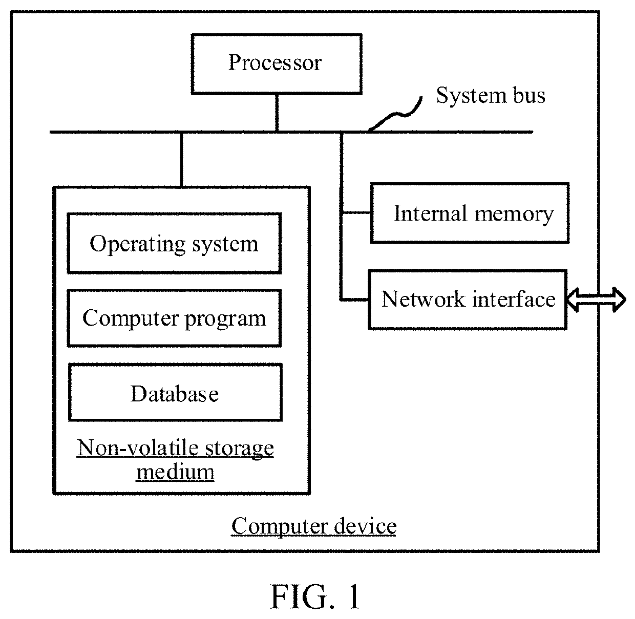
[0081]FIG. 1 is a diagram of an exemplary application environment of a tissue nodule detection method and / or a tissue nodule detection model training method according to an embodiment. The tissue nodule detection method and / or the tissue nodule detection model training method may be applied to a computer-assisted cancer diagnostic system. As shown in FIG. 1, the tissue nodule detection method and / or the tissue nodule detection model training method is applied to a computer device. The computer device may be a terminal or a server. The terminal may be a desktop device or a mobile terminal. The serve...


PUM


Abstract

This application relates to a tissue nodule detection and tissue nodule detection model training method, apparatus, device, storage medium and system. The method for training a tissue nodule detection model includes: obtaining source domain data and target domain data, the source domain data comprising a source domain image and an image annotation, the target domain data comprising a target image, and the image annotation being used for indicating location information of a tissue nodule in the source domain image; performing feature extraction on the source domain image using a neural network model to obtain a source domain sampling feature, performing feature extraction on the target image using the neural network model to obtain a target sampling feature, and determining a model result according to the source domain sampling feature using the neural network model; determining a distance parameter between the source domain data and the target domain data according to the source domain sampling feature and the target sampling feature, the distance parameter being a parameter describing a magnitude of a data difference between the source domain data and the target domain data; determining, according to the model result and the image annotation, a loss function value corresponding to the source domain image; and training the neural network model to obtain a tissue nodule detection model by iteratively reducing a combination of the loss function value and the distance parameter. In this way, the detection accuracy can be improved.

Description

RELATED APPLICATION[0001]This application is a continuation application of the International PCT Application No. PCT / CN2019 / 115822, filed with the China National Intellectual Property Administration, PRC on Nov. 5, 2019 which claims priority to Chinese Patent Application No. 201811326267.4, entitled “TISSUE NODULE DETECTION METHOD AND APPARATUS, TISSUE NODULE DETECTION MODEL TRAINING METHOD AND APPARATUS, DEVICE, AND SYSTEM” and filed with the China National Intellectual Property Administration, PRC on Nov. 8, 2018, which are incorporated herein by reference in their entireties.FIELD OF THE TECHNOLOGY[0002]This application relates to the field of medical image processing technologies, and in particular, to a method, an apparatus, and a computer device for training a tissue nodule detection model, and a method, an apparatus, a computer device, and a system for tissue nodule detection.BACKGROUND OF THE DISCLOSURE[0003]Canceration of tissues is a leading cause of death, and therefore e...

Claims


Application Information

Patent Timeline
IPC IPC(8): G06T7/00G06K9/62G06V10/764
CPCG06T7/0012G06K9/6232G06T2207/30024G06T2207/20084G06T2207/20081G06K9/6215G06T2207/30096G06T2207/10081G06V10/462G06N3/045G06T2207/30064G06N20/10G06N3/08G16H50/20G16H30/40G06T7/11G06T2207/10088G06V20/695G06V20/698G06V10/82G06V10/764G06V10/7753G06F18/22G06F18/213
Inventor CHENG, CHENSUN, ZHONGQIANCHEN, ZHAOYANG, WEI
Owner TENCENT TECH (SHENZHEN) CO LTD
Why Eureka
Try Eureka