In vivo tissue engineering devices, methods and regenerative and cellular medicine employing scaffolds made of absorbable material

a tissue engineering and scaffold technology, applied in tissue regeneration, medical science, prosthesis, etc., can solve the problems of insufficient stent insertion, insufficient stent insertion, tissue necrosis and capsular contracture, etc., to prevent pooling of fat, promote vascularization, and improve vascularization

Active Publication Date: 2021-08-19
BARD SHANNON LTD
View PDF6 Cites 8 Cited by
  • Summary
  • Abstract
  • Description
  • Claims
  • Application Information

AI Technical Summary

Benefits of technology

[0009]The present invention overcomes the disadvantages of the prior art by configuring a scaffold made of porous absorbable material to provide physical support for a large volume of tissue, including autologous fat, in a plurality of tissue engineering chambers arranged around a core which can be hollow to accommodate blood vessels. In one embodiment, the tissue engineering device of the present invention employs a scaffold made of a mesh absorbable material formed of a mono-filament polymer knitted mesh which provides a biologic scaffold matrix for cell attachment and protein organization. The matrix is invaded by capillaries, in a granulation type process that transforms the mesh into engineered fascia. The absorbable mesh material structure has a plurality of partially open tissue engineering chambers. The chambers can be formed by folds in one or more sheets of absorbable material and arranged radially around a central core. The chambers can be segmented and / sub-segmented to increase the surface area of the scaffold. The tissue engineering device of the present invention has a shape to be implanted in an anatomical space in fascia of the human body, and the tissue engineering chambers have an aggregate surface area greater than the fascia surface area in the anatomical space. The anatomical space can be a defect created during surgery in superficial fascia, for example in the breast, or in deep fascia adjacent an organ, for example the pancreas, kidneys or liver, when the tissue engineering device is employed for recellularization with functional parenchymal cells. With respect to the pancreas, the tissue engineering device of the present invention can be used to replace insulin-producing islet beta cells destroyed in some types of diabetes. With respect to the liver, the tissue engineering device of the present invention can be used to produce a plentiful source of hepatocytes for regenerating liver tissue and treating metabolic diseases.
[0012]The tissue engineering devices and methods of the present invention employ a scaffold made of absorbable material. In one embodiment, the present invention employs an open-pore knit pattern of absorbable material to encourage rapid tissue ingrowth through the micro-pores of the scaffold.
[0013]The scaffold employed in the tissue engineering device of the present invention permits lipoaspirate fatty tissue injected in the tissue engineering chambers to be mixed with the absorbable material thus holding the micro-globules of liposuctioned fat in place in a three-dimensional, scattered fashion to promote vascularization and prevent pooling of the fat which otherwise could lead to necrosis. Additionally, the tissue engineering chambers can be, prior to insertion in the body, coated with substances known to encourage tissue regeneration and then coated with selected tissue cells such as pancreatic islet cells, hepatic cells, or other cells as well as with stem cells and exosomes genetically altered to contain genes for treatment of patient illnesses. The radially arranged tissue engineering chambers provide good neovascularization with mononuclear inflammatory cells and multi-nucleic giant cells as well as adipogenesis on the absorbable material surfaces, in the absorbable material and between the layers of folded absorbable material.
[0014]The number of tissue engineering chambers can vary but are normally somewhere between eight and ten thus dividing the greater overall volume of the space for tissue expansion into smaller, subunit spaces. The joining together of the tissue engineering chambers around a central core adds to the stability of the scaffold while minimizing the required mesh absorbable material.
[0021]Accordingly, it can be seen that the tissue engineering device of the present invention presents a building block for in vivo organ development (engineering) or supplementation in that the tissue engineering device provides mesh to engineer a supportive fascia which brings vascularity to a large three-dimensional space and stromal cells that are the supportive filler and functional cells of a specific organ in the parenchyma.
[0022]Along with the above noted advantages of the present invention in tissue engineering, the present invention has the additional advantages of being used as, essentially, a bio-hybrid organ for hormone replacement therapy to obviate the need for hormone pellets. That is, instead of implanting slowly dissolving pellets to release a set amount of estrogen or testosterone, the tissue engineering device of the present invention can be used to colonize cells that are a part of human body circulation to release the hormones naturally which is particularly advantageous in patients whose increased age has reduced their hormone levels. Similarly, the tissue engineering device of the present invention can be implanted in individuals with hypothyroidism to be the locus for a colony of transplanted cells that produce thyroid hormone. When the tissue engineering device of the present invention is used as a bio-hybrid organ, endocrine cells are generated with the individual's hypothalamus and pituitary glands controlling the amount of hormone production in accordance with their normal function. A pancreatic bio-hybrid tissue engineering device according to the present invention can also allow an islet cell colony to evade attack by the immune system in individuals with Type 1 diabetes. CRISPR technology can be used to take autologous islet cells and disable their NLRC5 gene, and the islet cells can then be implanted in the tissue engineering device allowing the device to function as a bio-hybrid endocrine organ without being attacked by the immune system. Tissue engineering devices of the present invention implanted in the breast do not present the complications of capsular contracture and infection frequently associated with silicone implants used for breast reconstruction in that adipogenesis is enhanced by capillaries from the superficial fascia growing into the open, porous surfaces of the scaffold which have received the small globules of aspirated fat. Thus, healthy tissue with good vascularity is created to avoid infection. When the tissue engineering device of the present invention is implanted following lumpectomy, the spherical-type shape fills the defect and promotes gradual healing of the lumpectomy space without scar contracture while the scaffold acts as a stent preventing wound contracture and scarring and promoting M2 regenerative healing of the lumpectomy defect. The round surface of the spherical-like scaffold minimizes damage to delicate tissue, prevents extrusion and, more completely, approximates the lumpectomy defect than prior art devices.

Problems solved by technology

These prior art breast implants have many disadvantages and frequently cause tissue necrosis and capsular contracture.
The result is that the device does not fill the surgical defect in many instances and, thus, does not achieve a consistently high quality aesthetic appearance which is the outcome desired following lumpectomy.
Additionally, the device does not adequately stent the defect in smaller breasted, thin women and in such cases does not promote gradual healing of the lumpectomy space without scar contracture.
The prior art attempts to improve fat survival have not been successful from a practical standpoint and have had many disadvantages, primarily the need to remove the structures after the surgical procedure.
Unfortunately, these attempts and associated therapies have not succeeded in clinical practice, largely due to the problem of developing vascular supply to the large volumes of tissue required for these therapies.
Prior art attempts to accomplish in vivo tissue engineering have failed to take advantage of the self-organizing properties of healing tissues.
This activity requires physical support and a blood supply, on a fractal order, and the prior art has failed to provide these requirements.
The attempts have not beneficially affected the function (parenchyma) of the organ due to the inability to integrate fully functioning vascular architectures into the engineered construct.

Method used


Image

  • In vivo tissue engineering devices, methods and regenerative and cellular medicine employing scaffolds made of absorbable material
  • In vivo tissue engineering devices, methods and regenerative and cellular medicine employing scaffolds made of absorbable material
  • In vivo tissue engineering devices, methods and regenerative and cellular medicine employing scaffolds made of absorbable material

Examples


Embodiment Construction

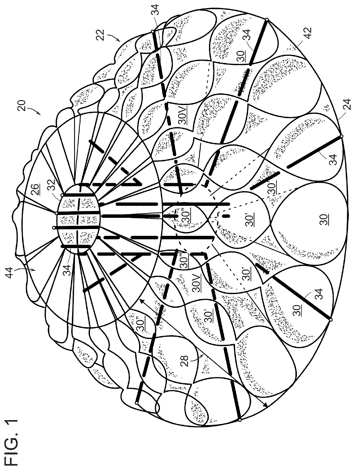
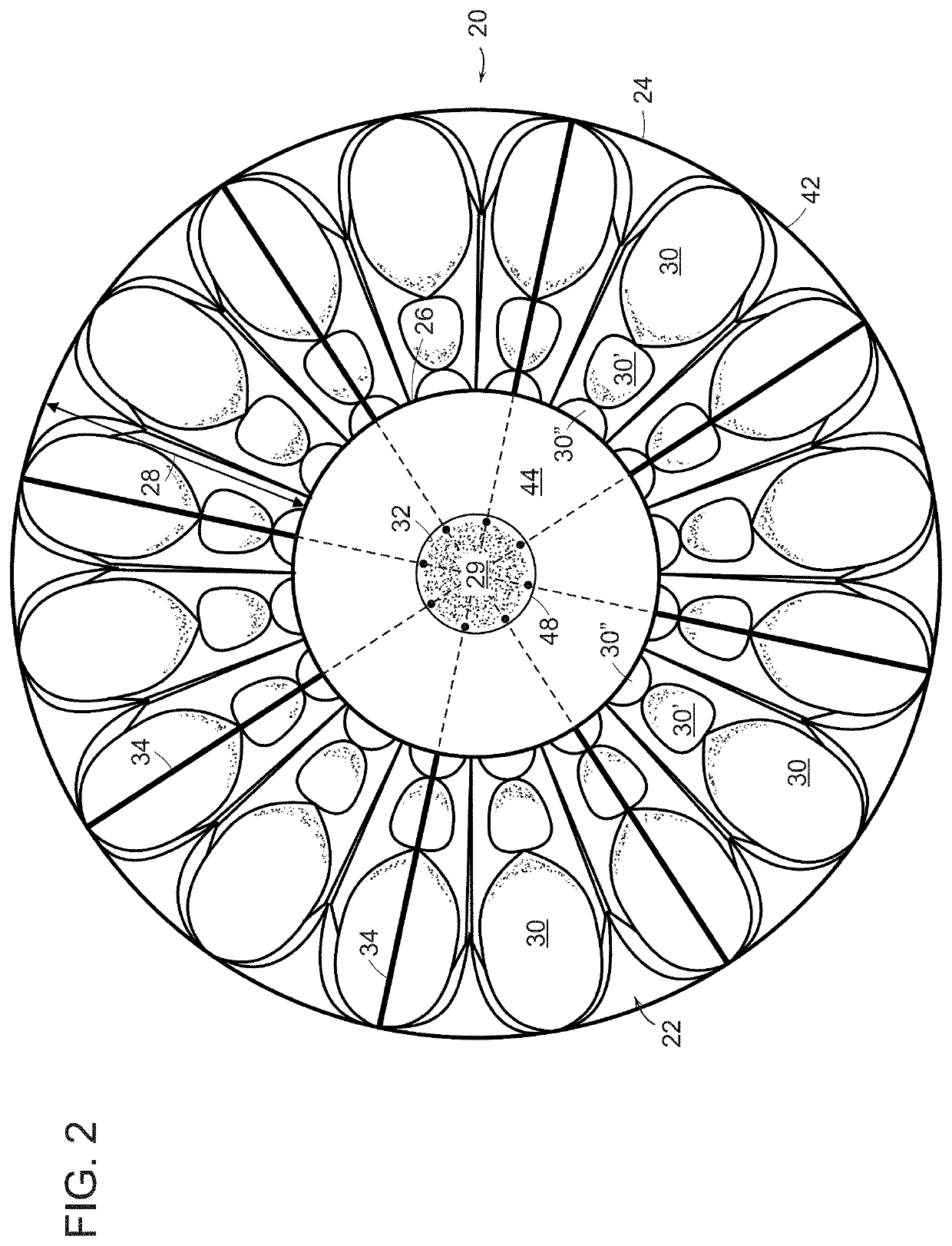
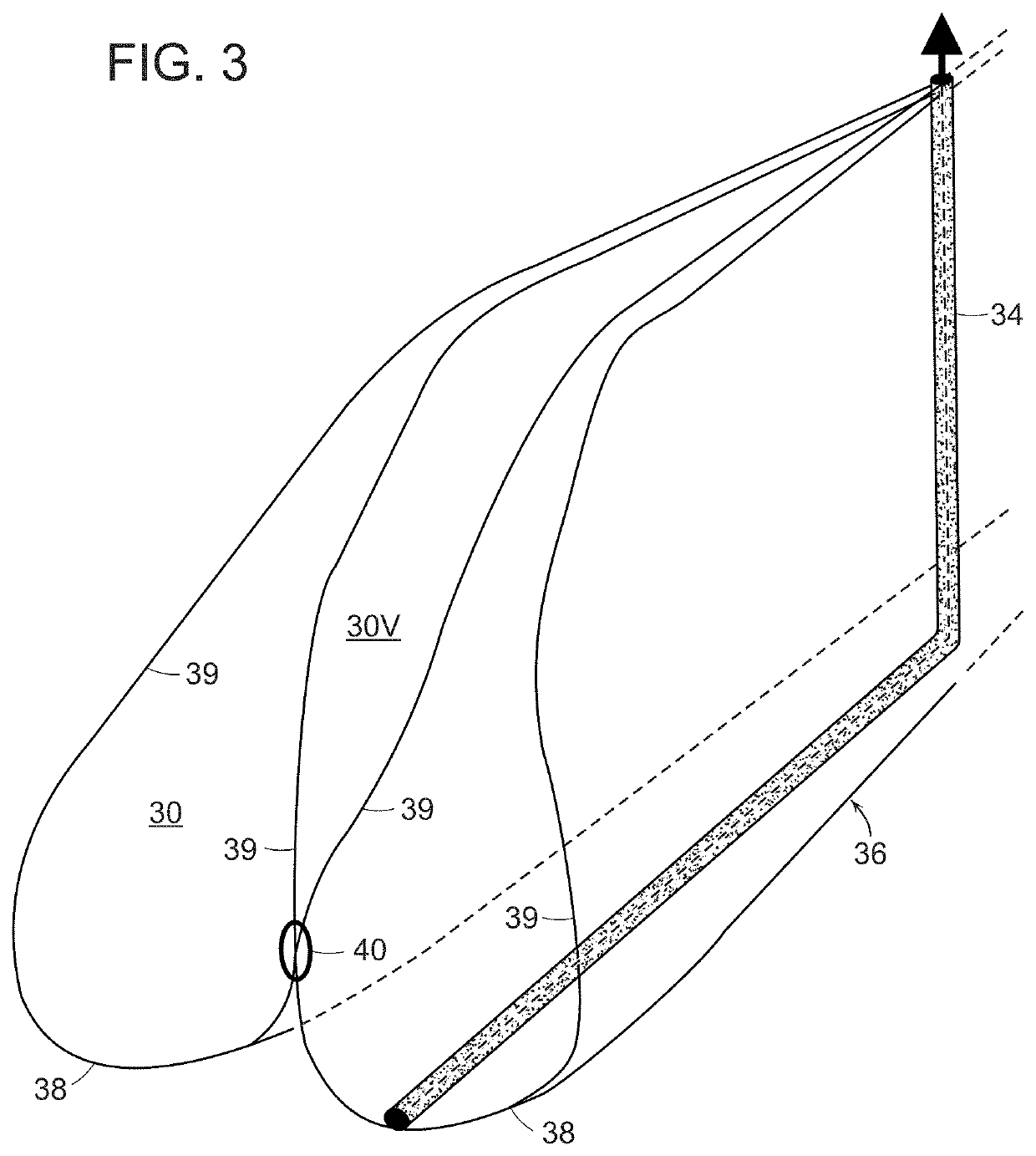
[0035]An in vivo tissue engineering device 20 according to the present invention is shown in FIGS. 1 and 2 and includes a scaffold 22 made of one or more sheets of mesh absorbable material which is, by nature, porous. The scaffold has a wide base or proximal portion 24, a narrower apex or distal portion 26 and a tapering, sidewall 28 extending between the base portion and the apex portion. The sidewall 28 is formed by a plurality of partially open, tissue engineering chambers 30 such that the sidewall has a rugose configuration formed by pleats or folds of the one or more sheets of absorbable material. The scaffold has a hollow inner region 29 which can be formed by a central core 32 extending between the base portion and the apex portion. If required by size, volume and stability, a plurality of L-shaped tubular struts 34 made of absorbable material can be radially arranged on the scaffold with legs extending through the core and bent 90° to terminate adjacent the perimeter of the ...


PUM

PropertyMeasurementUnit
volumeaaaaaaaaaa
diameteraaaaaaaaaa
thicknessaaaaaaaaaa

Abstract

Tissue engineering devices and methods employing scaffolds made of absorbable material for use in the human body for tissue genesis and regenerative and cellular medicine including breast reconstruction and cosmetic and aesthetic procedures and supplementing organ function in vivo.

Description

RELATED APPLICATIONS[0001]This application is a continuation of U.S. patent application Ser. No. 16 / 827,030, filed Mar. 23, 2020, which is a continuation-in-part of U.S. patent application Ser. No. 15 / 918,538, filed Mar. 12, 2018, now U.S. Pat. No. 10,595,986, each of which is incorporated herein by reference in its entirety.BACKGROUND OF THE INVENTIONField of the Invention[0002]The present invention relates to tissue engineering devices and methods employing scaffolds made of absorbable material implanted in the human body and use of such devices and methods in breast procedures, such as breast reconstruction, augmentation, mastopexy and reduction, in various cosmetic and aesthetic procedures involving tissue shaping and adipogenesis and in regenerative and cellular medicine to enhance and supplement organ function in vivo.Brief Discussion of Related Art[0003]Breast implants are commonly used to replace breast tissue that has been removed due to cancer and are also commonly used fo...

Claims


Application Information

Patent Timeline
Patent Type & Authority Applications(United States)
IPC IPC(8): A61F2/12A61L27/36
CPCA61F2/12A61F2002/0081A61L27/3604A61L2430/04A61L27/56A61L27/58A61L27/50A61L27/18A61F2210/0004A61F2002/0086A61F2230/0067A61F2/022A61F2230/0069A61F2230/0071C08L67/04A61F2240/002A61F2250/0023
Inventor REHNKE, ROBERT D.
Owner BARD SHANNON LTD
Why Eureka
Try Eureka