Imaging biomarkers based on ratio between diffusion and perfusion

a biomarker and ratio technology, applied in the field of magnetic resonance imaging, can solve the problem that the diffusion findings of the classical diffusion are not as readily apparen

Active Publication Date: 2021-08-19
OREGON HEALTH & SCI UNIV
View PDF14 Cites 1 Cited by
  • Summary
  • Abstract
  • Description
  • Claims
  • Application Information

AI Technical Summary

Problems solved by technology

In the neonatal population, the classic diffusion findings may not be as readily apparent due to the high water content and immature morphology of the brain.

Method used


Image

  • Imaging biomarkers based on ratio between diffusion and perfusion
  • Imaging biomarkers based on ratio between diffusion and perfusion
  • Imaging biomarkers based on ratio between diffusion and perfusion

Examples


example methodology

[0030]The present inventors retrospectively identified and reviewed medical records for all patients imaged at an institution between 2002 and 2019 whose brain MRI report contained the words “anoxic” or “hypoxic” using an institutional Radiology Search Engine. Anoxic patients were included if their MRI study contained diagnostic ASL perfusion and DWI sequences. Patients were excluded if there was severe motion artifact or the ASL or DWI sequence was otherwise non-diagnostic. Age matched controls imaged with ASL and DWI without a history of anoxic or hypoxic injury were also collected and analyzed. Similar to the anoxic group, control patients were excluded if the ASL or DWI sequence was non-diagnostic. In addition, patients with prior surgeries, intracranial hemorrhage, and ischemic injuries of any age were excluded. Patient age, cause of anoxic injury, and time from anoxic injury to imaging were recorded.

[0031]ASL CBF maps are generated using the single phase pulsed ASL clinical se...

example statistical

Analysis

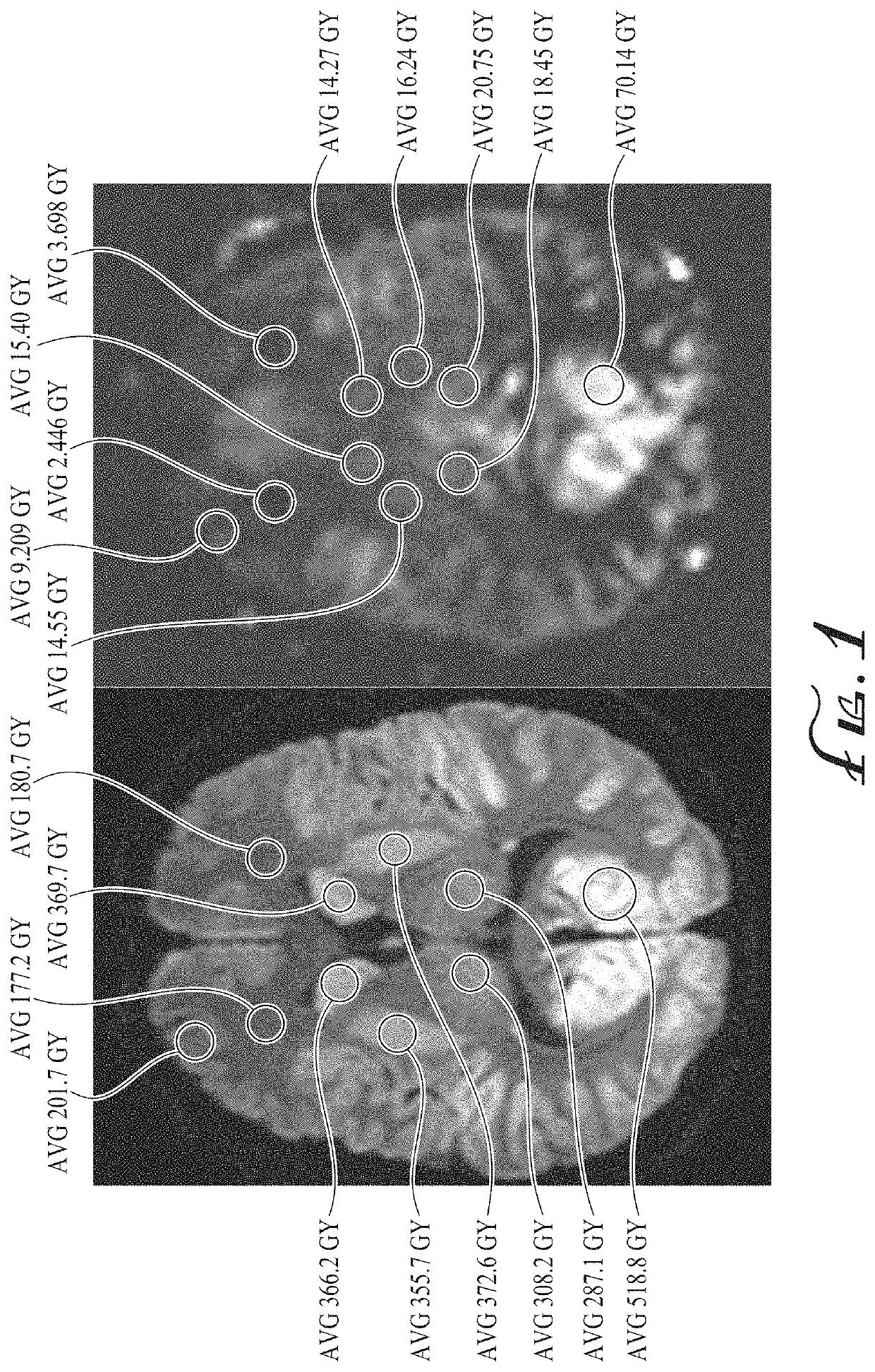
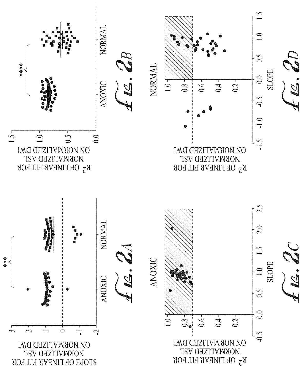
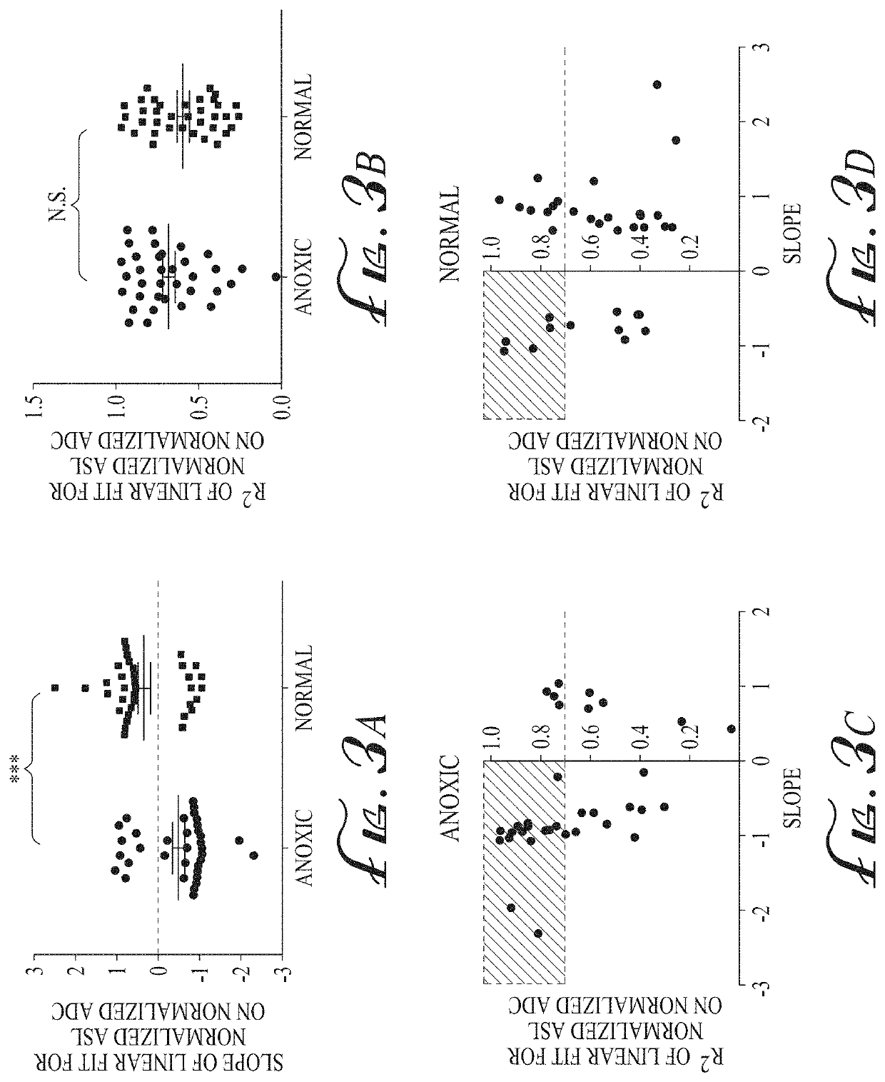
[0035]ASL perfusion, DWI, and ADC measurements for each ROI are normalized using the ROI signal intensity in the cerebellum or the brain stem, for grey matter and white matter, respectively. A linear best fit was performed between the normalized DWI and the normalized ASL values and the slope recorded, allowing up to 25% outlier identification. Graphs of the best-fitted line slopes and their respective R2 values are generated with a pre-determined R2 cutoff of 0.70. A similar analysis is performed using the normalized ADC measurements.

[0036]The relationship between the estimated time interval from inciting event to imaging and the linear regression slopes as well as R2s is examined to determine the dependence on the timing of imaging. Calculations are also made to determine the dependence on the number of ROI measurements. Two random raw (non-normalized) DWI and ASL ROls are selected for each patient and the slope was calculated using the two points. Combinations of two ROI ...

example experimental

Results

[0045]45 patients with anoxic injuries were initially identified using our institutional Radiology Search Engine from 2002 to 2019 with two-reader confirmation. Nine patients were excluded due to non-diagnostic imaging sequences; one patient was excluded due to remote injury (imaging was obtained 253 days after initial inciting event). The remaining 35 patients with anoxic injuries had imaging ranging from 0 to 14 days after the reported inciting event with mean of 3.8±0.5 days. 36 age-matched patients with a normal MRI had imaging performed between September 2010 and May 2019. Patient characteristics of the anoxic group and the normal group are shown in Table 1.

TABLE 1Patient characteristics of the anoxic injurygroup and the normal control group.AnoxicNormalVariable(n = 35)(n = 36)p valueAge (years)21.1 ± 4.426.5 ± 2.70.03Gender (Female)16 (45.7%)15 (41.7%)0.73Survived13 (37.1%)Interval to imaging (days) 3.8 ± 0.5Received CPR15 (42.9%)CPR time (minutes)22.7 ± 4.3Witnessed Se...


PUM


Abstract

The ratio of arterial spin labeled (ASL) perfusion to diffusion weighted imaging (DWI) is generally homogeneous in the anoxic / hypoxic injury population. Conversely, the ratio is more heterogeneous in the non-anoxic / hypoxic population. By plotting these ratios in a graphical format in the form of an axial color map of the brain—referred to as a normalized diffusion to perfusion (NDP) ratio colormap—it may be determined whether a patient has suffered from an anoxic / hypoxic injury. Thus, the anoxic and non-anoxic injury patients will have, respectively, homogenous and heterogeneous color maps. Anoxic brain injury patients have a global homogeneously positive relationship between qualitative ASL perfusion and diffusion weighted signal such that areas of restricted diffusion show significantly increased ASL perfusion signal, which may be attributable to BBB integrity. The NDP ratio colormap provides an imaging biomarker to differentiate anoxic brain injury from normal controls and to potentially assess BBB integrity.

Description

RELATED APPLICATION[0001]This application claims priority benefit of U.S. Provisional Patent Application No. 62 / 977,083, filed Feb. 14, 2020, which is hereby incorporated by reference in its entirety.TECHNICAL FIELD[0002]This disclosure relates generally to magnetic resonance imaging with diffusion restriction and arterial spin labeling and, more particularly, to brain imaging maps.BACKGROUND INFORMATION[0003]Anoxic brain injury is a result of prolonged hypoxia. Anoxic or hypoxic brain injury has several classic, well-described imaging findings including symmetric diffusion restriction in the basal ganglia and cortex. In the neonatal population, the classic diffusion findings may not be as readily apparent due to the high water content and immature morphology of the brain.SUMMARY OF THE DISCLOSURE[0004]In one aspect, a method for generating a diffusion to perfusion (DP) ratio brain image, includes receiving magnetic resonance imaging (MRI) data, the MRI data including perfusion and ...

Claims


Application Information

Patent Timeline
Patent Type & Authority Applications(United States)
IPC IPC(8): G01R33/563G16H30/20A61B5/00
CPCG01R33/56366A61B5/0042G16H30/20G01R33/56341G01R33/5608G16H50/20G16H30/40
Inventor POLLOCK, JEFFREY MICHAELLI, NINGCHENG
Owner OREGON HEALTH & SCI UNIV
Why Eureka
Try Eureka