
Bacteria-specific labeled substrtates as imaging biomarkers to diagnose, locate, and monitor infections

a microorganism and imaging biomarker technology, applied in the field of microorganism and microorganism culture, can solve the problems of inability to diagnose infectious diseases using standard diagnostic tools, inability to perform brain biopsies or biopsies in patients with bleeding risk, and inability to perform brain biopsies or biopsies

Active Publication Date: 2022-03-08
THE JOHN HOPKINS UNIV SCHOOL OF MEDICINE
View PDF3 Cites 0 Cited by
  • Summary
  • Abstract
  • Description
  • Claims
  • Application Information

AI Technical Summary

Benefits of technology

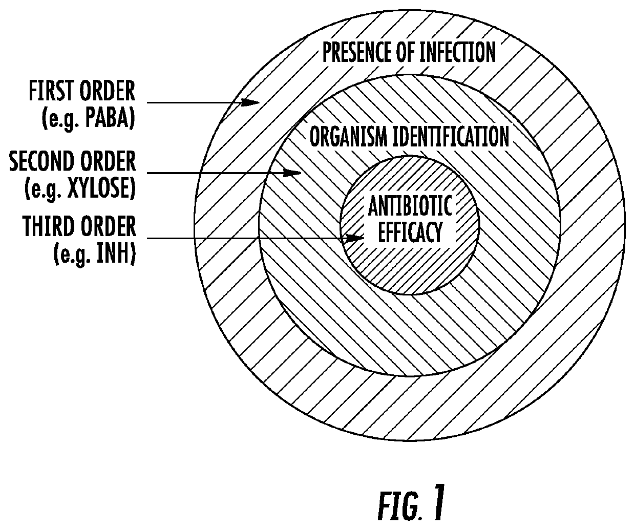
The present invention provides a method for detecting the presence of infectious organisms in a mammalian host by administering a therapeutically effective amount of a compound comprising a labeled substrate that is solely utilized by the metabolism of the infectious organisms. The label of the substrate is a detectable moiety, such as radionuclides, near-infrared dyes, or fluorescent dyes. The labeled substrate is allowed to take up by the infectious organisms for a sufficient period of time, and the presence or absence of the organisms is determined by detecting and quantifying the labeled substrate in the host. This method can be used to detect the location of the infectious organisms in the host and to assess the efficacy of antibiotic therapy.

Problems solved by technology

Diagnosis of infectious diseases by standard diagnostic tools, namely microscopy and microbiologic culture are often limited due to difficult sampling of obscure or deep sites of infection such as bone, cardiac, central nervous system, pulmonary or intra-abdominal locations.
This approach is both labor intensive and time consuming, and is subject to the uncertainties of incorrect sampling and contamination.
It may also be dangerous (e.g. brain biopsies or biopsies in patients with bleeding risk) or sometimes impossible (e.g. patients who are at high risk for complications secondary to anesthesia).
Complimentary techniques, such as cross sectional imaging by computed tomography or magnetic resonance imaging may provide a site of interest for invasive tissue sampling, but reveal structural abnormalities which are often a late occurrence in the disease process.
Furthermore, normal structure may be perturbed following surgical procedures, and indolent infections may not produce significant morphologic changes.
Nuclear medicine imaging techniques, such as 18F-fluorodeoxyglucose positron emission tomography have the potential to noninvasively determine the location and severity of infection as well as the response to therapy, but unfortunately are dependent upon host white blood cell function, and cannot differentiate between cancer, sterile inflammation or infection.

Method used


Image

  • Bacteria-specific labeled substrtates as imaging biomarkers to diagnose, locate, and monitor infections
  • Bacteria-specific labeled substrtates as imaging biomarkers to diagnose, locate, and monitor infections
  • Bacteria-specific labeled substrtates as imaging biomarkers to diagnose, locate, and monitor infections

Examples


example 1

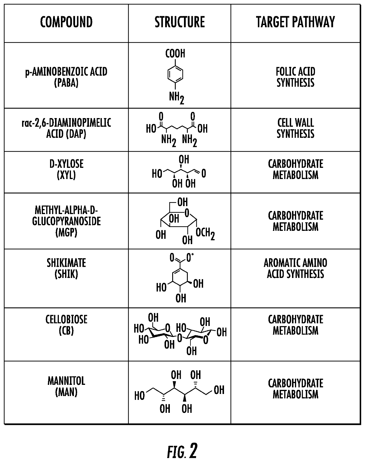
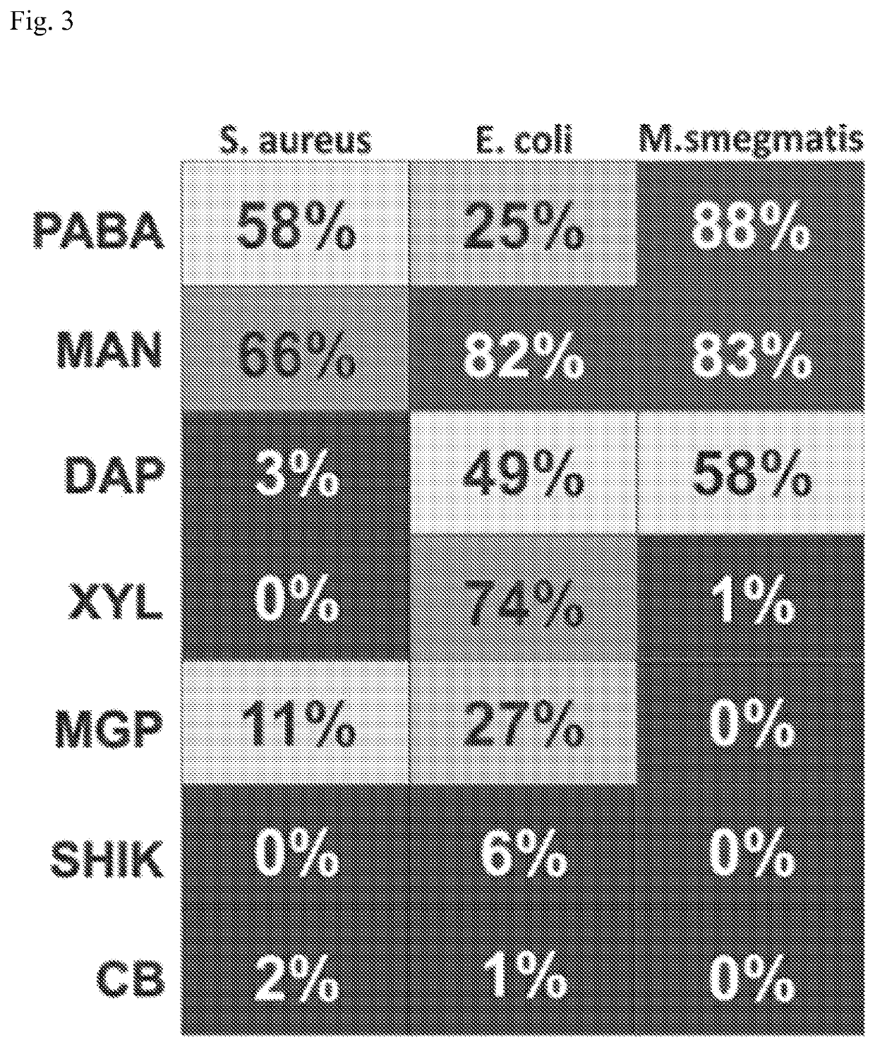
[0104]Seven (3%) compounds (FIG. 2) passed all 3 selection criteria; of these 1 was a known substrate for TK similar to FIAU (which also validated the screen), while the remaining 6 compounds were novel and tested for intracellular bacterial accumulation in model bacteria representing three important pathogen classes: Staphylococcus aureus (gram-positive), Escherichia coli (gram negative), or Mycobacterium smegmatis (mycobacteria). Intracellular uptake assays were performed at least in triplicate and indicate the percent of the total radioactivity (added to the culture), that was found in the bacterial pellet (after several steps of washing) (FIG. 3). Heat-killed bacteria were used as negative controls and no uptake was noted in any species. Intriguingly, some compounds were taken up differentially by the 3 different species of bacteria (e.g. XYL for E. coli alone, DAP for E. coli and M. smegmatis) indicating that the present inventive imaging methods can identify other bacterial sp...

example 2

[0105]A mycobacterial lead compound (PABA) was also evaluated in Mycobacterium tuberculosis and demonstrated significant (96.3% at 18-hours) and rapid (32.9% at 2-hours) accumulation. No significant accumulation was noted in host-cells (J774 macrophages) at the corresponding time-points, indicating that the uptake is bacteria-specific (FIG. 4A). Moreover, addition of excess unlabelled tracer blocked accumulation of M. tuberculosis-associated activity, indicating that PABA uptake was saturable and specific (FIG. 4B). PABA uptake was also measured in S. aureus and E. coli cultures and was also saturable and specific (FIG. 4C).

example 3

[0106][14C] D-mannitol was another of the promising molecules identified by the methods of the present invention. It accumulated rapidly and in significant amounts in all three models bacteria tested. Intracellular uptake was 66%, 82% and 83% in S. aureus, E. coli and M. smegmatis respectively after only 2 hours of incubation (FIG. 3). No uptake was observed in heat killed bacteria. Moreover, uptake in macrophage-like cells (J774) and Human Brain Microvascular Endothelial Cells (HBMEC) was <5% uptake at 2 hours.


PUM

PropertyMeasurementUnit
concentrationsaaaaaaaaaa
incubation timeaaaaaaaaaa
timeaaaaaaaaaa

Abstract

The methods of the present invention exploit unique biochemical pathways present within infectious organisms to develop small molecule metabolic tracers. Labeled substrates created using these inventive methods were created. The labeled substrates can be used to determine whether a subject is infected with an infectious organism by imaging means, and with use of two or more such labeled substrates, methods of differentiating gram negative infection from gram positive infection, and methods of localizing and quantifying infectious disease burden are provided. The methods of the present invention can assist in the clinical decision to begin empiric antibiotic therapy, determine its efficacy, as well as the choice of antibacterial agents.

Description

CROSS-REFERENCE TO RELATED APPLICATIONS[0001]This application is a 35 U.S.C. § 371 U.S. national entry of International Application PCT / US2013 / 059897, having an international filing date of Sep. 16, 2013, which claims the benefit of U.S. Provisional Patent Application Nos. 61 / 701,041, filed on Sep. 14, 2012, and 61 / 765,925, filed on Feb. 18, 2013, the content of each of the aforementioned applications is herein incorporated by reference in their entirety.STATEMENT OF GOVERNMENTAL INTEREST[0002]This invention was made with government support under grant number OD006492 and EB020539, awarded by the National Institutes of Health. The government has certain rights in the invention.BACKGROUND OF THE INVENTION[0003]Diagnosis of infectious diseases by standard diagnostic tools, namely microscopy and microbiologic culture are often limited due to difficult sampling of obscure or deep sites of infection such as bone, cardiac, central nervous system, pulmonary or intra-abdominal locations. Ra...

Claims


Application Information

Patent Timeline
Patent Type & Authority Patents(United States)
IPC IPC(8): G01N33/569
CPCG01N33/56944G01N33/5695G01N33/56916G01N33/56938G01N2800/26A61K51/0402A61K51/0491
Inventor JAIN, SANJAY K.POMPER, MARTIN GILBERTWEINSTEIN, EDWARD A.ORDONEZ, ALVAROKLUNK, MARIAH
Owner THE JOHN HOPKINS UNIV SCHOOL OF MEDICINE
Learn More