Mapping brain perivascular spaces

a brain perivascular space and fully automated technology, applied in the field of brain perivascular space fully automated mapping, can solve the problems of less effort made to enhance the visibility of pvs through postprocessing means, laborious process and prone to errors, and achieve the effect of accurately quantifying pvs in the brain and increasing pvs visibility

Pending Publication Date: 2021-08-19
UNIV OF SOUTHERN CALIFORNIA
View PDF4 Cites 0 Cited by
  • Summary
  • Abstract
  • Description
  • Claims
  • Application Information

AI Technical Summary

Benefits of technology

[0007]Described herein are systems and methods for mapping brain PVS using various imaging techniques. The imaging techniques may include PVS mapping using T1-weighted (T1w) and T2w images. The technique may increase PVS visibility on MRI and accurately quantify PVS in the brain. The imaging techniques may further include biophysical modeling of PVS fluid. The techn

Problems solved by technology

This process can be laborious and prone to error, and, as such, efforts to improve efficiency and accuracy have been made by using a wide range of automatic or semi-automatic segmentatio

Method used


Image

  • Mapping brain perivascular spaces
  • Mapping brain perivascular spaces
  • Mapping brain perivascular spaces

Examples


Embodiment Construction

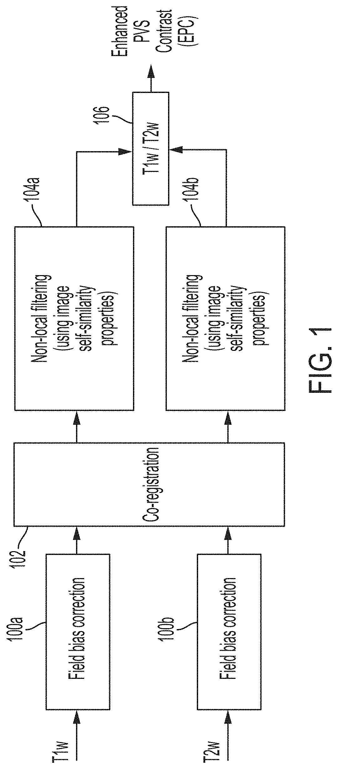
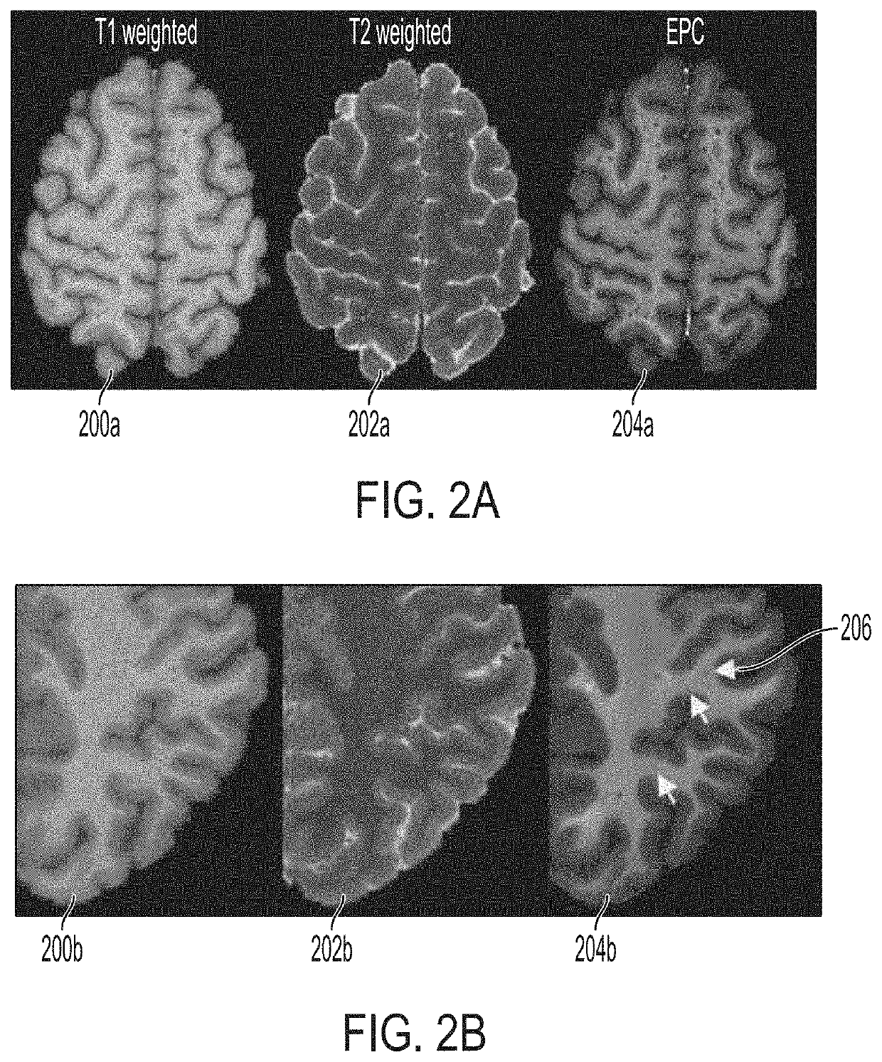
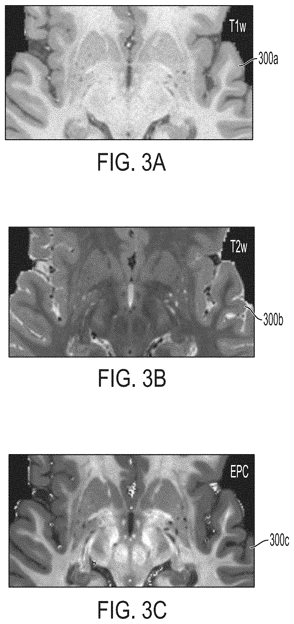
[0044]The systems and methods described herein may automatically map brain PVS using MRI scans. The mapping of brain PVS may quantify brain clearance pathways which may be used as a clinical biomarker or a prognosis marker for various neurological diseases and / or may be used to guide neurosurgery. In some embodiments, the systems and methods may include obtaining two images of a brain of a patient having different modalities (e.g., Tw1, Tw2, EPC) and combine the two images to advantageously generate and display 3-D map of the PVS within the brain. The systems and methods may further include advantageously generating and providing to an operator statistics of the brain PVS indicating, correlating with, or relating to a neurological disease condition. Additional image sequences (e.g. FLAIR) may be advantageous to discern PVS from pathological changes (e.g., white matter hyperintensities). Multi-modal approach for PVS quantification may further improve misclassification of vessels. In ...


PUM


Abstract

Systems and methods for mapping brain perivascular spaces. A system may include a memory to store one or more images of a brain of a patient. The system may further include a processor coupled to the memory. The processor may be configured to obtain a first and a second image of the brain. The processor may be further configured to combine the first image and the second image to preserve and magnify structures including the brain perivascular spaces within the image of the brain. The processor may be further configured to determine the brain perivascular spaces within the combined image of the brain of the patient. The processor may be further configured to generate a three-dimensional (3-D) map of the perivascular spaces. The system may further include a display configured to display the perivascular spaces to an operator.

Description

CROSS REFERENCE TO RELATED APPLICATIONS[0001]This application claims the benefit and priority of U.S. Provisional Application Ser. No. 62 / 976,696, entitled “Mapping Brain Perivascular Spaces,” filed on Feb. 14, 2020, the contents of which are hereby incorporated by reference in its entirety herein.STATEMENT AS TO FEDERALLY SPONSORED RESEARCH[0002]This invention was made with government support under grant numbers 2P41EB015922-21, 1P01AG052350-01, R01NS100973 and USC ADRC 5P50AG005142 awarded by the National Institute of Health (NIH). The government has certain rights in this invention.BACKGROUND1. Field[0003]The present disclosure relates to systems and methods for a fully automated mapping of brain perivascular spaces, particularly, using magnetic resonance image (MRI) scans.2. Description of the Related Art[0004]Imaging perivascular spaces (PVS), also known as Virchow-Robin spaces, has significant clinical value. PVS are pial-lined, fluid-filled structures that accompany vessels e...

Claims


Application Information

Patent Timeline
IPC IPC(8): G06T7/00G01R33/50G01R33/56G06T7/11G01R33/563
CPCG06T7/0012G01R33/50G01R33/5608G06T7/11G01R33/56341G06T2207/10092G06T2207/30016G06T2207/30096G06T2207/10028G06T2207/20024G06T2207/20084G06T2207/20221G01R33/5602G06T7/136G06T2207/10088
Inventor SEPEHRBAND, FARSHIDCHOUPAN, JEIRAN
Owner UNIV OF SOUTHERN CALIFORNIA
Why Eureka
Try Eureka