Interventional device with an ultrasound transducer

a technology of ultrasound transducer and intervention device, which is applied in the direction of ultrasonic/sonic/infrasonic image/data processing, ultrasonic/sonic/infrasonic diagnostics, and catheters. it can solve the problems of thin layer of air trapped between, affecting the performance of the transducer, etc., and achieves smooth introduction of the interventional device into the body , the effect of reducing the ingress of moistur

Pending Publication Date: 2021-10-07
B BRAUN MELSUNGEN AG +1
View PDF1 Cites 0 Cited by
  • Summary
  • Abstract
  • Description
  • Claims
  • Application Information

AI Technical Summary

Benefits of technology

The protective tube helps prevent moisture from getting inside the ultrasound transducer and makes the device smoother as it is inserted into the body. The use of a heat shrink material ensures a reliable manufacturing process. An adhesive layer between the ultrasound transducer and the protective tube improves the ultrasound transducer's performance by reducing the likelihood of air bubbles between them, which can cause sensitivity and signal strength issues. The adhesive layer also prevents moisture from reaching the ultrasound transducer and maintains its performance over time.

Problems solved by technology

Some known solutions to this problem may suffer from the ingress of moisture in the vicinity of the ultrasound transducer, which may affect transducer performance.
Other known solutions to this problem may suffer from the interventional device having an irregular topology, particularly in the vicinity of the ultrasound transducer.
It has been found that when such a protective tube is disposed over the ultrasound transducer, typically a thin layer of air is trapped between the ultrasound transducer and the protective tube.

Method used


Image

  • Interventional device with an ultrasound transducer
  • Interventional device with an ultrasound transducer
  • Interventional device with an ultrasound transducer

Examples


Embodiment Construction

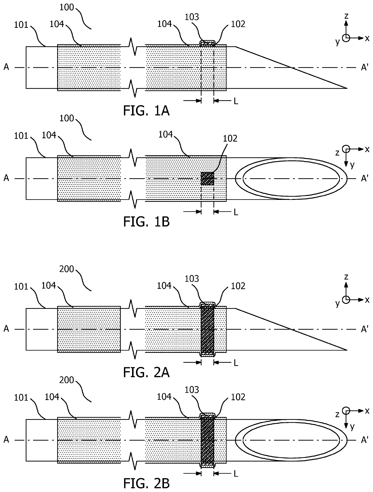
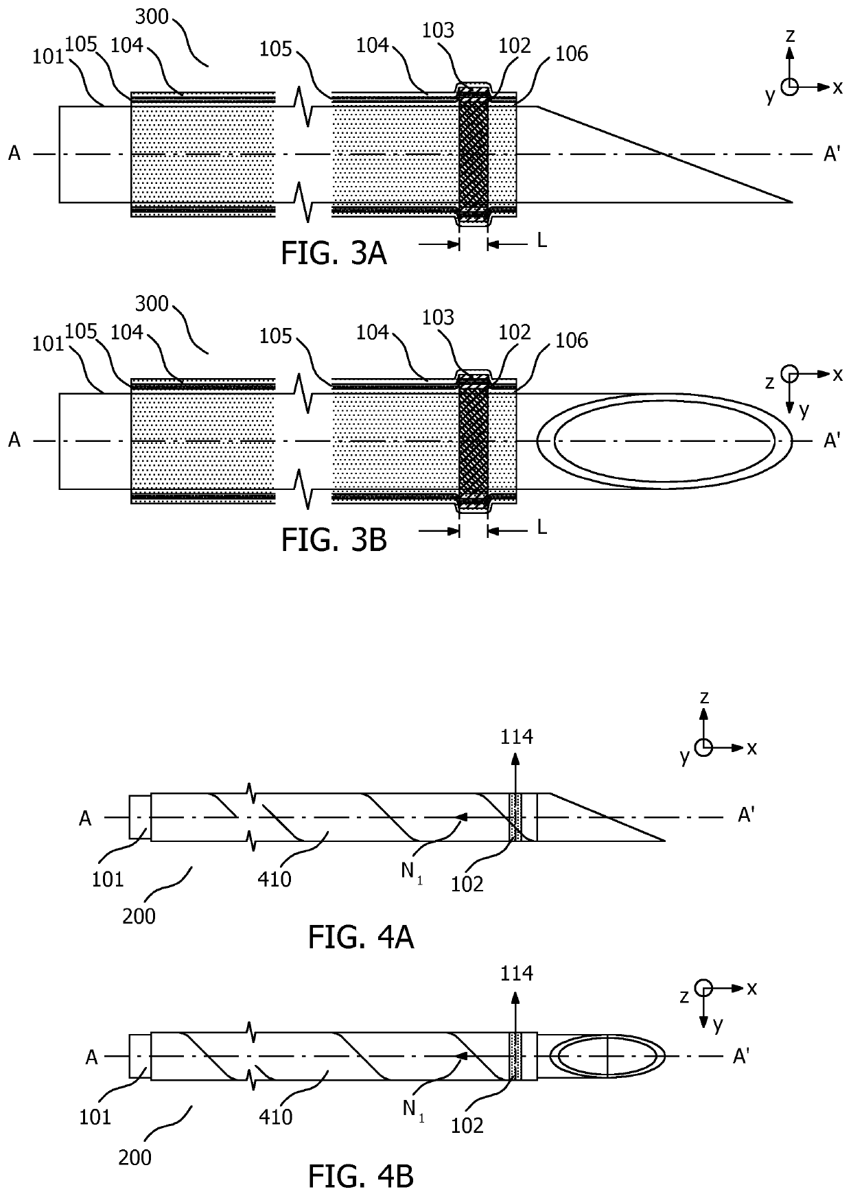
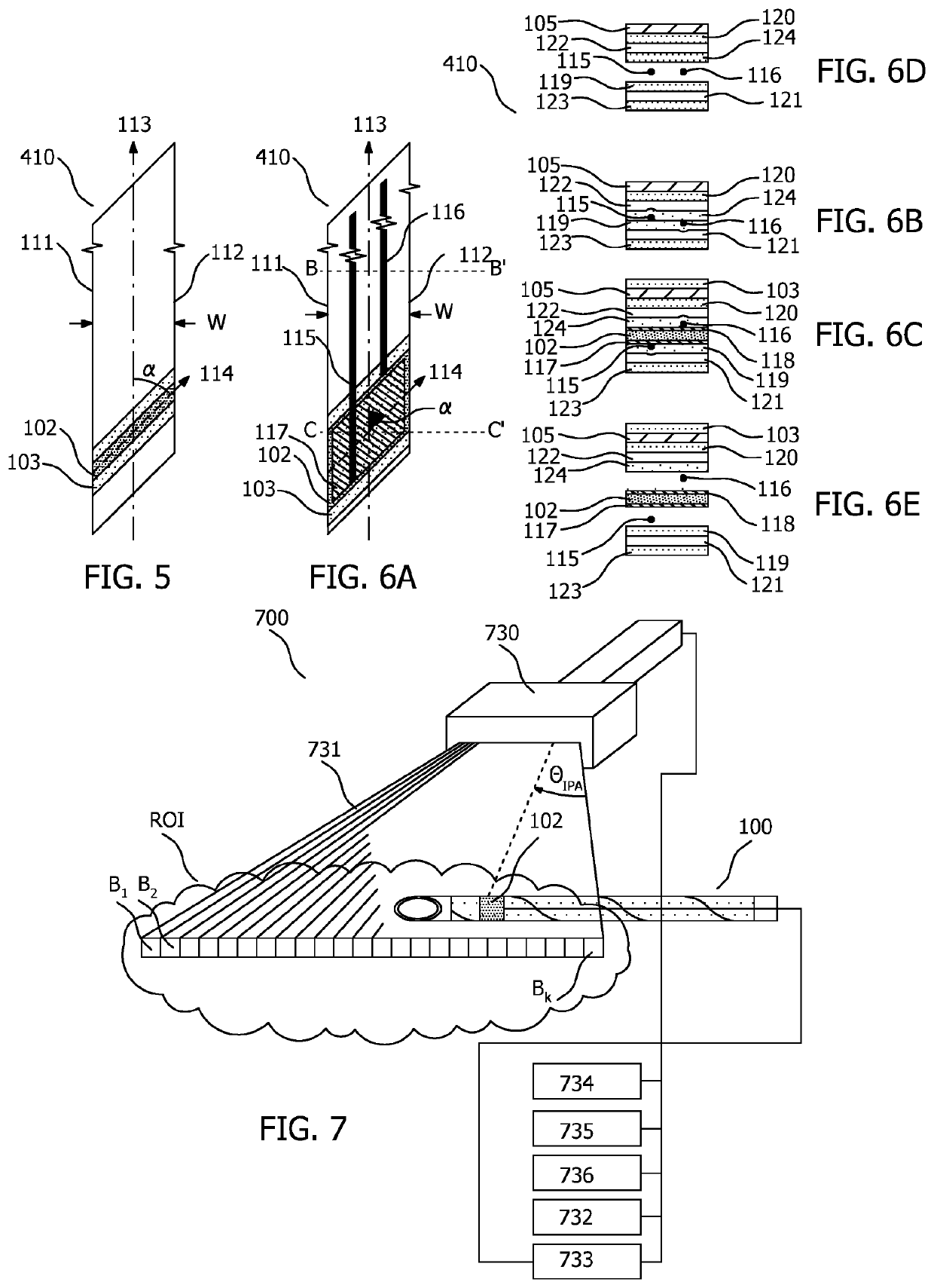
[0023]In order to illustrate the principles of the present invention an interventional device in the form of a medical needle is described with particular reference to an exemplary position tracking application in which the positon of an ultrasound detector on the needle is determined respective the ultrasound field of a beamforming ultrasound imaging system. It is however to be appreciated that the invention may also be used in other application areas that employ ultrasound transducers such as ultrasound imaging and treatment applications. It is also to be appreciated that whilst reference is made to an ultrasound transducer in the form of an ultrasound detector, the ultrasound transducer may alternatively be an ultrasound emitter, or indeed be capable of both detecting and emitting ultrasound signals, or indeed comprise both an ultrasound emitter and an ultrasound detector. The invention also finds application with other interventional devices than a medical needle, including with...


PUM


Abstract

An interventional device (100, 200, 300) includes an elongate shaft (101) having a longitudinal axis A-A′, an ultrasound transducer (102), an adhesive layer (103), and a protective tube (104) formed from a protective tube (104) formed from a heat-shrink material. The ultrasound transducer (102) is disposed on the elongate shaft (101) such that the ultrasound transducer (102) has an axial extent L along the longitudinal axis A-A′, At least along the axial extent L of the adhesive layer (103) is disposed between the ultrasound transducer (102) and the protective tube (104) surrounds the ultrasound transducer (102) and the adhesive layer (103) is disposed between the ultrasound transducer (102) and the protective tube (104).

Description

FIELD OF THE INVENTION[0001]The invention relates to an interventional device with an ultrasound transducer. The interventional device may be used in various interventional procedures in the medical field. In one contemplated application the ultrasound transducer is an ultrasound detector used to track a position of the interventional device respective an ultrasound field of an ultrasound imaging probe.BACKGROUND OF THE INVENTION[0002]Interventional procedures in the medical field increasingly use ultrasound to gain more information about, or to treat, a patient's anatomy. In this regard, ultrasound devices may be equipped with an ultrasound transducer, defined herein as an ultrasound detector, or an ultrasound emitter, or a device that is capable of both detecting and emitting ultrasound signals, for use in sensing and actuation applications such as tracking, imaging, or treatment.[0003]In one exemplary application described in more detail in document “A Non-disruptive Technology f...

Claims


Application Information

Patent Timeline
Patent Type & Authority Applications(United States)
IPC IPC(8): A61B8/08H01L41/313H01L41/23A61B17/34H10N30/02H10N30/073H10N30/87
CPCA61B8/0841H01L41/313H01L41/053A61B17/3403A61B2017/3413H01L41/23A61B2090/3929A61M25/0108H10N30/02H10N30/073H10N30/88
Inventor STAPERT, HENDRIK ROELOFLIJTEN, GERARDUS FRANCISCUS CORNELIS MARIADE WIJS, WILLEM-JAN ARENDSHIBAYAMA, YUICHITHORENZ, MATHIAS
Owner B BRAUN MELSUNGEN AG
Why Eureka
Try Eureka