

Method for identification of different categories of biopsy sample images
a biopsy sample and image identification technology, applied in the field of biopsy sample image screening and image identification, can solve the problems of time-consuming process and increased risk of prostatic adenocarcinoma, and achieve the effect of rapid screening of tissue biopsies
- Summary
- Abstract
- Description
- Claims
- Application Information
AI Technical Summary
Benefits of technology
Problems solved by technology
Method used
Image

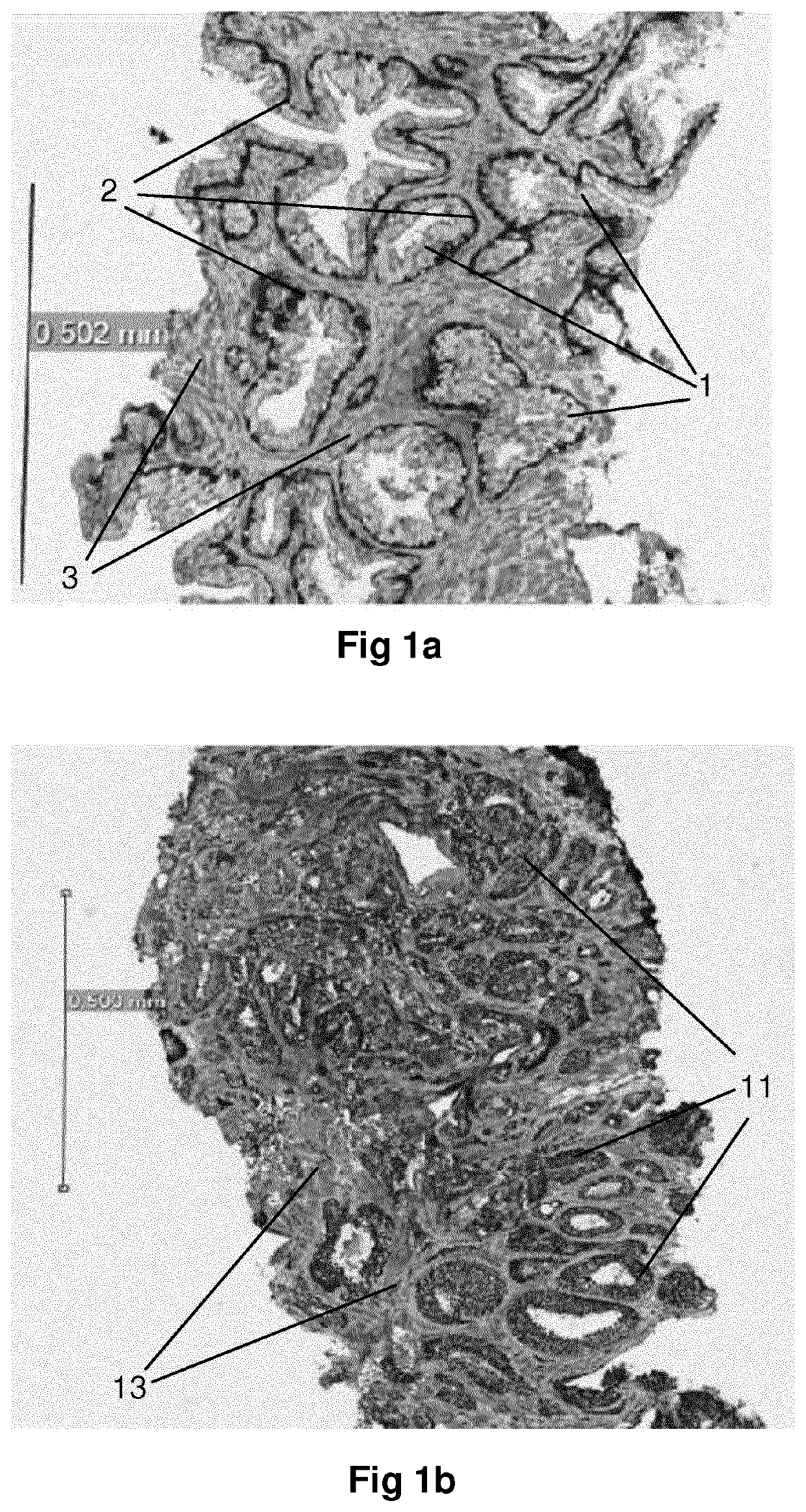
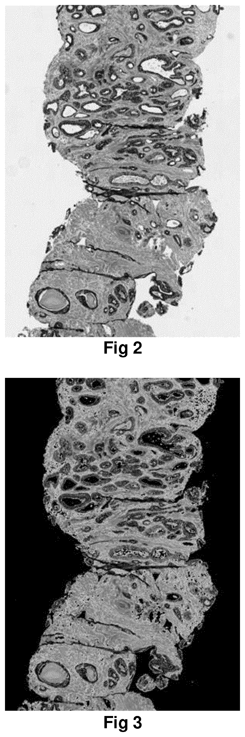
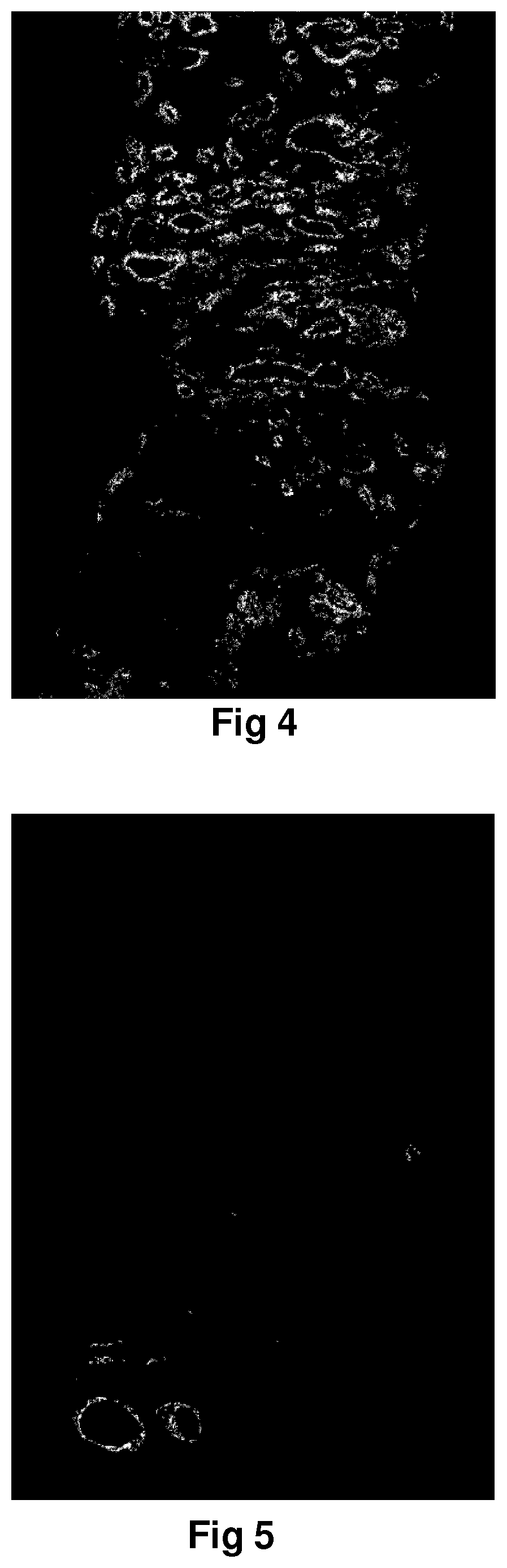
Examples
example a
[0100]Weighted pixel calculation will yield an malign indication value for the sample to be 4 / 100=0.04. The maximum malign indication value is 1.0 if all pixels are malign.
example b
[0101]Weighted anomaly calculation is obtained by assigning each pixel a weight from 0-255. This weight is inversely proportional to the distance between its owner pixel and the red-pixel (many false cancer-colour pixels happen closest to blood). A mathematical formula is used to determine the weight of each pixel:
wd=(d*a1)−a2,
where a1 and a2 are constants and d is the distance between the malign pixel and the red pixel. In this example a1=3.8 and a2=10. However, other constants may be used depending on use-case,
[0102]The first malign pixel has a distance of 10 pixels to the red pixel, and the weight for the first pixel is w1=(10*3.8)−10=28. The second pixel has a distance of 20 pixels to the red pixel, and the weight for the second pixel is w2=(20*3.8)−10=66. The third pixel has a distance of 50 pixels to the red pixel, and the weight for the third pixel is w3=(50*3.8)−10=180. The fourth pixel has a distance of 100 pixels to the red pixel, and the weight for the second pixel is w4=...
example c
[0106]The method of Example B may be performed with updated / other numbers:
[0107]Weighted anomaly calculation is obtained by assigning each pixel a weight from 0-255. This weight is inversely proportional to the distance between its owner pixel and the red-pixel (many false cancer-colour pixels happen closest to blood), A mathematical formula is used to determine the weight of each pixel:
wd=(d*a)−a2,
where a1 and a2 are constants and d is the distance between the malign pixel and the red pixel. In this example a1=7.0 and a2=10. However, other constants may be used depending on use-case.
[0108]The first malign pixel has a distance of 10 pixels to the red pixel, and the weight for the first pixel is w1=(10*7.0)−10=60. The second pixel has a distance of 20 pixels to the red pixel, and the weight for the second pixel is w2=(20*7.0)−10=130. The third pixel has a distance of 50 pixels to the red pixel, and the weight for the third pixel is w3=(30*7.0)−10=200. The fourth pixel has a distance ...
PUM
 Abstract
Description
Claims
Application Information