Guided lung coverage and automated detection using ultrasound devices

a technology of ultrasound device and lung, applied in the field of improved medical imaging system and method, can solve the problems of affecting the ultrasound image quality, affecting the accuracy of the operator, and affecting so as to limit the scope of the claimed subject matter

Pending Publication Date: 2022-03-24
GE PRECISION HEALTHCARE LLC
View PDF5 Cites 1 Cited by
  • Summary
  • Abstract
  • Description
  • Claims
  • Application Information

AI Technical Summary

Problems solved by technology

Any error on part of the operator would result in an image containing excessive disturbance or noise.
Therefore, training of the operator and her experience in handling the ultrasound imaging devices will affect the ultrasound image quality.
The operator may be skilled in acquiring the good quality images of the certain portion of the body due to her experience in obtaining the images of that body portion, but the operator may not be skilled to obtain the high-quality images of the entire subject body.
This may be due to the lack of the imaging experience or lack of the anatomical knowledge of the entire body.
Also, placing the ultrasound probe at the appropriate body portion affects the quality of the image.
Physical parameters of the subject body like obesity, bone density, height, chest or abdominal size will create a challenging situation for the operator to correctly position the ultrasound probe for obtaining the complete image of the region of interest.
Any wrong placement of the ultrasound probe would result in incomplete imaging of the regions of interest.
Lack of scanning expertise in operating the ultrasound devices will result in generating the images with poor quality resulting in poor assessment of the subject condition.
This may cause loss of several invaluable lives.
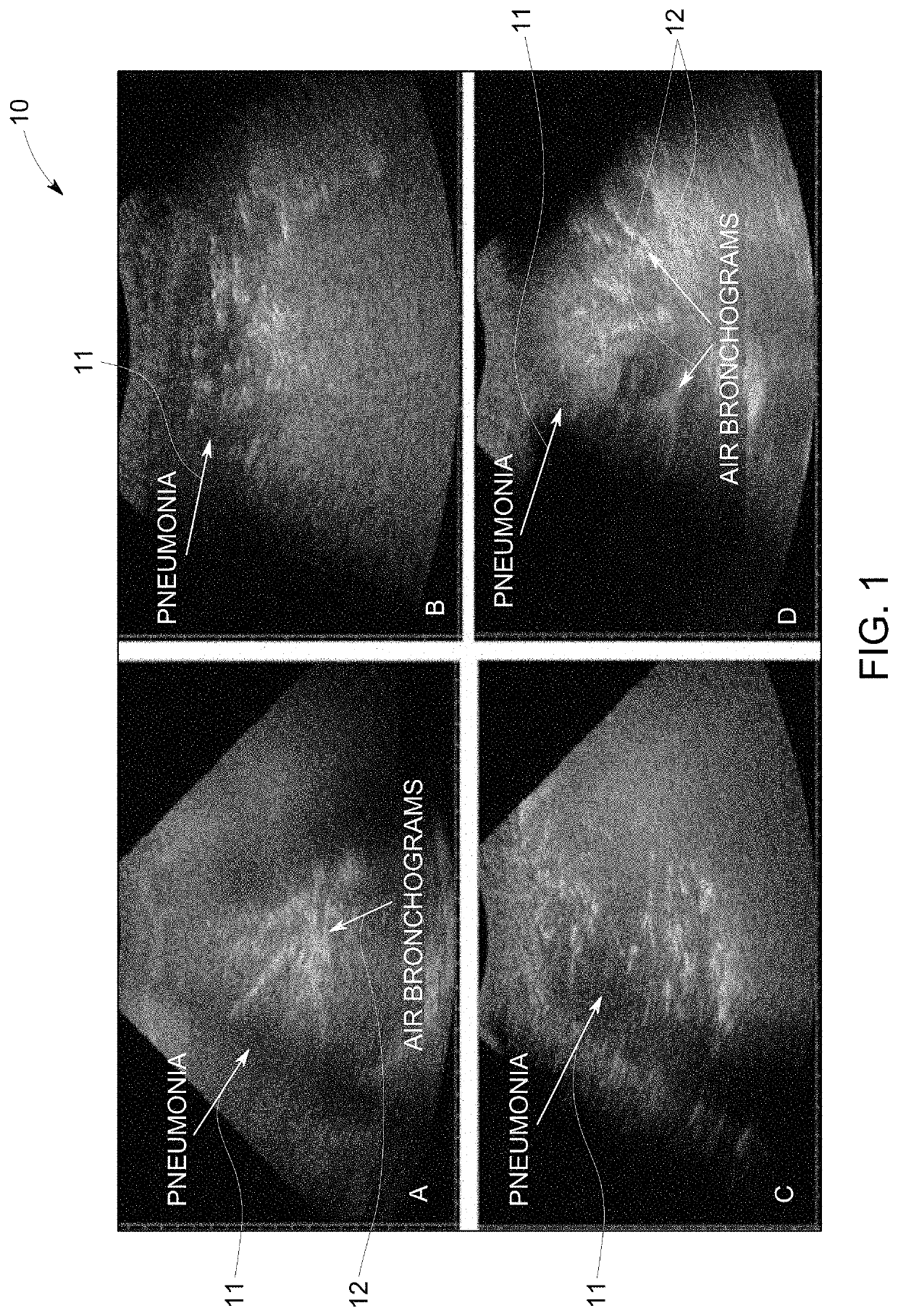
Ultrasound has been shown to detect the pleural effusion and the lung consolidation but requires substantial skill on the part of the operator to maneuver the probe.
Since the field of view (FoV) of the ultrasound is very limited, it is not possible to know if all the Covid-19 affected portions or hotspots within the lung of a subject have been covered during the lung examination.

Method used


Image

  • Guided lung coverage and automated detection using ultrasound devices
  • Guided lung coverage and automated detection using ultrasound devices
  • Guided lung coverage and automated detection using ultrasound devices

Examples


Embodiment Construction

[0020]In the following specification and the claims, reference will be made to a few terms, which shall be defined to have the following meanings.

[0021]The singular forms “a”, “an”, and “the” include plural references unless the context clearly dictates otherwise.

[0022]As used herein, the term “non-transitory computer-readable media” is intended to be representative of any tangible computer-based device implemented in any method or technology for short-term and long-term storage of information, such as, computer-readable instructions, data structures, program modules and sub-modules, or other data in any device. Therefore, the methods described herein may be encoded as executable instructions embodied in a tangible, non-transitory, computer readable medium, including, without limitation, a storage device and / or a memory device. Such instructions, when executed by a processor, cause the processor to perform at least a portion of the methods described herein. Moreover, as used herein,...


PUM


Abstract

Systems and methods for guided lung coverage and automated detection using ultrasound devices are disclosed. The method for guided coverage and automated detection of pathologies of a subject includes positioning an ultrasound probe on a region of the subject body to be imaged. The method includes capturing a video of the subject and processing the video to generate a torso image of the subject and identify location of the ultrasound probe on the subject body. The method includes registering the video to an anatomical atlas to generate a mask of the region of the subject body comprising a plurality of sub-regions of the subject body to be imaged and superimposing the mask over the torso image. The method further includes displaying an indicator corresponding to a location of the each of the plurality of the sub-regions on the torso image. The method further includes displaying the relative position of the ultrasound probe with respect to the indicator corresponding to the location of the each of the plurality of plurality of sub-regions of the subject body to be imaged.

Description

FIELD OF THE INVENTION[0001]This disclosure relates generally to improved medical imaging systems and methods, and more particularly to systems and methods for guided lung coverage and automated detection of the pathologies by adjusting the ultrasound probe position on the subject body.BACKGROUND OF THE INVENTION[0002]Various medical imaging systems and methods are used to obtain the images of the affected regions of the subject for diagnosing the medical condition. Ultrasound imaging is a known medical imaging technique used for imaging the different body parts like joints, muscles vessels and pregnant woman (known as obstetric ultrasound). Ultrasound imaging offers several advantages over the other imaging techniques as ultrasound is a real-time imaging technique that provides live stream of images. Commercially, the ultrasound devices are available in various configurations and the portable ultrasound devices are used for capturing the relatively bigger regions of the subject ana...

Claims


Application Information

Patent Timeline
Patent Type & Authority Applications(United States)
IPC IPC(8): A61B8/00A61B8/08G06T7/00G06K9/22G06T7/70G06T11/00
CPCA61B8/4263A61B8/463A61B8/5223G06T7/0012G06T2207/10016G06T7/70G06T11/00G06T2207/30061G06T2207/10132G06K9/22A61B8/085A61B8/08A61B8/4411A61B8/4416A61B8/4427A61B8/4472A61B8/462A61B8/565G06T2207/20081G06T2207/20084G06V10/25G06V2201/031G06V10/82G06T11/60G06V10/17A61B8/4245
Inventor MELAPUDI, VIKRAMALADAHALLI, CHANDAN KUMAR MALLAPPASHRIRAM, KRISHNA SEETHARAM
Owner GE PRECISION HEALTHCARE LLC
Why Eureka
Try Eureka