Process for classification of glioma

a glioma and brain technology, applied in the field of tumor brain classification, can solve the problems of inability to complete surgical removal, high variability of interpretation, and inability to completely resected ii to iv gliomas, so as to reduce the number of subgroups and achieve the same prognosis significance.

Pending Publication Date: 2022-04-21
HOSPICES CIVILS DE LYON +1
View PDF0 Cites 0 Cited by
  • Summary
  • Abstract
  • Description
  • Claims
  • Application Information

AI Technical Summary

Benefits of technology

[0136]The TeloDIAG has the advantage to reduce the number of subgroups and to keep the same prognosis significance. Note that the crossing of th

Problems solved by technology

Because of their diffusely infiltrating nature, grade II to IV gliomas cannot be completely resected and are not curable by surgical excision.
The extremely infiltrative nature of this tumor makes complete surgical removal impossible.
Histopathological classification is the basis of the World Health Organization (WHO) classification; however, it suffers from a high variability of interpretation from one practician to another.
Consequently, therapeutic strategies may be wrongly chosen, if the glioma is incorrectly classified.
If none of these markers is retrieved, no guideline is available for the treatment of said

Method used


Image

  • Process for classification of glioma
  • Process for classification of glioma
  • Process for classification of glioma

Examples


example 1

Reaction

[0354]Rolling circle amplification of C-circle is performed as described in (Henson et al., 2009) and (Henson et al., 2014). Briefly, 3.2 μl of total genomic DNA (5 ng / μL) were incubated for 18 h at 30° C. with 3.75 units of φ29 DNA polymerase (New England Biolabs) (0.375 μL of 10 U / μL), in 0.2 μg / μL of BSA, 0.1% Tween, 4 μM DTT (Dithiothreitol), 1 mM dNTP, 1 μL of 10×NEB buffer. Enzyme is heat-inactivated at 65° C. for 20 min. The same reaction is performed without the enzyme φ29 (φ−).

[0355]For each experiment, two internal controls are added, namely TA and ALT. TA and ALT correspond to total genomic DNA extracted from HeLa (ALT−) and U2OS (ALT+) cell lines respectively.

[0356]The 10 μL of φ− and φ+ reactions are then diluted by adding 30 μL of water (molecular biology grade), 5 μL are used to performed each qPCR reaction.

example 2

riment

[0357]TeloPCR

[0358]Oligonucleotides for the qPCR reaction have been previously described in (Gil et al., 2004) and (Lau et al., 2013) and are listed below

[0359]The sequence for oligonucleotides used in the qPCR are presented in table 2:

SEQ ID5′-CGGTTTGTTTGGforwardNO: 3GTTTGGGTTTGGGTTTel1aTGGGTTTGGGTT-3′SEQ ID5′-GGCTTGCCTTACreverseNO: 4CCTTACCCTTACCCTprimerTACCCTTACCCT-3′tel2BSEQ ID5′-CAGCAAGTGGGARPL0 / 36B4NO: 5AGGTGTAATCC-3′forwardprimerSEQ ID5′-CCATTCTATCATRPL0 / 36B4NO: 6CAACGGGTACAA-3′reverseprimer

[0360]Telo-PCR and qPCR against 36B4 are run in duplicate for each condition φ− and φ+, on a 480 Light Cycler Thermocycler (Roche, Houwald, Luxembourg), in 1× final LightCycler® DNA Master SYBR Green I (10 μL), 200 nM final of TeloPCR-specific primers or 300 nMm final of 36B4-specific primers. Details of thermocycling conditions are detailed below

[0361]For each qPCR, the exact conditions are summarized in tables 3 and 4 below:

TABLE 3TeloPCRAnalysis ModeCyclesSegmentTarget Température...

example 3

ysis, Normalization and Classification

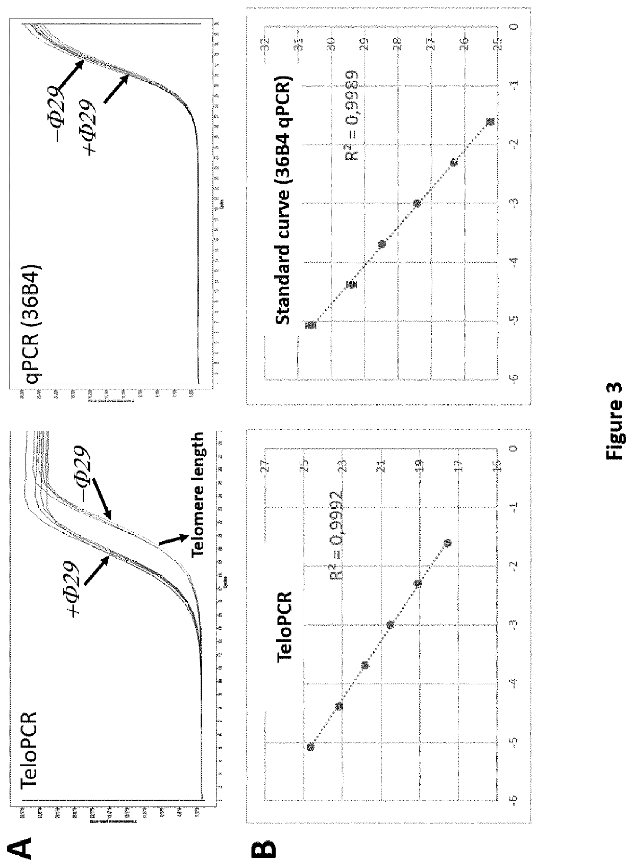
[0366]The fluorescence in logarithmic scale is analyzed as a function of PCR cycle, the threshold is determined by the second derivative method (all experiments). Intersection between the threshold of amplification curve gives the CT for each reaction. The fluorescence channel corresponding to the TeloPCR is the following: SYBR Green (465-510). For the dTeloPCR, two channels are analyzed: SYBR Green (465-510) for the telomeric sequence and CY5 (618-660) for RPLP0 / 36B4.

[0367]Efficiency of TeloPCR (Etelo) and 36B4 (E36B4) PCR are respectively 1.70111 and 1.9672.

[0368]For each reaction, the following value is calculated: Etelo−CT / E36B4−CT, and annotated as φ+ and φ− as a function of the initial circle reaction.

[0369]φ− correspond to the telomere length (T-Length)

[0370]Cr correspond to the Circle score and is calculated as follow: φ+ / φ−

[0371]The difference of T-Length between the two internal controls (U2OS and HeLa) A is calculated and correspond ...


PUM

PropertyMeasurementUnit
Timeaaaaaaaaaa
Lengthaaaaaaaaaa

Abstract

The invention relates to an in vitro process for classifying a glioma, comprising the following steps: a. Measuring at least, from a glioma patient biological sample, the Alternative Lengthening of Telomeres (ALT) status of said glioma; b. Optionally, determining the isocitrate dehydrogenase genes mutation status (IDH status) of said glioma; c. Based on the data obtained in steps (a) and optionally (b) and, if available, on the histological grade of said glioma, classifying said glioma in one of the five following classes: oligodendroglioma-like, glioblastoma IDHwt-like, glioblastoma IDHmt-like, low-grade astrocytoma-like, and other gliomas.

Description

FIELD OF THE INVENTION[0001]The present invention concerns the classification of tumor brains and the choice of therapeutic options useful for treating patients with tumor brains, based on said classification.BACKGROUND OF THE INVENTION[0002]A glioma is a type of tumor deriving from the glial cells of the brain or the spine. Gliomas represent about 30% of all brain and central nervous system tumors, and 80% of the malignant brain tumors.[0003]Malignant gliomas are graded from grade II to grade IV, although benign gliomas are designated as gliomas of grade I.[0004]According to the 2016 WHO classification (4th Edition) of tumors of the central nervous system (CNS), summarized in (Louis et al., 2016), malignant tumors of the CNS are classified according to immuno-histological and molecular criteria into different categories:[0005]diffuse astrocytic and oligodendroglial tumors;[0006]others astrocytic tumors;[0007]ependymal tumors;[0008]choroid plexus tumors;[0009]neuronal and mixed neur...

Claims


Application Information

Patent Timeline
IPC IPC(8): C12Q1/6886
CPCC12Q1/6886C12Q2600/158C12Q2600/118C12Q2600/112
Inventor PONCET, DELPHINE
Owner HOSPICES CIVILS DE LYON
Why Eureka
Try Eureka