X-ray and gamma imaging using a single radiation detector

Pending Publication Date: 2022-06-09
KONINKLJIJKE PHILIPS NV
View PDF0 Cites 0 Cited by
  • Summary
  • Abstract
  • Description
  • Claims
  • Application Information

AI Technical Summary

Benefits of technology

[0012]It is an object of the invention to allow for an accurate x-ray and

Problems solved by technology

However, due to the use of two detectors, such systems are very complex and expensive.
Detector assemblies including two detectors are itself quite complex and costly.
Further, detector assemblies including two detectors are heavier than conventional detector assemblies used in x-ray or gamma imaging, which include only a single detector.
However, x-ray imaging often requires a relatively high spatial resolution.
With such small pixel sizes, the detector may suffer from charge sharing and escape photons generated due to K-fluorescence (so called K-escape).
These effects particularly result in inaccuracies in the determination of the energies of the x-ray photons, which are also referred as spectral distortions.
However, these spectral distortions only affect a relatively small fraction of detection even

Method used


Image

  • X-ray and gamma imaging using a single radiation detector
  • X-ray and gamma imaging using a single radiation detector

Examples


Example

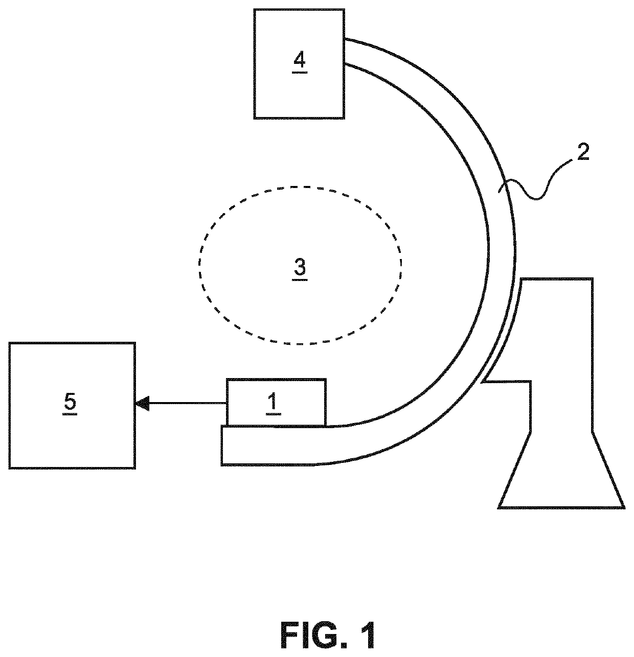
[0032]FIG. 1 schematically and exemplarily shows components of a combined x-ray and gamma imaging system, which includes a single direct conversion radiation detector 1 for detecting x-ray and gamma photons. The radiation detector 1 is configured as photon-counting spectral detector, which is capable of detecting individual photon events and assigning each detection event to one of a plurality of energy bins. The energy bins correspond to predetermined energy intervals. The system may particularly be used in medical applications in order to acquire three-dimensional x-ray and nuclear images of regions of patient bodies. However, the system may likewise be utilized in order to image other objects.

[0033]Since the radiation detector 1 is configured as a spectral detector, the system is capable of generating energy-selective x-ray images. For instance, these images allow for distinguishing between different materials of the object—this usually also referred to as material decomposition....


PUM


Abstract

The invention relates to a system for imaging an object in an x-ray imaging mode and in a gamma imaging mode. A radiation detector (1) of the system comprises a conversion unit (202) including a plurality of detector pixels (2061, . . . ,M) and generating for each detection event a detection signal indicative of an energy of the event, and a counting unit (203) including for each detector pixel (2061, . . . ,M) a plurality of comparators (209i; 1, . . . ,N) and associating each detection event to one of a plurality of predetermined energy bins based on the detection signals using the comparators (209i; 1, . . . ,N). In the x-ray imaging mode, the comparators (209i; 1, . . . ,N) of one pixel (2061, . . . ,M), and in the gamma imaging mode, the comparators (209i; 1, . . . ,N) of several pixels (2061, . . . ,M) are available for the association so that more energy bins are available in the gamma imaging mode than in the x-ray imaging mode.

Description

FIELD OF THE INVENTION[0001]The invention relates to the acquisition of x-ray and gamma images of objects. More specifically, the invention relates to an imaging system which is operable in an x-ray imaging mode to detect x-ray photons having traversed an object and which is operable in a gamma imaging mode to detect gamma photons emitted by the object. Further, the invention relates to an imaging method for imaging an object using such an imaging system.BACKGROUND OF THE INVENTION[0002]In various medical imaging procedures, it is beneficial to provide both an x-ray image and a nuclear image of a region of interest. The x-ray image typically provides structural information indicative of the anatomy of the region of interest. The nuclear image, defined herein to mean an image indicative of radiotracer distribution in an object, is generated based on detected gamma quanta. The nuclear image may, for example, be a gamma scintigraphy or a SPECT image and typically provides functional or...

Claims


Application Information

Patent Timeline
IPC IPC(8): A61B6/00
CPCA61B6/4258A61B6/4441A61B6/4241A61B6/4057G01T1/1611G01T1/1647
Inventor WIECZOREK, HERFRIED KARLSTEADMAN BOOKER, ROGERALVING, PETER LEX
Owner KONINKLJIJKE PHILIPS NV
Why Eureka
Try Eureka