Real-time detection of artifacts in ophthalmic images

a technology of artifact detection and ophthalmology, applied in image analysis, image enhancement, instruments, etc., can solve the problems of reducing the efficacy of such procedures, requiring additional surgical intervention, and poor patient outcomes

Pending Publication Date: 2022-07-14
ALCON INC
View PDF0 Cites 0 Cited by
  • Summary
  • Abstract
  • Description
  • Claims
  • Application Information

AI Technical Summary

Benefits of technology

[0002]Aspects of the present disclosure relate to systems and methods for detecting artifacts in image data used during surgical procedures, such as cataract surgery, which enables improved surgical outcomes for patients.

Problems solved by technology

However, artifacts present in imaging data of the patient's eye can lead to measurement errors that go unknown or unnoticed by a medical practitioner, and may consequently reduce the efficacy of such procedures and lead to poor patient outcomes.
Often, such outcomes require additional surgical intervention.

Method used


Image

  • Real-time detection of artifacts in ophthalmic images
  • Real-time detection of artifacts in ophthalmic images
  • Real-time detection of artifacts in ophthalmic images

Examples


example method

of Training a Machine Learning Model for Processing Image Data to Identify Artifacts Therein

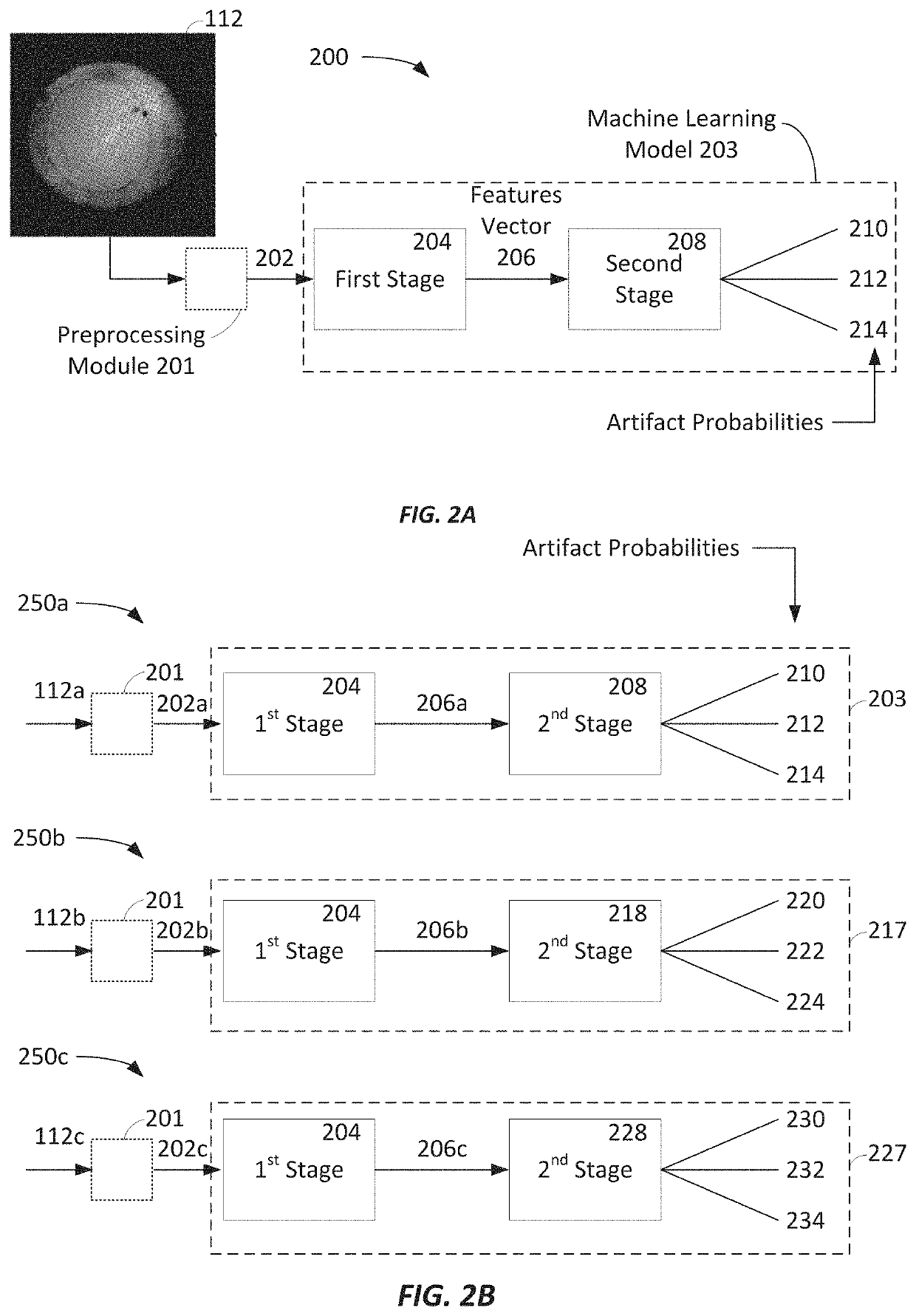
[0164]FIG. 6 depicts an example method 600 for training a machine learning model to identify digital images that include one or more artifacts. The training of the machine learning model may be completed by one or more of the controller 106 and / or the system 100 of FIG. 1 or a component or system external to the system 100. The method 600 may be used to train, for example, the machine learning models of FIGS. 2A and 2B. In some embodiments, the method 600 only trains the second stage 208, 218, and / or 228 (i.e., the classification stage) and the first stage 204 (i.e., the feature extraction stage) is not trained.

[0165]Method 600 starts at block 602 and, at block 604, begins with obtaining the images that will be used to train the machine learning model. In some instances, the images are obtained in real-time from an image capture devices (for example, the aberrometer 104 and / or one or more of ...

example processing

Systems

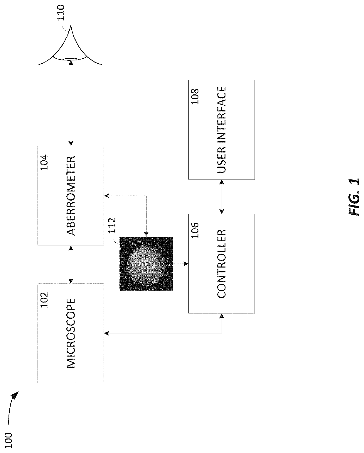
[0171]FIG. 7 is a diagram of an embodiment of a processing system or device. According to some embodiments, the processing system of FIG. 7 is representative of a computing system that may be included in one or more of intraoperative aberrometry systems that implement the machine learning model processing described herein, with reference to the aberrometer 104, the controller 106, the user interface 108, and / or the like. Specifically, the processing system of FIG. 7 may implement one or more of the machine learning models 203, 217, and 227 according to the data flows 200, 250a, 250b, and 250c and otherwise introduced and discussed herein to identify artifacts in image data of patient eye images, as shown in FIGS. 4A-4G, and / or the like.

[0172]FIG. 7 illustrates a computing system 700 where the components of system 700 are in electrical communication with each other. The system 700 includes a processor 710 and a system bus 705 that couples the various components. For example, t...

example clauses

[0200]Implementation examples are described in the following numbered clauses:

[0201]Clause 1: A system for processing image data from an intraoperative diagnostic device in real-time during an ophthalmic procedure, the system comprising: an image capture element configured to capture a grayscale image of a patient's eye from the intraoperative diagnostic device, the grayscale image having a first size; an image processing element configured to: obtain the grayscale image from the image capture element; scale the grayscale image from the first size to a second size; and preprocess the scaled grayscale image in preparation for classification; a two-stage classification model comprising: a feature extraction stage configured to process the scaled grayscale image and generate a feature vector based on the scaled grayscale image, and a classification stage configured to process the feature vector and generate an output vector based on the feature vector; wherein the image processing elem...


PUM


Abstract

Certain aspects of the present disclosure provide a system for processing image data from an intraoperative diagnostic device in real-time during an ophthalmic procedure. The system comprises an image capture element that captures a grayscale image of a first size and an image processing element that scales the grayscale image from the first size to a second size. The system also comprises a two-stage classification model comprising: a feature extraction stage to process the scaled grayscale image and generate a feature vector based on the scaled grayscale image and a classification stage to process the feature vector and generate an output vector. The image processing element is further configured to determine an image quality of the obtained grayscale image based on the output vector for display to an operator and the image quality of the obtained grayscale image indicates a probability that the obtained grayscale image includes an artifact.

Description

CROSS-REFERENCE TO RELATED APPLICATIONS[0001]The present application claims the benefit of and priority to U.S. Provisional Application No. 63 / 135,125, entitled “Real-Time Detection of Multilabel Image Artifacts in an Ophthalmic Instrument Using a Convolutional Neural Network / Deep Neural Network Model”, and filed on Jan. 8, 2021, the entirety of which is incorporated by reference herein.INTRODUCTION[0002]Aspects of the present disclosure relate to systems and methods for detecting artifacts in image data used during surgical procedures, such as cataract surgery, which enables improved surgical outcomes for patients.[0003]Cataract surgery generally involves replacing a natural lens of a patient's eye with an artificial intraocular lens (IOL). During cataract surgery, medical practitioners may utilize various image-based measurement systems to analyze the patient's eye in real-time and to assist with performing the cataract procedure—such as to ensure proper selection, placement, and ...

Claims


Application Information

Patent Timeline
Patent Type & Authority Applications(United States)
IPC IPC(8): A61B3/103A61B3/16G06T7/00G06T5/00
CPCA61B3/103A61B3/16G06T7/0012G06T2207/30041G06T2207/20084G06T2207/20081G06T5/001G06T2207/30168G06T2200/24G06T2207/10056G06T2207/10016
Inventor SARVER, EDWIN JAYHALL, MAX
Owner ALCON INC
Why Eureka
Try Eureka