Method and system for the automatic segmentation of white matter hyperintensities in magnetic resonance brain images

a magnetic resonance brain image and hyperintensity technology, applied in the field of neuroradiology, can solve the problems of insufficient robustness to offer the precision required in such a specific field of application, and the state of the art high-precision imaging solution for the automatic segmentation of magnetic resonance images, etc., to achieve greater robustness and better understanding.

Pending Publication Date: 2022-10-27
QUIBIM
View PDF0 Cites 3 Cited by
  • Summary
  • Abstract
  • Description
  • Claims
  • Application Information

AI Technical Summary

Benefits of technology

[0041]Due to all of the above, the present invention has technical features which involve a multitude of advantageous effects. For example, the arrangement of neural networks described in the present invention enables discerning between physiological hyperintensities and hyperintensities that could correspond to white matter injuries, which makes it possible for a specialist to concentrate their work on the areas segmented by the present invention and included in the image mask, or even obtain additional quantitative parameters based on the segmentation provided in order to describe the injury load (such as volume or number of injuries) of people with leukoaraiosis. The benefits extend for example to the optimisation of the time of the specialist and in an increase in their precision.
[0042]Additionally, the novel combination of convolutional neural networks of the present invention offers greater robustness to the segmentation than any other of the known solutions. It enables the images to be processed by extracting characteristics at different levels, specifically focusing on the complex task of segmenting white matter injuries, which vary the distribution thereof from one person to another and require a special configuration such as that of the present invention.BRIEF DESCRIPTION OF THE FIGURES
[0043]To complete the description of the invention and for the purpose of helping to better understand its features according to a preferred embodiment thereof, a set of drawings is attached in which the following figures are depicted in an illustrative and non-limiting manner:

Problems solved by technology

The state of the art includes some systems to help specialists based on artificial intelligence for the segmentation of the images, but they are not robust enough to offer the precision required in such a specific field of application.
Therefore, a high-precision imaging solution for the automatic segmentation of magnetic resonance images is lacking in the state of the art, which can help specialists in their subsequent diagnoses and studies of white matter injuries.

Method used


Image

  • Method and system for the automatic segmentation of white matter hyperintensities in magnetic resonance brain images
  • Method and system for the automatic segmentation of white matter hyperintensities in magnetic resonance brain images
  • Method and system for the automatic segmentation of white matter hyperintensities in magnetic resonance brain images

Examples


Embodiment Construction

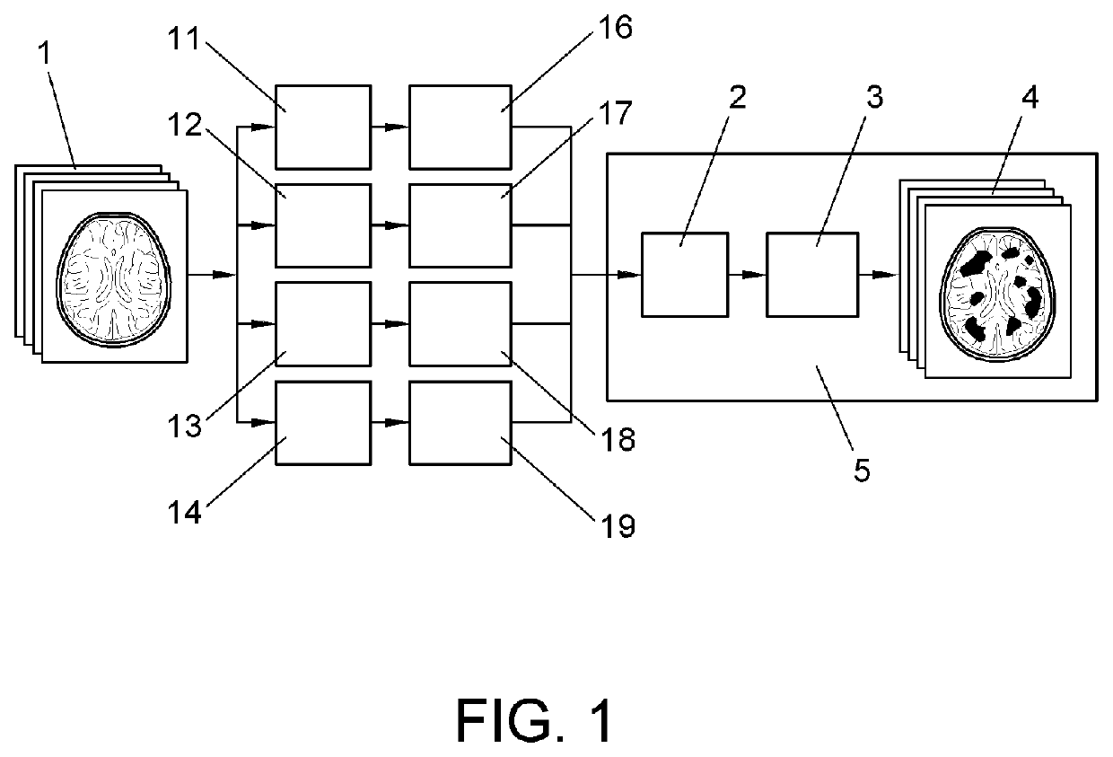
[0008]In order to achieve the objectives and avoid the drawbacks mentioned above, the present invention describes, in a first aspect, a method for segmenting white matter hyperintensities present in magnetic resonance brain images comprising:[0009]providing an array of previously trained convolutional neural networks with a magnetic resonance brain image;[0010]identifying the voxels of the image containing white matter hyperintensities;[0011]determining, for each of the convolutional neural networks and for each voxel, the probability that the identified hyperintensity corresponds to a previously defined pathological hyperintensity;[0012]calculating the average of all the probabilities determined for each voxel;[0013]comparing the averaged probabilities for each voxel with a pre-established threshold; and[0014]generating an image mask with the voxels that exceed the threshold.

[0015]Additionally, one of the embodiments of the invention envisages pre-processing of the provided image c...


PUM


Abstract

The present invention relates to a method and a system for the segmentation of white matter hyperintensities (WMHs) present in magnetic resonance brain images, comprising: providing an array of trained convolutional neural networks (CNNs) with a magnetic resonance brain image; determining, for each of the CNNs and for each voxel, the probability that the given voxel corresponds to a pathological hyperintensity; calculating the average of all the probabilities determined for each voxel; comparing the averaged probabilities for each voxel with a threshold; generating an image mask with the voxels that exceed the threshold.

Description

OBJECT OF THE INVENTION[0001]The present invention relates to the technical field of computer-assisted analysis of medical images and more specifically to the processing by means of convolutional neural networks of images obtained by magnetic resonance, for an automatic segmentation of the images based on the volume of leukoaraiosis, especially useful for the study of radiologists-neurologists on possible brain injuries.BACKGROUND OF THE INVENTION[0002]Currently, magnetic resonance imaging is a preliminary diagnostic tool commonly used to detect abnormalities in tissues. Specifically, in the field of neuroradiology, it is used by specialists to diagnose white matter injuries, among other things.[0003]White matter injuries are observed in the magnetic resonance images as hyperintensities in the sequence of fluid-attenuated inversion recovery (FLAIR), however, not all the hyperintensities observed correspond to injuries, but other physiological hyperintensities also appear, such as th...

Claims


Application Information

Patent Timeline
Patent Type & Authority Applications(United States)
IPC IPC(8): G06N3/04G06N3/08G16H30/40
CPCG06N3/0454G06N3/0481G06N3/08G16H30/40G06N3/048G06N3/0464G06N3/0455G06N3/09A61B5/055G06T7/0012G06T2207/10088G06T2207/20084G06T2207/20081G06T2207/30016G06T2207/30096G06N3/04G06N3/045
Inventor JIMÉNEZ PASTOR, ANA MARÍACAMACHO RAMOS, EDUARDOGARCÍA CASTRO, FABIOALBERICH BAYARRI, ÁNGELPUIG ALCÁNTARA, JOSEPBIARNES DURÁN, CARLESMARTÍ BONMATÍ, LUISPEDRAZA GUTIÉRREZ, SALVADOR
Owner QUIBIM
Why Eureka
Try Eureka