Automatic contextual segmentation for imaging bones for osteoporosis therapies

a technology of contextual segmentation and bone imaging, applied in the field of bone imaging, can solve problems such as adverse effects of these techniques

Inactive Publication Date: 2000-02-01
ELI LILLY & CO
View PDF6 Cites 65 Cited by
  • Summary
  • Abstract
  • Description
  • Claims
  • Application Information

AI Technical Summary

Problems solved by technology

In noncontextual segmentation, spatial relations among pixels or regions are ignored; though often computationally efficient, the performance of these techniques is adversely affected by noise in the image recording process and multiple features of similar intensity included in a scene.
One complicating factor for automatic segmentation is the presence of partial volume averaging near region transitions due to the low resolution of the QCT instrument.

Method used


Image

  • Automatic contextual segmentation for imaging bones for osteoporosis therapies
  • Automatic contextual segmentation for imaging bones for osteoporosis therapies
  • Automatic contextual segmentation for imaging bones for osteoporosis therapies

Examples


Embodiment Construction

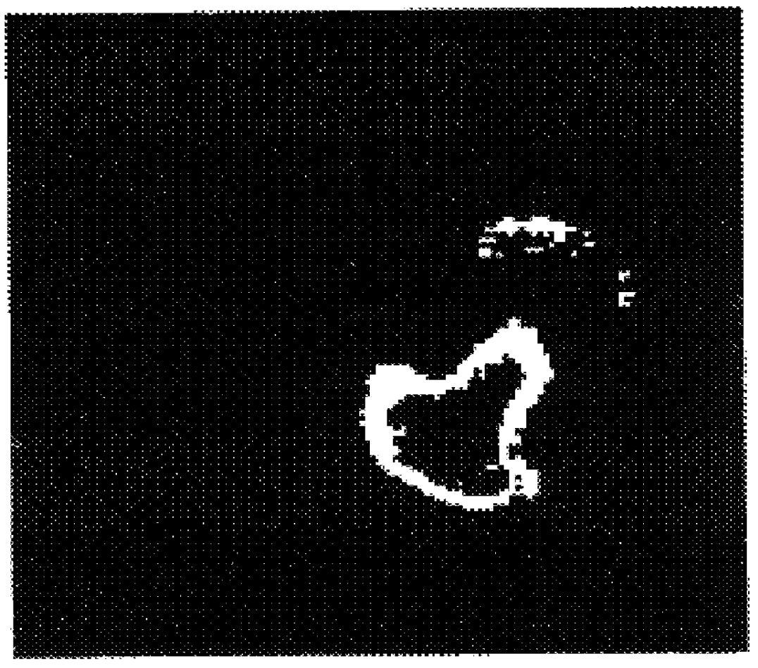
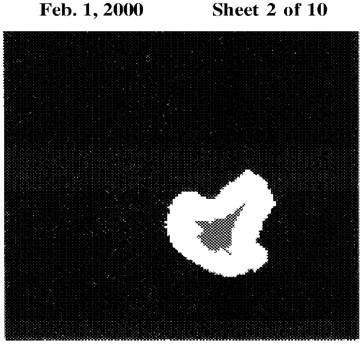
A schematic arrangement of the system of the present invention is illustrated in FIG. 12. Image processing system 120 includes imagining device 122, computer 124, and display device 126. Imaging device 122 may be a suitable medical imaging apparatus for obtaining an image of body tissue, such as an x-ray, MRI, or ultrasonic image machine. Computer 124 may be a general purpose computer with a specific program (such as the programs in the source code appendixes), or alternatively, computer 124 may be implemented as a dedicated computer with specific programming embedded in its memory, or as a discrete device implementing the functionality described below. Display device 126 may be a video monitor, a printer, or other output device capable of providing a visual image, alternatively if system 120 is used purely for measurement purposes, display device 126 may be a printer, LCD device, or audio speaker. Computer 124 executes certain algorithms on data supplied by imaging device 122 to pr...


PUM


Abstract

An automatic contextual segmentation method which can be used to identify features in QCT images of femora, tibiae and vertebrae. The principal advantages of this automatic approach over traditional techniques such as histomorphometry are, 1) the algorithms can be implemented in a fast, uniform, non-subjective manner across many images allowing unbiased comparisons of therapeutic efficacy, 2) much larger volumes in the region of interest can be analyzed, and 3) QCT can be used longitudinally. Two automatic contextual segmentation algorithms relate to a cortical bone algorithm (CBA) and a whole bone algorithm (WBA). These methods include a preprocessing step, a threshold selection step, a segmentation step satisfying logical constraints, a pixel wise label image updating step, and a feature extraction step; with the WBA including whole bone segmentation, cortical segmentation, spine segmentation, and centrum segmentation. The algorithms are constructed to provide successful segmentations for known classes of bones with known topological constraints.

Description

SOURCE CODE APPENDIXThis application includes two appendixes having (A) 36 and (B) 19 pages. A portion of the disclosure of this patent document contains material which is the subject to copyright protection. The copyright owner has no objection to the facsimile reproduction by anyone of the patent document or the patent disclosure, as it appears in the Patent and Trademark Office patent files or records, but otherwise reserves all copyright rights whatsoever.1. Field of the InventionThe present invention relates to image analysis with the conceptual interpretation of features identified in complex scenes. More specifically, the field of the invention is bone imaging.2. Prior ArtImage segmentation is used to identify relevant features in an image of body tissue such as an x-ray, MRI, or ultrasonic image. The data are gray levels, or a finite set of colors, on a rectangular lattice of picture elements (pixels), and correspond to digitized values of radiated intensity from some photoc...

Claims


Application Information

Patent Timeline
Patent Type & Authority Patents(United States)
IPC IPC(8): G06T5/00
CPCG06T7/0081G06T7/0091G06T2207/10072G06T2207/10116G06T2207/20141G06T2207/30008G06T7/11G06T7/155G06T7/187
Inventor HELTERBRAND, JEFFREY DONALDHIGGS, JR., RICHARD EARLIVERSEN, PHILIP WAYNE
Owner ELI LILLY & CO
Why Eureka
Try Eureka