Immunoassay for detection of mutant P53 polypeptide in serum

Inactive Publication Date: 2000-07-04
SIEMENS HEALTHCARE DIAGNOSTICS INC
View PDF9 Cites 19 Cited by
  • Summary
  • Abstract
  • Description
  • Claims
  • Application Information

AI Technical Summary

Problems solved by technology

In contrast, wild type p53 polypeptide does not accumulate and is not easily detected.
However, mutant p53 polypeptides could not be detected in any normal or premalignant tissues from these patients.
The literature described monoclonal antibodies which specifically recognize and bind mutant p53 polypeptides, however, such antibodies did not (1) detect mutant p53 polypepti

Method used


Image

  • Immunoassay for detection of mutant P53 polypeptide in serum
  • Immunoassay for detection of mutant P53 polypeptide in serum
  • Immunoassay for detection of mutant P53 polypeptide in serum

Examples


Example

EXAMPLE 1

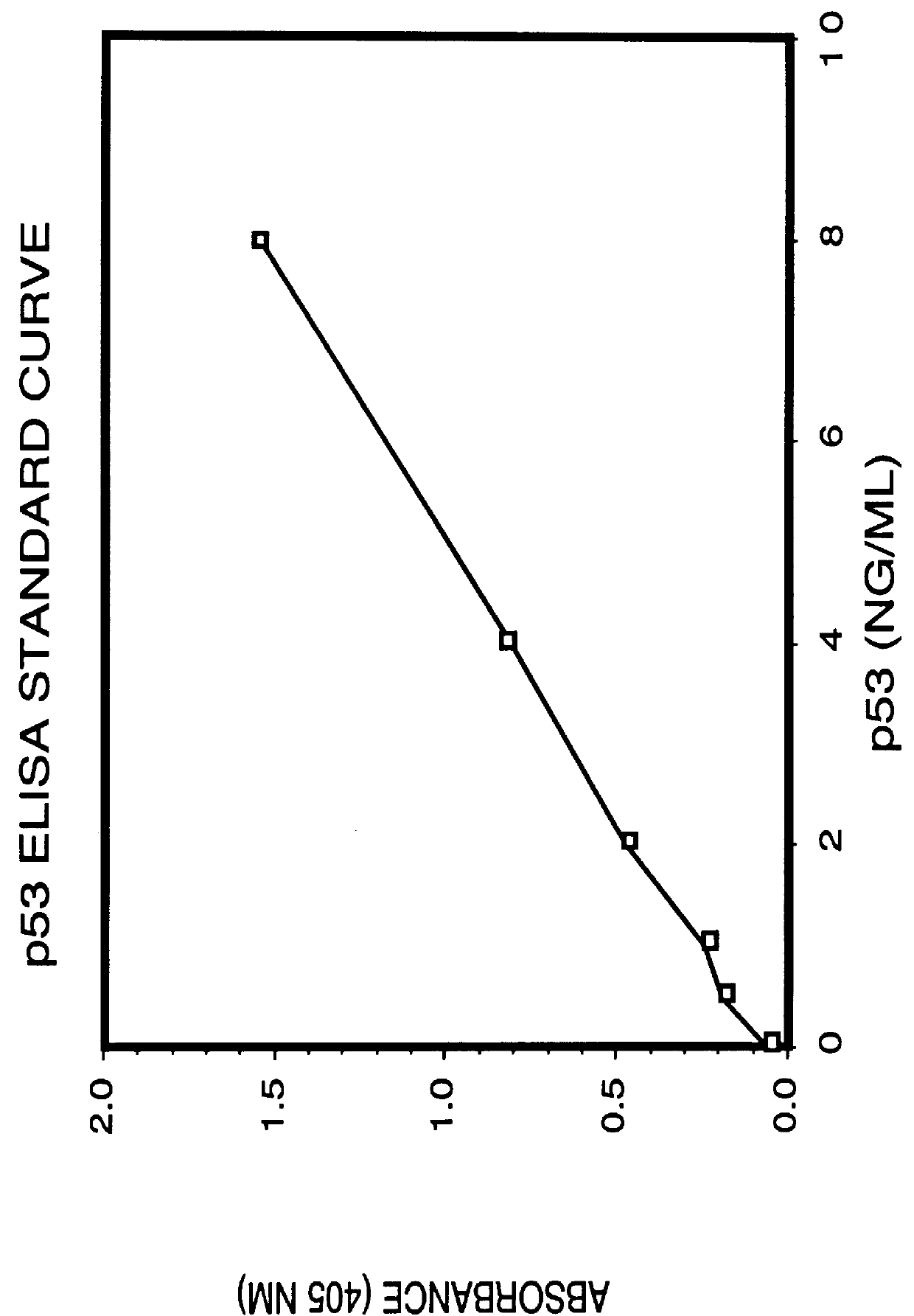
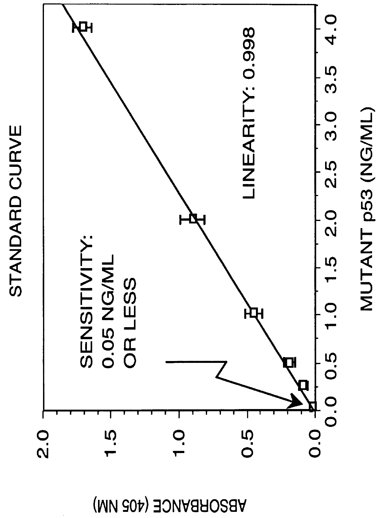
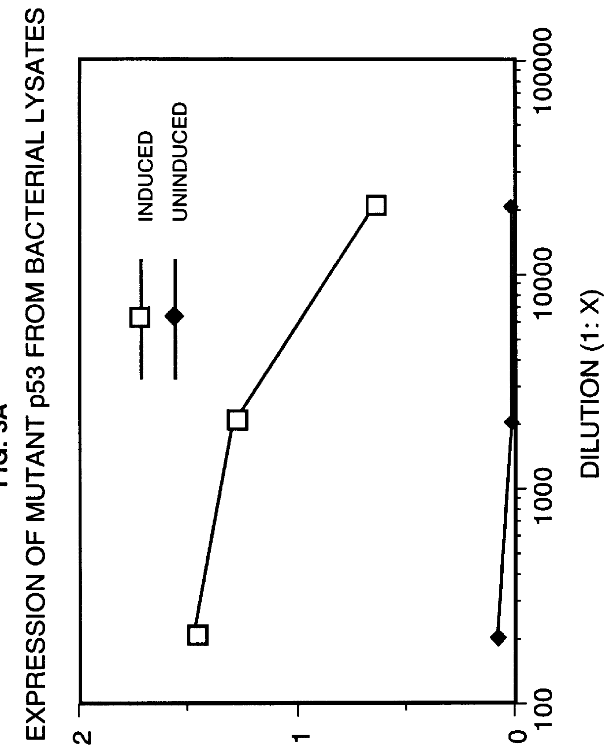
Two site sandwich ELISA assay. In a first example, purified monoclonal antibody from clone PAb1801 (5 .mu.g / ml in 0.1M sodium carbonate buffer, pH 9.0, 100 .mu.l / well), which binds to both wild-type and mutant p53 polypeptide, is adsorbed onto microtiter wells for 16 to 20 hours at 4.degree. C. The antibody solution is removed from the wells and any remaining protein-binding sites are blocked by incubating the wells with PBS containing 1% BSA at pH 7.2 for 2 hours at room temperature (approximately 23.degree. C.). After blocking, the wells are washed 4-times with 200 .mu.l of Wash Buffer (PBS containing 0.1% BSA and 0.05% Tween-20, pH 7.3) then appropriately marked wells are incubated with varying amounts of p53 polypeptide-.beta.-galactosidase fusion protein (standard), or the biological sample to be assayed, for 16-18 hours at 4.degree. C. (alternatively for 3 hours at 37.degree. C.). The plates are emptied by inverting over paper towels and the well washed 4-times as des...

Example

EXAMPLE 2

Immunoblotting Analysis. Sera samples or cell extracts to be analyzed are first subjected to SDS-PAGE as described hereinabove on 5-20% acrylamide gradient gels. After electrophoresis, transfer of proteins to nitrocellulose (Schleicher & Schuell, BA 85, 0.45 um) is performed. Proteins are transferred at 70 volts for 2-3 hours at 4.degree. C. The nitrocellulose is incubated in 0.5% non-fat powdered milk (Carnation) in Phosphate buffered saline pH 7.4 (PBS) containing 0.02% sodium azide for 1 hour at room temperature. The filter is then washed with PBS containing 0.1% Tween 20 (Bio-Rad Lab) and incubated with .sup.125 [I] labeled PAb240 monoclonal antibody (2.0.times.10.sup.6 CPM / ml) diluted in the same buffer for 1.5 hours at 37.degree. C. The filters are washed three times with PBS-Tween-20, dried and exposed to Kodak XR-2 film at -70.degree. C. using intensifying screens. Alternatively, the filter is incubated with PAb240 monoclonal antibody then washed with PBS and incuba...

Example

EXAMPLE 3

Immunoprecipitation of mutant p53 polypeptide with Immunoglobulin. One ml of sera is reacted for 15 hours at 4.degree. C. with 1 microgram of monoclonal antibody PAb240 and 50 .mu.l of a 10% (v / v) suspension of

Protein-A agarose (BRL) containing 0.175 mg Protein-A. The immunocomplexes are washed three times in PBS TDS and collected by centrifugation at 1000 RPM for 10 minutes. The immunoprecipitates are resolved by electrophoresis using a 5-20% acrylamide SDS-PAGE. The resolved proteins are electrophoretically transferred (blotted) to nylon, nitrocellulose or other suitable membrane support. Blotted membranes are blocked for 1 h in a solution containing PBS and 1% to 10% BSA or casein in order to block nonspecific protein binding sites. The blocked membranes are incubated with radiolabeled antibodies directed against mutant p53 polypeptides (either monoclonal or polyclonal) then subjected to autoradiography. Alternatively, the antibodies directed against mutant p53 polypepti...


PUM


Abstract

The invention provides a method for diagnosing in a subject a neoplastic condition which comprises (a) obtaining from the subject a sample of a biological fluid; and (b) detecting the presence in the sample of a mutant p53 polypeptide encoded by an activated p53 oncogene, the presence of the mutant p53 polypeptide in the sample indicating that the subject has the neoplastic condition.

Description

BACKGROUND OF THE INVENTIONThroughout this application various publications are referenced by arabic numbers within parentheses. Full citations for these publications may be found at the end of the specification immediately preceding the claims. The disclosures of these publications in their entireties are hereby incorporated by reference into this application in order to more fully describe the state of the art to which this invention pertains.Cellular oncogenes are normal genes which have been conserved throughoutevolution and are believed to have normal functional roles in the cell. In their non-activated (wild-type) state such cellular oncogenes are sometimes referred to as proto-oncogenes. Proto-oncogenes are not oncogenic or tumorigenic until they are activated in some way. A number of different genetic mechanisms may cause the somatic mutation of oncogenes that results in the activated oncogenes found in tumor cells. These include point mutations, translocations, gene rearran...

Claims


Application Information

Patent Timeline
IPC IPC(8): G01N33/536G01N33/53G01N33/543G01N33/537G01N33/574
CPCG01N33/53G01N33/537G01N33/543G01N33/574G01N33/57484G01N2333/4748G01N2800/52G01N33/57449
Inventor REYNOLDS, JR., FREDERICK H.ZEHEB, RONSTEPHENSON, JOHN R.SORVILLO, JOHN M.
Owner SIEMENS HEALTHCARE DIAGNOSTICS INC
Why Eureka
Try Eureka