
Use of dimly fluorescing nucleic acid dyes in the identification of nucleated cells

Inactive Publication Date: 2001-12-11
BECTON DICKINSON & CO
View PDF6 Cites 34 Cited by
  • Summary
  • Abstract
  • Description
  • Claims
  • Application Information

AI Technical Summary

Benefits of technology

The dye is used at or near saturating concentration to permit simple identification of the nucleated cells.

Problems solved by technology

The importance of such identification is particularly seen in that nucleated cells preparations often contain debris or other matter that can contribute to the overall noise, thereby decreasing the system's sensitivity.
However, when immunofluorescent markers are used, this approach suffers from the drawback that fluorescence signals from such nucleic acid dyes generally overlap and are much brighter than those of immunofluorescent dyes, and thus, tend to interfere with the observation of the immunofluorescent signal.
While nucleic acid dyes which have emission spectra which do not overlap the emission spectra of immunofluorescent dyes are known, they are not practical for use in many instances
However, this procedure suffers from the drawback that the useful range of cell concentrations is greatly reduced as the fluorescence intensity in such circumstances is highly dependent on the number of cells in the sample.
Further, when lower concentration of dye are used, the small amount of dye adsorbed onto the container surface also has a significant effect on the overall amount of dye available for staining nucleic acids, further affecting the sensitivity.

Method used


Image

  • Use of dimly fluorescing nucleic acid dyes in the identification of nucleated cells
  • Use of dimly fluorescing nucleic acid dyes in the identification of nucleated cells
  • Use of dimly fluorescing nucleic acid dyes in the identification of nucleated cells

Examples


example 1

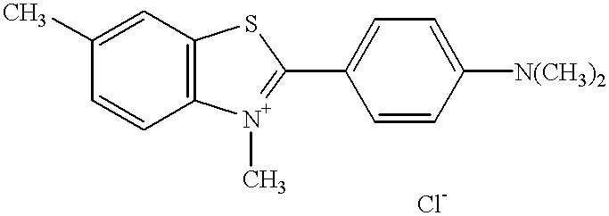
Thioflavin T or another fluorescent nucleic acid dye, SY-VI-24, were used to stain whole blood or diluted whole blood by admixing 50 .mu.L whole blood, 20 .mu.L dye solution, and a phycoerythrin tagged anti-CD4 monoclonal antibody (CD4-PE). After staining the sample we diluted with approximately 0.5 ml of FACS.RTM. Lysing Solution.

Each sample was analyzed on a FACSort flow cytometer at an excitation wavelength of 488 mn and a 530 / 30 filter for green fluorescence and 585 / 42 filter for yellow fluorescence.

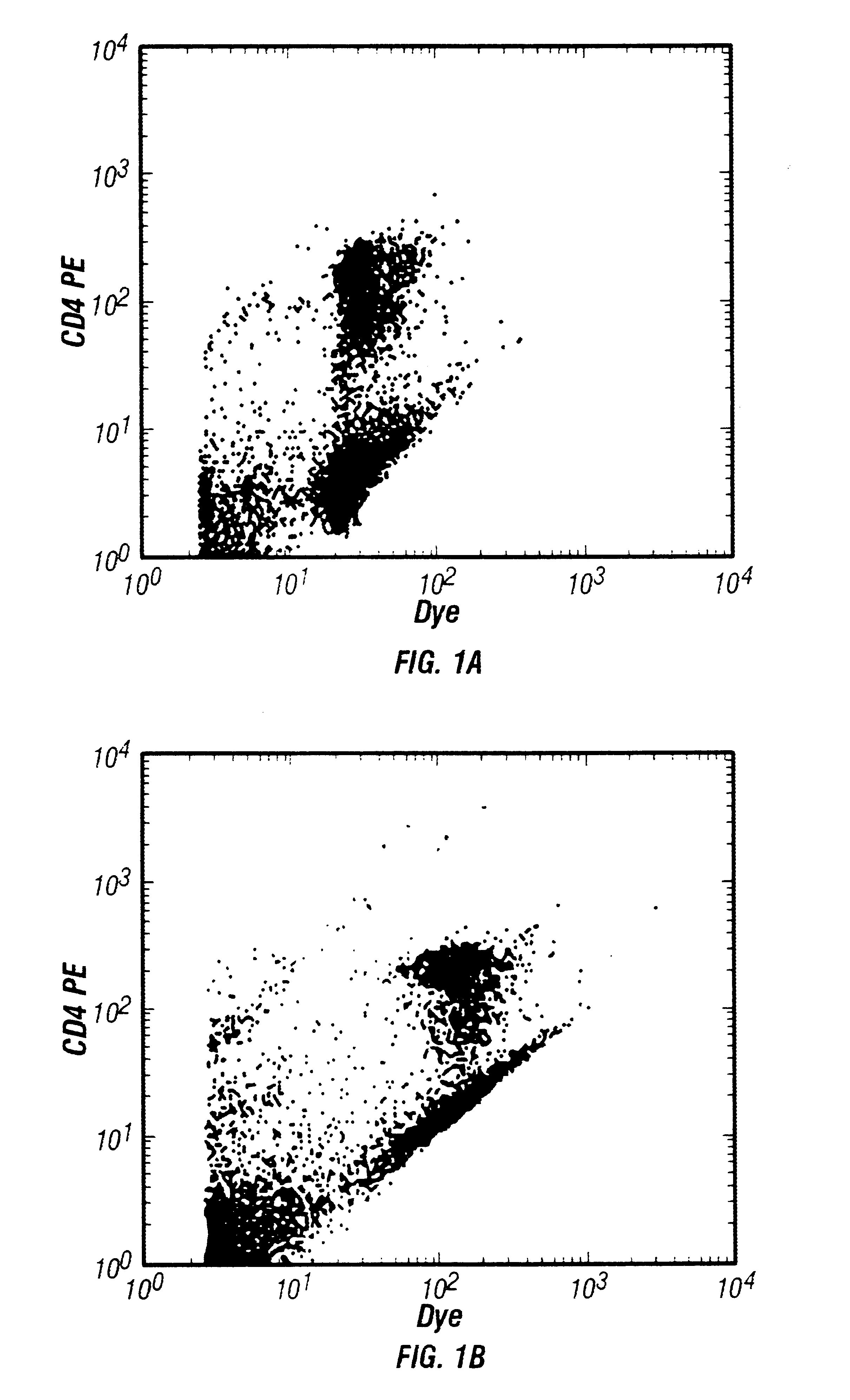
FIG. 3 illustrates dot plots of CD4-PE versus thioflavin T as a function of blood concentration using whole blood and blood diluted 1:10 and 1:100. Briefly, using subsaturating amounts of SY-VI-24 as the nucleic acid dye, a 10 fold decrease in blood cell concentration caused a 10 fold increase in fluorescence from the dye. When lymphocytes were stained simultaneously with SY-VI-24 and CD4 antibody conjugated to the yellow fluorescing tag phycoerythrin (PE), increased fluorescence fro...

example 2

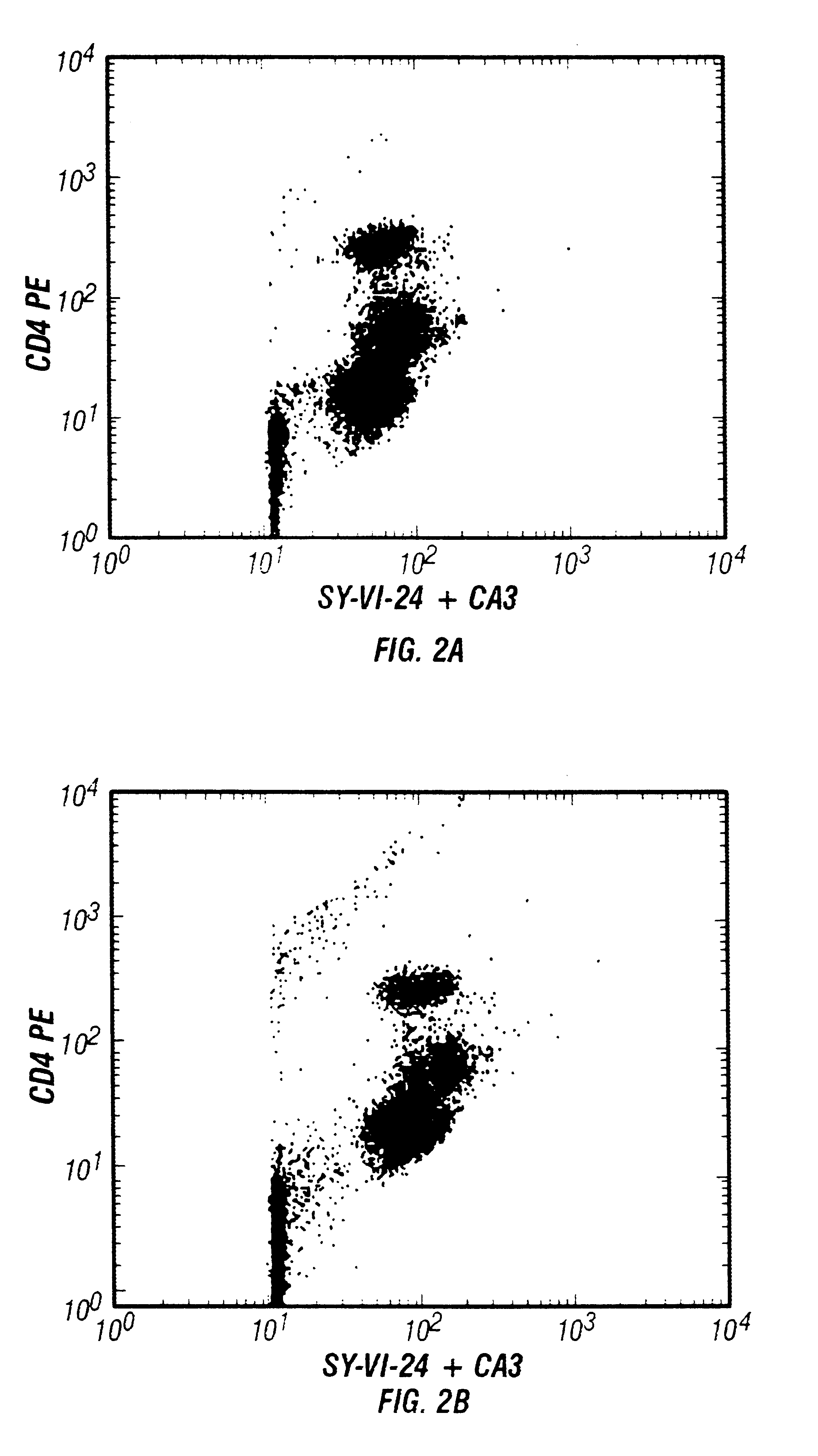
The above experiment was repeated using a nucleic acid dye mixture, SY-VI-24 and chromycin A3 (CA3). Each test used 25 .mu.g CA3 and 1 ng SY-VI-24, at saturating concentrations. The results are shown in FIG. 2.

FIG. 2 shows dot plots of CD4-PE versus nucleic acid stain mixture as a function of blood cell concentration. Panels A-C in FIG. 2 show dot plots of CD4-PE versus nucleic acid dye fluorescence due to the mixture of SY-VI-24 and chromycin A3 (CA3). Results with whole blood are shown in FIG. 2A, and with blood diluted 1:8 and 1:32 before staining in FIGS. 2B and 2C, respectively. There is almost no difference in the results with blood cell concentration varied over this 32 fold range.

FIG. 3 shows similar results where a single dye, thioflavin T, is used at essentially saturating concentration to stain whole blood (FIG. 3A) or the same dye concentration is used to stain blood diluted 1:10 or 1:100 (FIGS. 3B and 3C respectively). Again, the results do not change significantly when...


PUM

PropertyMeasurementUnit
Wavelengthaaaaaaaaaa
Concentrationaaaaaaaaaa
Wavelengthaaaaaaaaaa

Abstract

This invention presents improved methodology for identification of nucleated cells in flow cytometric analysis when immunofluorescent dyes are also used. Briefly, in the method nucleic acids are stained with a fluorescent dye which can then be used to identify the nucleated cells by measurement of fluorescence on a flow cytometer. The improvement presented by this invention is the use of a saturating (or near saturating) amount of a nucleic acid dye, or mixture of dyes, which gives low fluorescence at excitation conditions, so as not to greatly interfere with the signals of the immunofluorescent dyes.

Description

The identification of nucleated cells in multi-parameter flow cytometric analysis is important in a wide array of procedures. The importance of such identification is particularly seen in that nucleated cells preparations often contain debris or other matter that can contribute to the overall noise, thereby decreasing the system's sensitivity.Such cells are often identified by a nucleic acid dye which stains nucleic acids within the nucleus, permitting easy detection. However, when immunofluorescent markers are used, this approach suffers from the drawback that fluorescence signals from such nucleic acid dyes generally overlap and are much brighter than those of immunofluorescent dyes, and thus, tend to interfere with the observation of the immunofluorescent signal. While nucleic acid dyes which have emission spectra which do not overlap the emission spectra of immunofluorescent dyes are known, they are not practical for use in many instancesTo remedy this, researchers have tried to...

Claims


Application Information

Patent Timeline
IPC IPC(8): C12Q1/68G01N21/64G01N33/58G01N33/569
CPCC12Q1/6816G01N21/6428G01N33/56972G01N33/582C12Q2565/625C12Q2563/107
Inventor HOFFMAN, ROBERT A.FREY, THOMAS
Owner BECTON DICKINSON & CO
Learn More