Circumferential transillumination of anatomic junctions using light energy

Inactive Publication Date: 2003-02-04
STRYKER CORP
View PDF16 Cites 81 Cited by
  • Summary
  • Abstract
  • Description
  • Claims
  • Application Information

AI Technical Summary

Benefits of technology

It is an object of the present invention to provide a device which circumferentially transilluminates a junction between anatomic structures using visible light or infrared light or combination of visible and infrared light so as to precisely locate such junction and warn the surgeon of imminent approach thereto.

Problems solved by technology

No prior materials known to the applicants disclose the concept of employing visible or infrared light or a combination of both to circumferentially transilluminate junctions between living structures such as the cervico-vaginal junction.

Method used


Image

  • Circumferential transillumination of anatomic junctions using light energy
  • Circumferential transillumination of anatomic junctions using light energy
  • Circumferential transillumination of anatomic junctions using light energy

Examples


Embodiment Construction

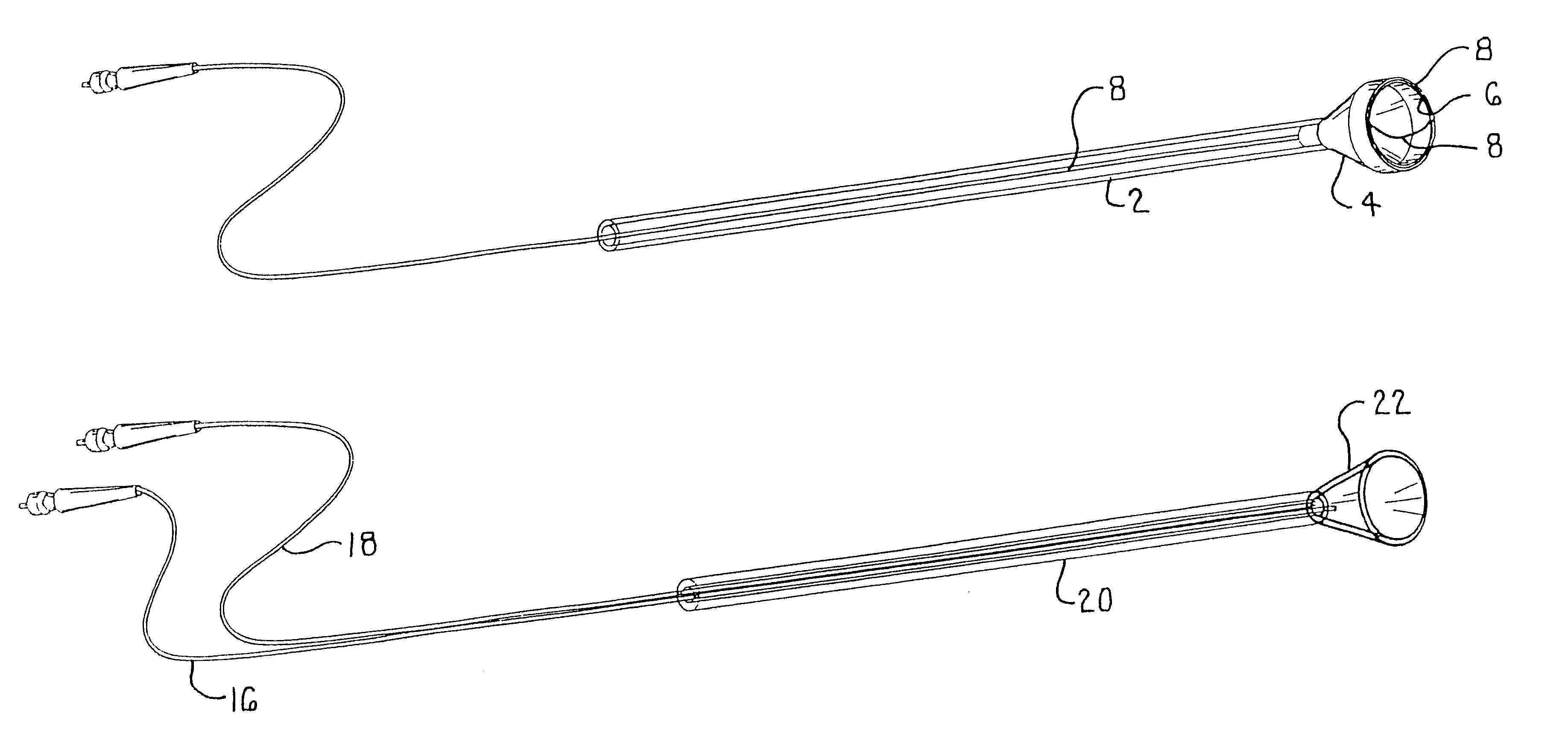
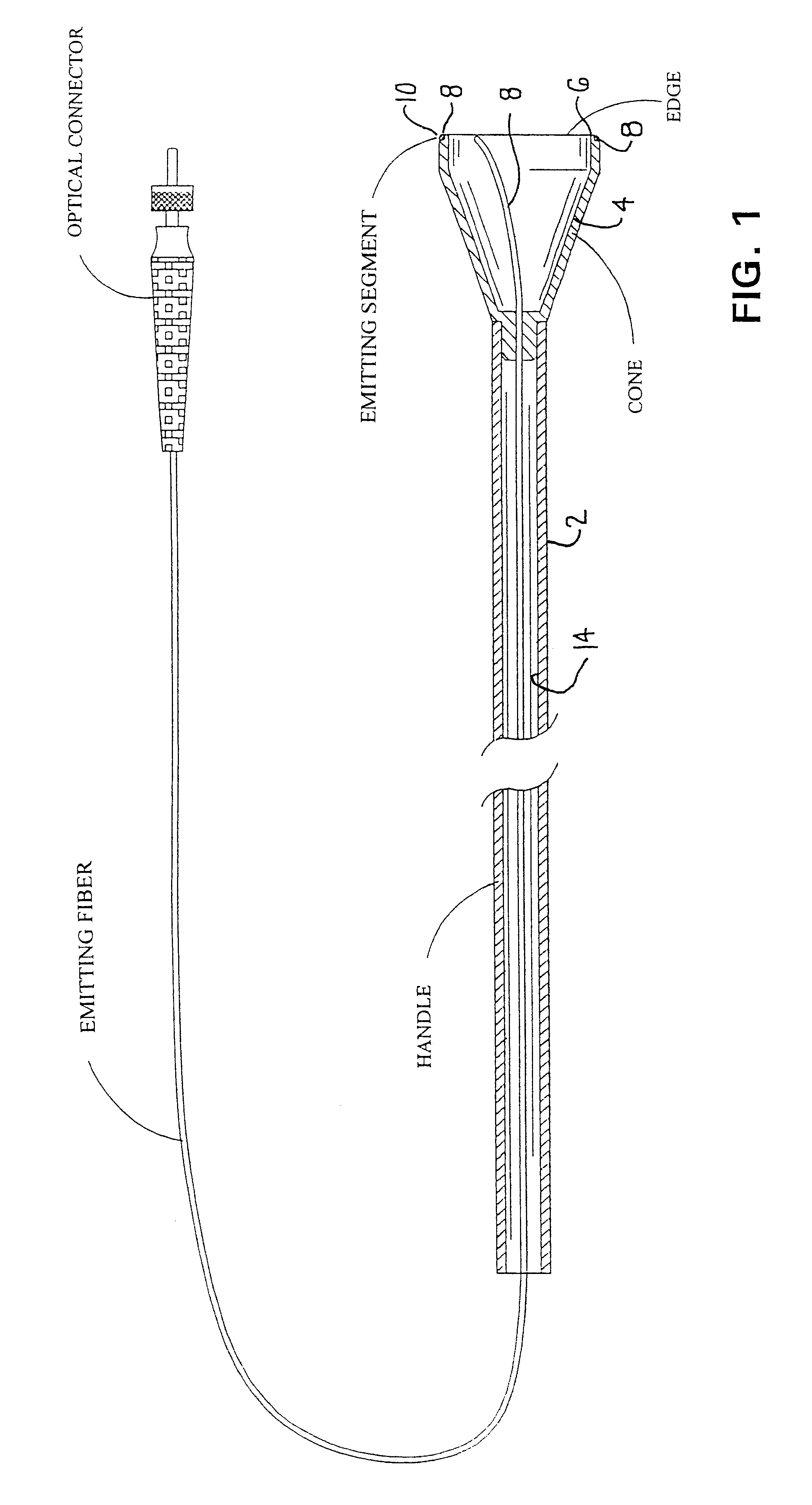
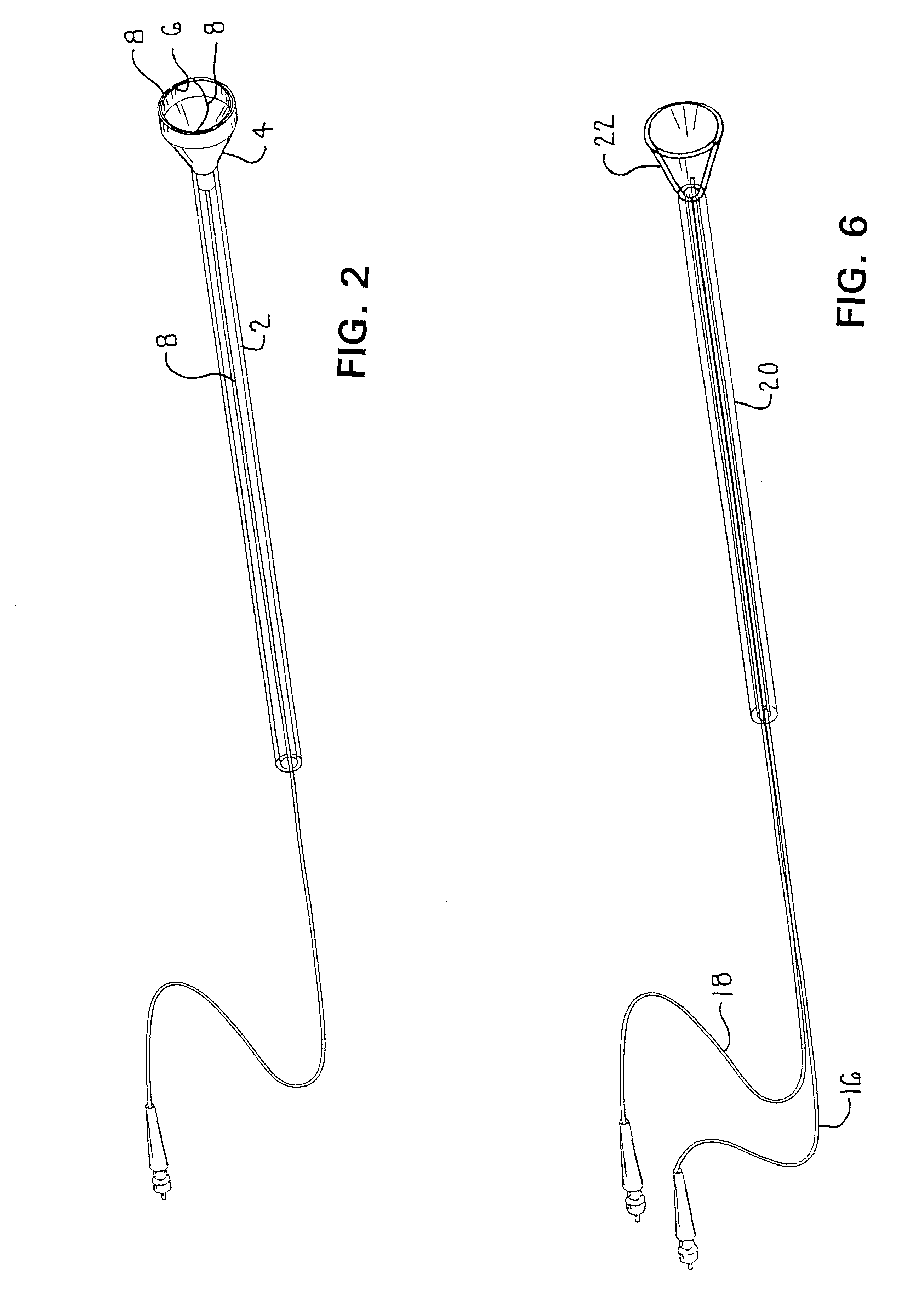
Referring to FIGS. 1, 2, 3 and 4 of the accompanying drawings, the CJT in accordance with the invention comprises a handle 2 attached to the virtual apex of a hollow cone 4. The distal aspect of the cone has an edge 6 to receive an emitting fiber 8. The distal portion of the emitting fiber (ESKA SK-40) is scored at 10 to form the emitting segment as shown in FIG. 4. The region 10 of the fiber is circumferentially bonded to the edge 6 of the cone 4 using epoxy or the like. The emitting fiber has an optical connector 12 (SMA-905) fitted at its proximal end. The emitting fiber is secured to the inside wall 14 of the cone 4 as it passes out of the handle. The emitting segment bonded to the edge of the cone allows light to exit the wall of the end of the emitting fiber 8 resulting in a circumferential light emitting junctional transilluminator.

The emitting segment can be selectively abraded or scored so as to allow light to be emitted such that the entire cervico-vaginal junction (circum...


PUM


Abstract

Apparatus is provided that provides a ring of light around a junction to be viewed; the ring of light being bright enough to be seen on the other side of the junction. Such an arrangement may be used in severing an anatomic body, such as the cervix, from the vagina whereby a surgeon viewing the structure from the pelvic cavity has the junction outlined in light brought into contact with the junction through the vagina.

Description

The present invention relates to methods and apparatus for circumferential transillumination of junctions between anatomic structures of a living body to allow surgeons to identify these junctions and guide incisions during surgical procedures and more specifically assisting in locating and / or avoiding such junctions.Separating the cervix from the vagina during surgical procedures may be difficult. McCartney and Johnson (1995) reported on the use of a vaginal tube to separate the uterus from the vagina during laparoscopic hysterectomy which overcomes this difficulty by placing a wide bore plastic tube in the vagina to expose the cervico-vaginal junction and stretch the vaginal fornices. The stretched vagina falls away from the cervix as the surgeon attempts to locate the most distal aspect of the tube as it protrudes into the pelvic cavity. Palpation using an instrument during laparoscopy is frequently used to discover the distal aspect of the tube at which time the surgeon employs ...

Claims


Application Information

Patent Timeline
Patent Type & Authority Patents(United States)
IPC IPC(8): A61B1/307A61B17/42A61B19/00
CPCA61B1/00167A61B1/0607A61B1/07A61B1/307A61B2017/4225A61B2019/5206A61B2090/306A61B1/0625
Inventor FONTENOT, MARK G.FEINBERG, RICHARDVINING, JIM
Owner STRYKER CORP
Why Eureka
Try Eureka