Integrated imaging apparatus

a technology of integrated imaging and imaging equipment, applied in the field of imaging equipment, can solve the problems of few techniques that can be distinguished, few techniques that can be used, and the raw spectral or imaging measurement seldom reveals directly the property of clinical interes

Inactive Publication Date: 2003-10-28
HYPERMED
View PDF8 Cites 176 Cited by
  • Summary
  • Abstract
  • Description
  • Claims
  • Application Information

AI Technical Summary

Problems solved by technology

However, the volume of information contained in spectroscopic images can make standard data processing techniques cumbersome.
Furthermore, there are few techniques that can demarcate which regions of a spectroscopic image contain similar spectra without a priori knowledge of either the spectral data or the sample's composition.
There are few techniques that can demarcate which regions of a sample contain similar substances without a priori knowledge of the sample's composition.
While it is now clear that both spectroscopy and spectroscopic imaging can play roles in providing medically relevant information, the raw spectral or imaging measurement seldom reveals directly the property of clinical interest.
For example using spectroscopy, one cannot easily determine whether the tissue is cancerous, or determine blood glucose concentrations and the adequacy of tissue perfusion.
The very reason why multiple scans are useful is what makes the registration process difficult.
As each modality images tissue differently and has its own artifacts and noise characteristics, accurately modeling the intensity relationship between the scans, and subsequently aligning them, is difficult.
The main drawback of this method is that the markers must remain attached to the patient at the same positions throughout all image acquisitions.
For applications such as change detection over months or years, this registration method is not suitable.
In general, methods of feature extraction such as intensity thresholding or edge detection do not work well on medical scans, due to non-linear gain fields and highly textured structures.
Without the ability to accurately localize corresponding features in the images, alignment in this manner is difficult.
Therefore, two images that have a simple linear intensity relationship may be straightforward to register, but do not provide any additional information than one scan by itself.
On the other hand, if the images are completely independent (e.g. no intensity relationship exists between them), then they cannot be registered using voxel-based methods.
In general, explicitly computing the function F that relates two imaging modalities is difficult and under-constrained.
However, when this information is available, the prior joint intensity model provides the registration algorithm with additional guidance which results in convergence on the correct alignment more quickly, more reliably and from more remote initial starting points.

Method used


Image

  • Integrated imaging apparatus
  • Integrated imaging apparatus

Examples


Embodiment Construction

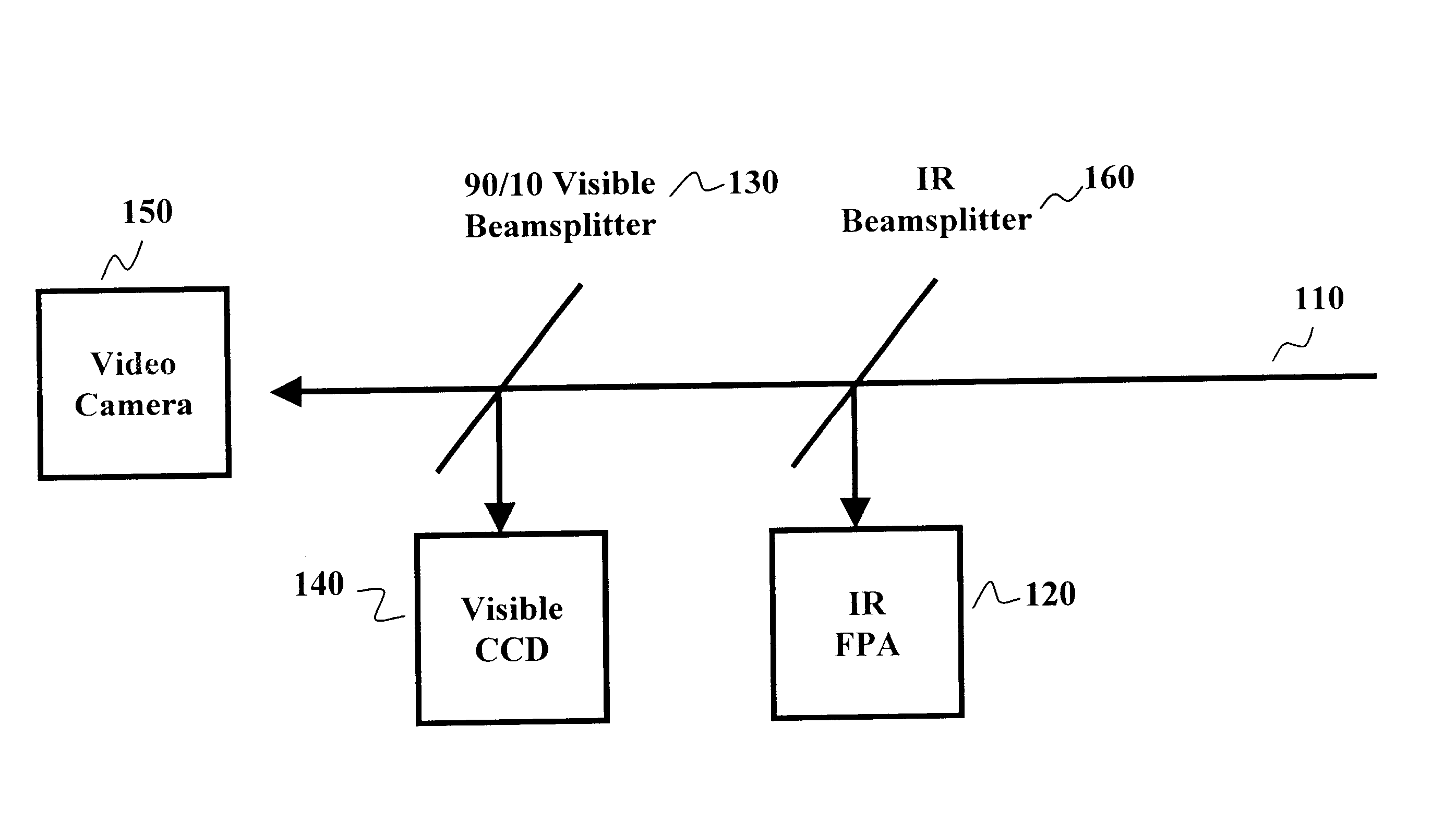
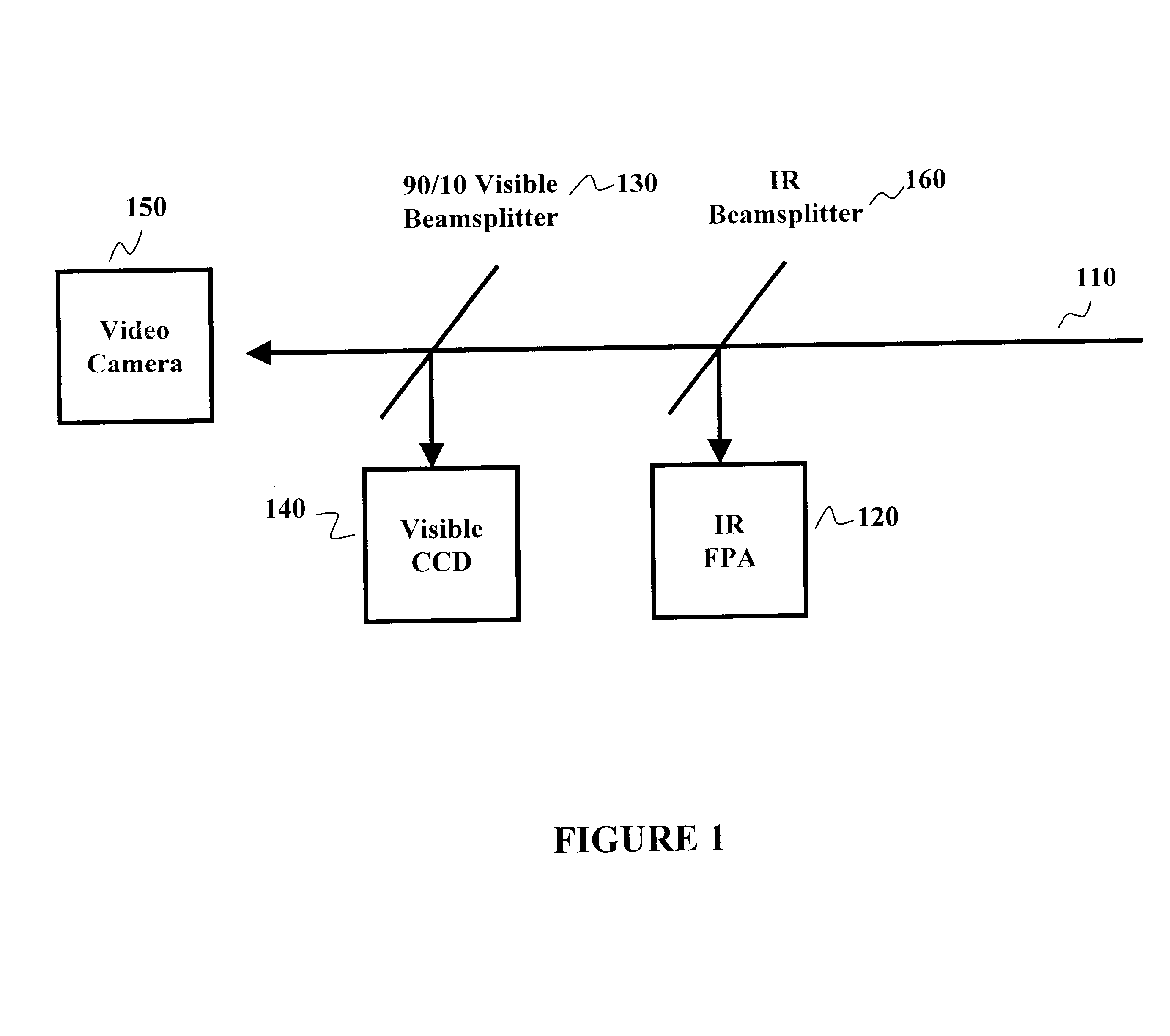
As embodied and broadly described herein, the present invention is directed to an imaging apparatus and methods for performing real-time or near real-time assessment and monitoring. Embodiments of the device are useful in a plurality of settings including surgery, clinical procedures, tissue assessment, diagnostic procedures, forensic, health monitoring and medical evaluations.

It has been surprisingly found that the pairing of hyperspectral imaging data with data obtained from other single-image imaging methodologies, (examples of which include thermal imaging or fluorescence imaging) provides a sensitive and accurate assessment measure of a physiological condition. This is particularly appealing in terms of tissue assessment in that both thermal perfusion assessments and various multi-modal tissue signatures which incorporate things such as oxyhemoglobin / deoxyhemoglobin ratios and other indices of tissue physiology, pathology or function are interrelated. By fusing data from multip...


PUM


Abstract

The invention is directed to imaging methods for performing real-time or near real-time assessment and monitoring. Embodiments of these methods are useful in a plurality of settings including surgery, clinical procedures, tissue assessment, diagnostic procedures, forensic, health monitoring and medical evaluations.

Description

1. Field of the InventionThe invention is directed to an imaging apparatus and methods for performing assessment and monitoring with interpreted imaging. Embodiments of the invention are particularly useful in surgery, clinical procedures, tissue assessment, diagnostic procedures, health monitoring, and medical evaluations.2. Description of the BackgroundSpectroscopy, whether it is visible, near infrared, infrared or Raman, is an enormously powerful tool for the analysis of biomedical samples. The medical community, however, has a definite preference for imaging methods, as exemplified by methods such as MRI and CT scanning as well as standard X-ray photography and ultrasound imaging. This is entirely understandable as many factors need to be taken into account for a physician to make a clinical diagnosis. Imaging methods potentially can provide far more information to a physician than their non-imaging counterparts. With this medical reality in mind, there has been considerable eff...

Claims


Application Information

Patent Timeline
Patent Type & Authority Patents(United States)
IPC IPC(8): A61B5/00A61B5/103A61B10/00A61B5/145A61B5/1455G06T1/00
CPCA61B5/0059A61B5/0071A61B5/0073A61B5/0077A61B5/015A61B5/14535A61B5/445A61B5/415A61B5/418A61B5/442A61B5/444A61B5/1455A61B5/441
Inventor FREEMAN, JENNY E.HOPMEIER, MICHAEL J.LEVENTEN, M.MANSFIELD, JAMESLEWIS, EDGAR NEIL
Owner HYPERMED
Why Eureka
Try Eureka