

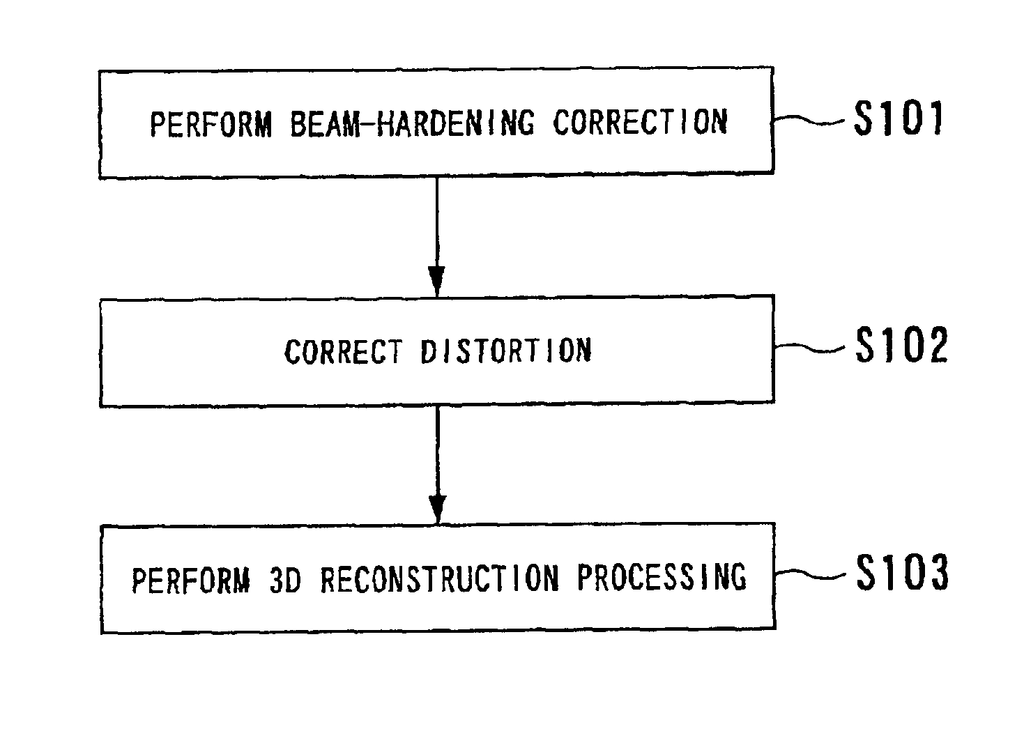
Image processing involving correction of beam hardening
a beam hardening and image processing technology, applied in the field of image processing apparatus and medical imaging modality, can solve the problems of no correction technique for artifacts caused by contrast agents, no correction techniques have been reported yet, and no effect of blood vessel artifacts
- Summary
- Abstract
- Description
- Claims
- Application Information
AI Technical Summary
Benefits of technology
Problems solved by technology
Method used
Image

Examples
first embodiment
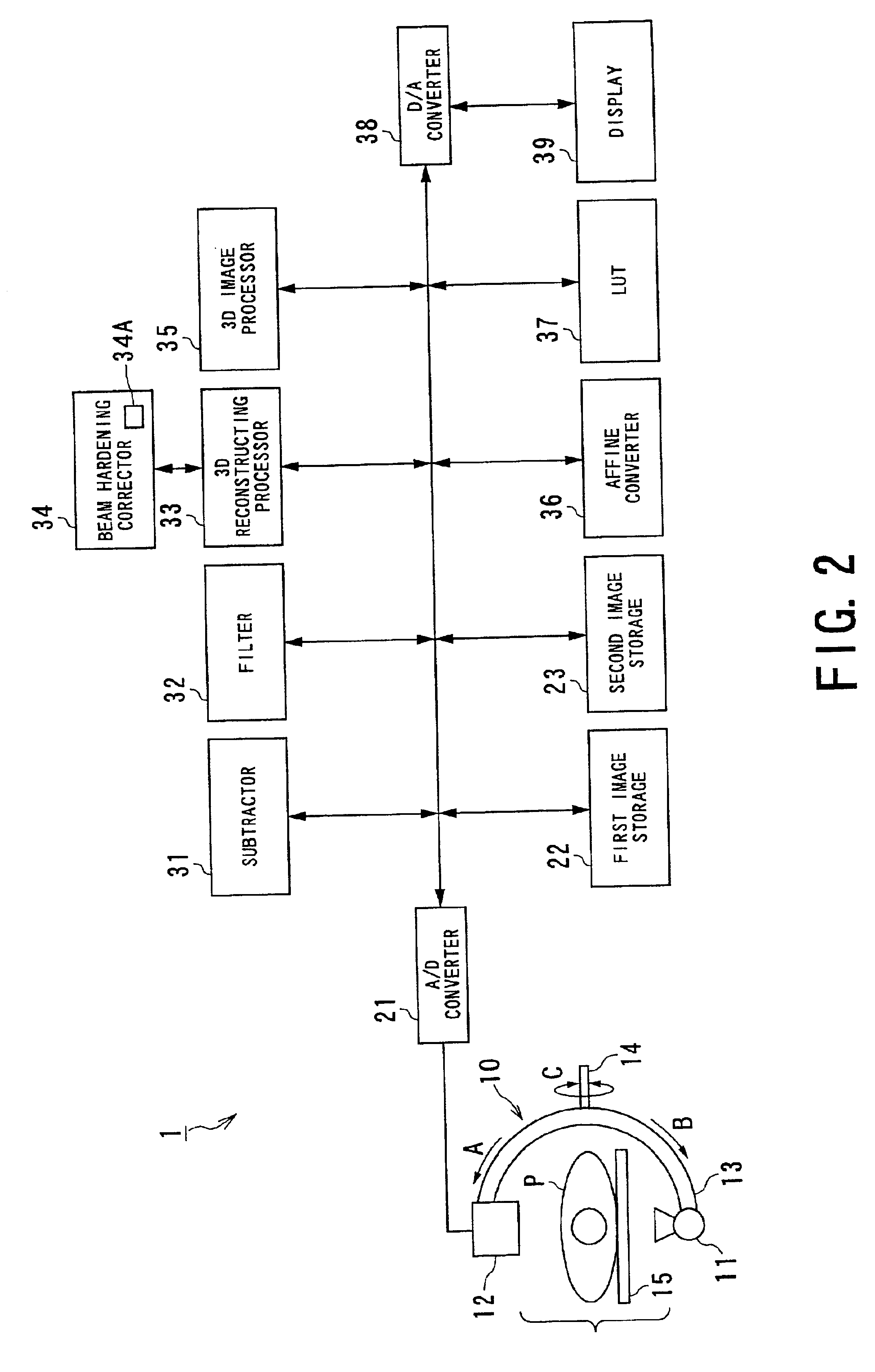
Referring to FIGS. 2 to 6, a first embodiment of the medical modality according to the present invention will now be described, in which the image processing apparatus of the present invention is functionally incorporated in the imaging modality. In this first embodiment, the medical modality is reduced into practice as a three-dimensional (3D) angiography system.
The 3D-angiography system according to the present embodiment is able to perform processing including the production of images on the basis of two modes consisting of a DA (Digital angiography) mode and a DSA (Digital Subtraction Angiography) mode. The DA mode allows the system to perform X-ray imaging with a contrast agent to acquire an X-ray image that includes flows of the contrast agent administered into an object and to display and store the acquired image. On the other hand, under the DSA mode, both of an X-ray mask image acquired without injecting a contrast agent and an X-ray image (contrast-enhanced image or live i...
second embodiment
A second embodiment of the present invention will now be described. In the following, the constituents substantially similar or identical to those in the first embodiment will be omitted from the description, only different constituents being described.
In the first embodiment, the beam-hardening phenomenon has been corrected using the plural correction tables 34A each of which memorizes correction data collected to each value of voltage to be applied to the X-ray tube. Instead of this, the second embodiment adopts a correction table memorizing correction data that is set to only a representative of a certain range of voltages to be applied to the X-ray tube.
Practically, the beam hardening corrector 34 shown in FIG. 2 is configured so that it has a correction table 34A that memorizes correcting data that is set to only a representative voltage selected from a certain range of voltages to be applied to the X-ray tube. In this case, however, it is required that the voltage of the X-ray...
third embodiment
A third embodiment of the present invention will now be described, which relates to a further configuration of the correction table 34A.
Although the first embodiment takes only the influences of a contrast agent into account, some other factors, such as locations (a bone or a soft tissue), may have a large influence on measured DSA data. For example, in the case of measuring the head, its bone influences the measurement largely. By contrast, in measuring the abdomen, the soft tissue plays as an influential factor.
In the present embodiment, the beam-hardening corrector 34 shown in FIG. 2 has one or more correction tables 34A, in which correction data for the bone and / or the soft tissue, each having typical amount of densities, are stored.
The one or more correction tables 34A are divided into one for measuring the head, another for measuring the abdomen, and others, depending on which part of an object is imaged. Each of such categorized correction tables 34A is further prepared for e...
PUM
 Abstract
Description
Claims
Application Information