Method for treating a sphincter

Inactive Publication Date: 2005-03-15
CURON MEDICAL +1
View PDF182 Cites 127 Cited by
  • Summary
  • Abstract
  • Description
  • Claims
  • Application Information

AI Technical Summary

Benefits of technology

One aspect of the invention provides a method for treating a sphincter. The method provides a polymer material having a liquid state. The method also provides a catheter having a distal end, a tissue piercing device carried by the distal end, and an energy delivery device coupled to the tissue piercing device. The tissue piercing device has a lumen. The method introduces the catheter into an esophagus and pierces an exterior sphincter tissu

Problems solved by technology

These contents are highly acidic and potentially injurious to the esophagus resulting in a number of possible complications of varying medical severity.
On a chronic basis, GERD subjects the esophagus to ulcer formation, or esophagitis and may result in more severe complications including esophageal obstruction, significant blood loss and perforation of the esophagus.
However, while pharmacologic agents may provide short term relief, they do not address the underlying cause of LES dysfunction.
Although the operation has a high rate of success, it is an open abdominal procedure with the usual risks of abdominal surgery including: postoperative infection, herniation at the operative site, internal hemorrhage and perforation of the esophagus or of the cardia.
This rate of complication drives up both the medical cost and convalescence period for the procedure and may exclude portions of certain patient populations (e.g., the elderly and immuno-compromised).
However, this procedure is still traumatic to the LES

Method used


Image

  • Method for treating a sphincter
  • Method for treating a sphincter
  • Method for treating a sphincter

Examples


Embodiment Construction

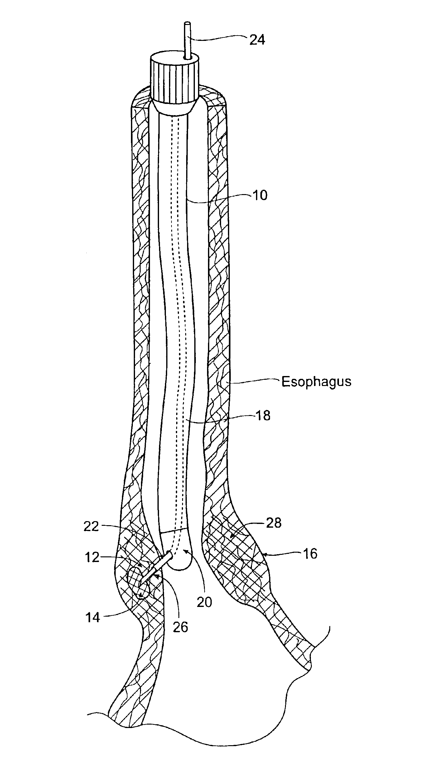
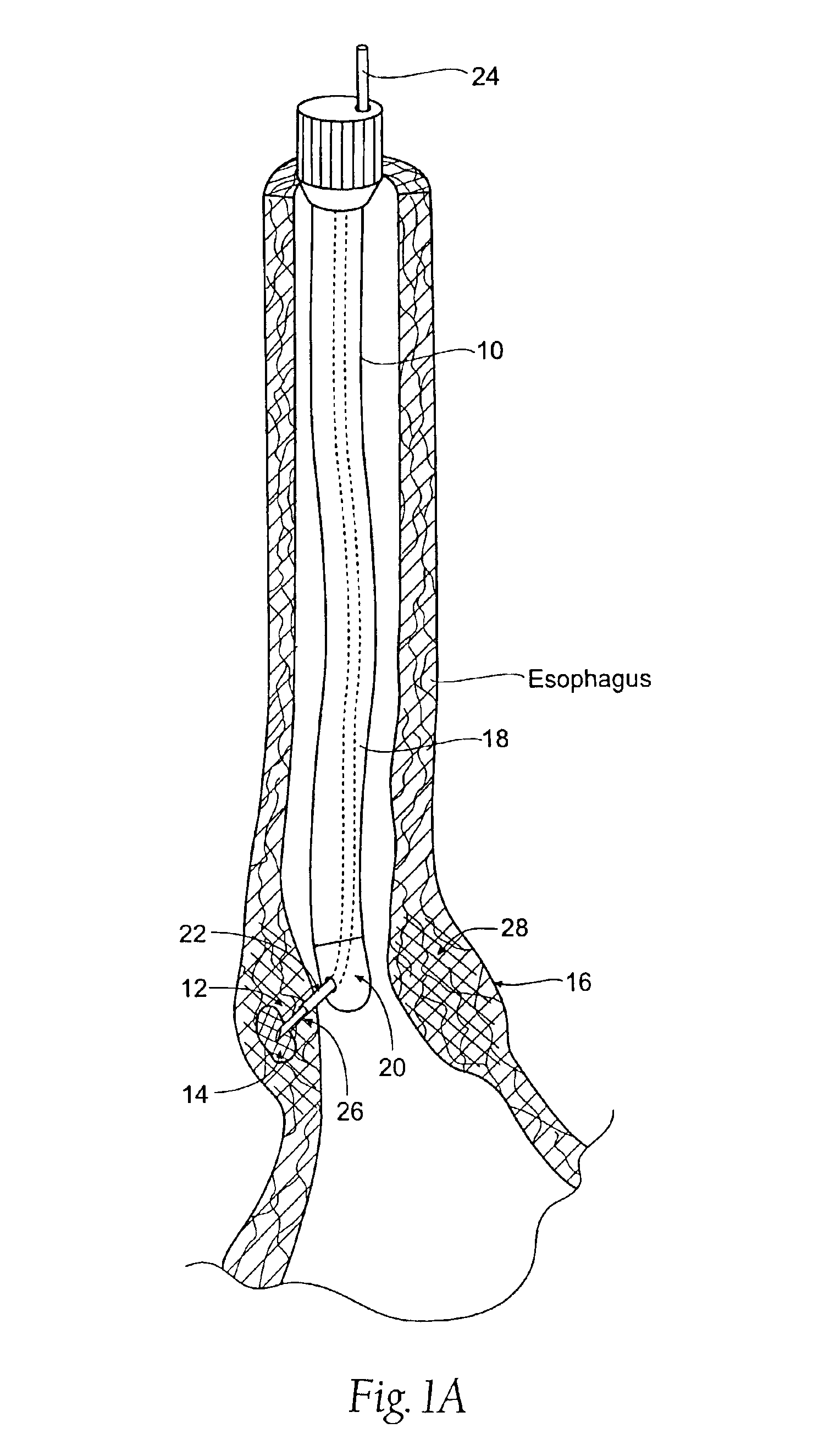
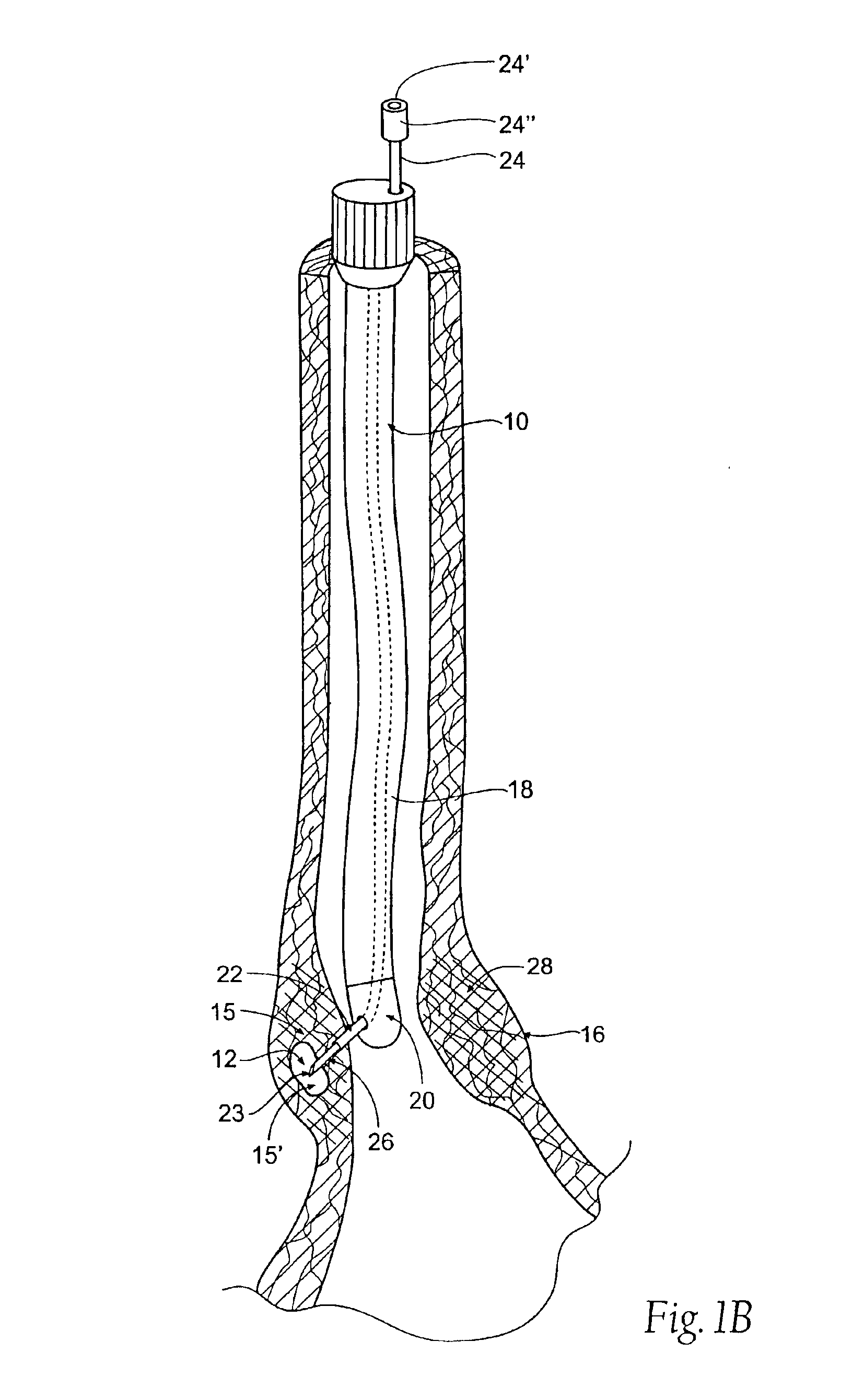
Referring now to FIGS. 1A, 1B and 2, one embodiment of a sphincter treatment apparatus 10 delivers energy to a target tissue site 12, also called treatment site 12, to produce cell necrosis 14 in a sphincter 16, such as the lower esophageal sphincter (LES). In this embodiment, sphincter treatment apparatus 10 comprises a flexible elongate shaft 18, also called introducer 18, or catheter 18, with a distal extremity 20, also called catheter end 20, in turn coupled with one or more energy delivery devices 22. Energy delivery devices 22 are coupled to a guide wire 24 also called cable 24 and are also configured to be coupled to a power source. Energy delivery device 22 is coupled to a tissue piercing device 26, which can also be the distal end 26 of energy delivery device 22. Energy delivery device 22 and tissue piercing device 26 may both have a continuous internal lumen 23 that is fluidically coupled to a fluid lumen 24′ in guide wire 24. Energy delivery device 22 and tissue piercing ...


PUM


Abstract

A method for treating a sphincter provides a polymer material having a liquid state. The method also provides a catheter having a distal end, a tissue piercing device carried by the distal end, and an energy delivery device coupled to the tissue piercing device. The tissue piercing device has a lumen. The method introduces the catheter into an esophagus and pierces an exterior sphincter tissue surface within with the tissue piercing device. The method advances the tissue piercing device into an interior sphincter tissue site and conveys the polymer material while in a liquid state through the lumen into the interior sphincter tissue site. The method delivers energy to the tissue piercing device to transform the polymer material into a less liquid state within the interior sphincter tissue site, to thereby remodel the sphincter.

Description

FIELD OF THE INVENTIONThis invention relates generally to an apparatus to treat sphincters, and more particularly to an apparatus to treat esophageal sphincters.DESCRIPTION OF RELATED ARTGastroesophageal reflux disease (GERD) is a common gastroesophageal disorder in which the stomach contents are ejected into the lower esophagus due to a dysfunction of the lower esophageal sphincter (LES). These contents are highly acidic and potentially injurious to the esophagus resulting in a number of possible complications of varying medical severity. The reported incidence of GERD in the U.S. is as high as 10% of the population (Castell D O; Johnston B T: Gastroesophageal Reflux Disease: Current Strategies For Patient Management. Arch Fam Med, 5(4):221-7; (1996 April)).Acute symptoms of GERD include heartburn, pulmonary disorders and chest pain. On a chronic basis, GERD subjects the esophagus to ulcer formation, or esophagitis and may result in more severe complications including esophageal ob...

Claims


Application Information

Patent Timeline
IPC IPC(8): A61B18/14A61B18/12A61B18/18A61B18/00
CPCA61B18/1492A61B18/1477A61B18/1206A61B2218/002A61B18/18A61B2018/00011A61B2018/00029A61B2018/00047A61B2018/00214A61B2018/0022A61B2018/00494A61B2018/00553A61B2018/00666A61B2018/00678A61B2018/00684A61B2018/00702A61B2018/00708A61B2018/00726A61B2018/00744A61B2018/00791A61B2018/00875A61B2018/124A61B2018/1253A61B2018/126A61B2018/1425
Inventor EDWARDS, STUART DUTLEY, DAVID SLAX, RONALD GGAISER, JOHN
Owner CURON MEDICAL
Why Eureka
Try Eureka