Magnetic resonance imaging (MRI) with continuous table motion

a magnetic resonance imaging and table motion technology, applied in the field of magnetic resonance imaging, can solve the problems of inability to precisely monitor the gradual distribution of contrast agents, and inability to accurately monitor the distribution of angiographic images

Active Publication Date: 2005-03-22
SIEMENS HEALTHCARE GMBH
View PDF5 Cites 17 Cited by
  • Summary
  • Abstract
  • Description
  • Claims
  • Application Information

AI Technical Summary

Benefits of technology

An object of the present invention is to provide a method for angiograp

Problems solved by technology

In spite of this defining feature, angiographic images (i.e., images of the blood vessels in the human body, particularly those in the organs with sufficient blood supply) are limited in terms of their contrast in the generic MR imaging process; these limits, how

Method used


Image

  • Magnetic resonance imaging (MRI) with continuous table motion
  • Magnetic resonance imaging (MRI) with continuous table motion
  • Magnetic resonance imaging (MRI) with continuous table motion

Examples


Embodiment Construction

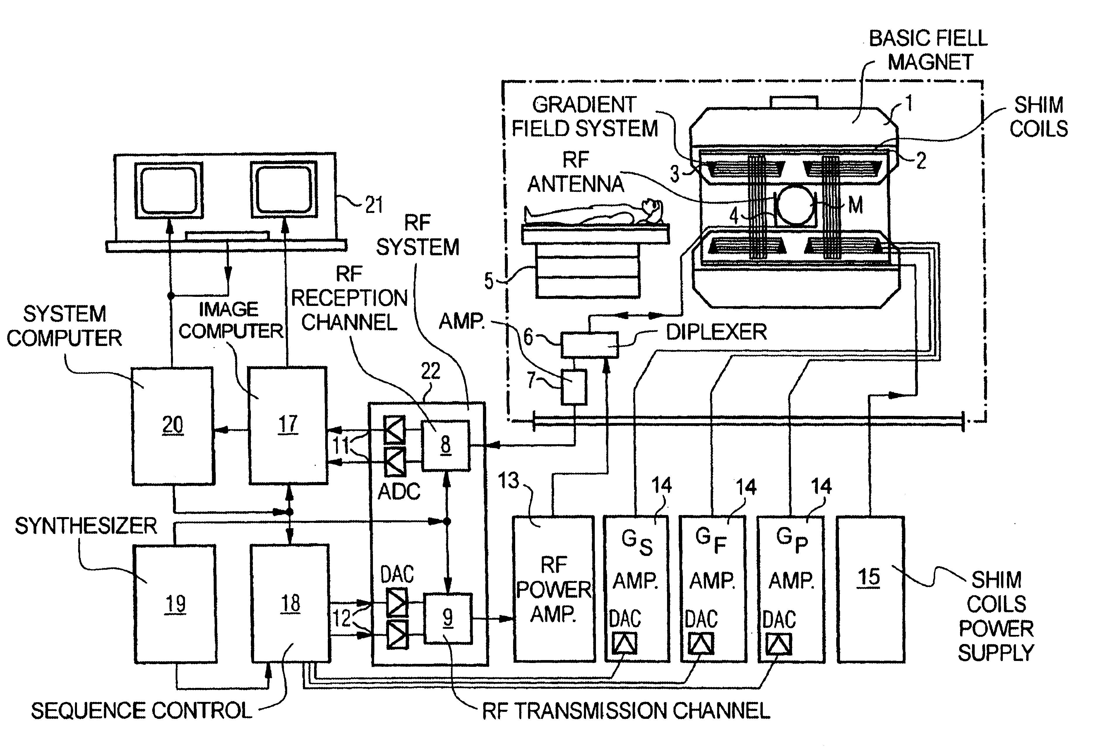
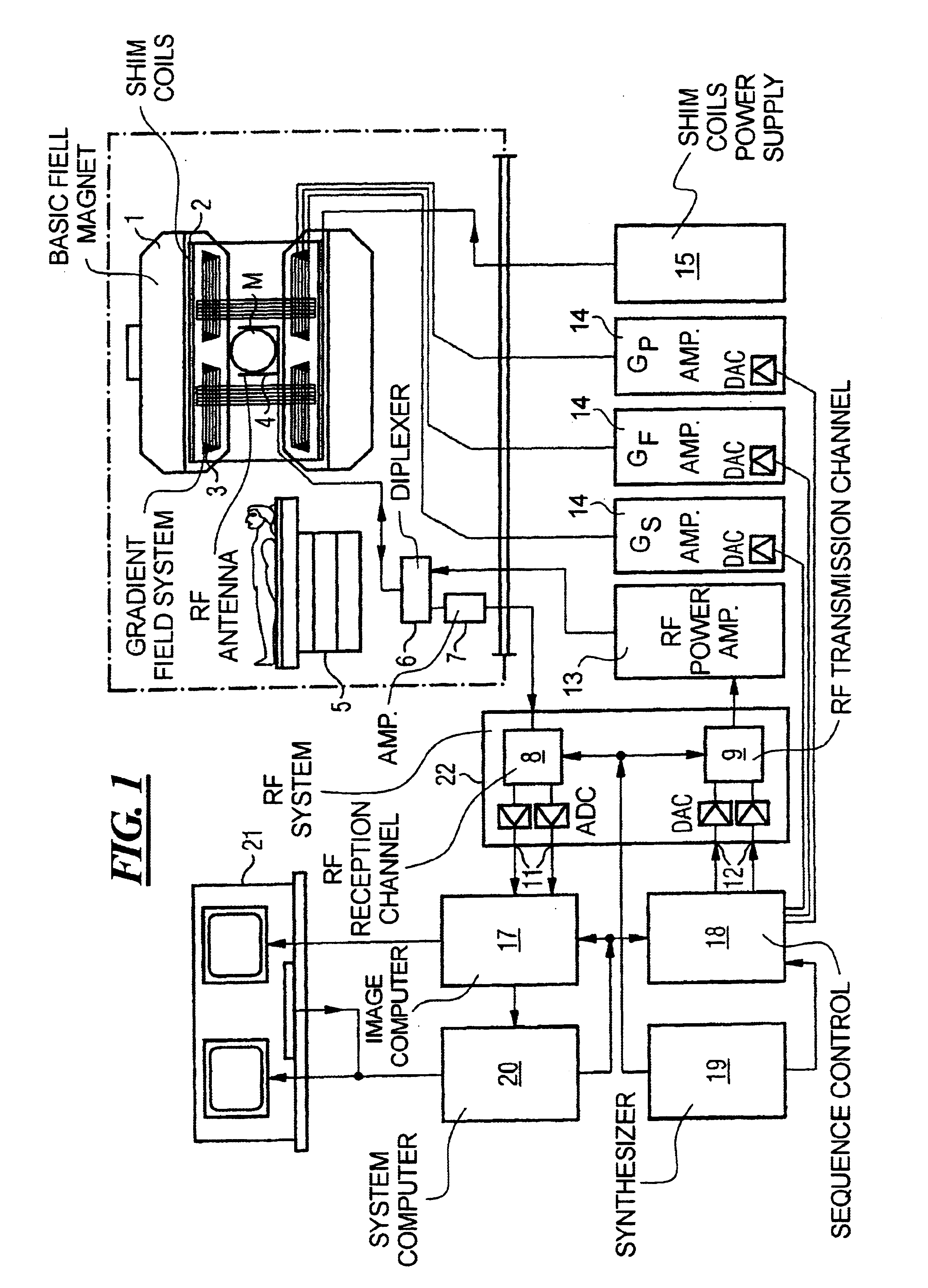
FIG. 1 is a schematic illustration of a magnetic resonance imaging apparatus operable with an improved projection angiography acquisition technique according to the present invention. The arrangement of the components of the magnetic resonance imaging apparatus is the same as that of a conventional magnetic resonance imaging apparatus, but it is operable in a non-conventional manner, as described below. A basic field magnet 1 generates a strong, time invariant, basic magnetic field that polarizes, i.e., aligns the nuclear spins in the zone of the object, such as a part of the human body, to be scanned. The high degree of homogeneity of the magnetic field required for magnetic resonance imaging is defined within a sphere-like volume M, into which the relevant region of the patient's body must be brought. In order to meet the homogeneity requirements, and especially to eliminate any time invariant influences, the shim plates (made of a ferromagnetic material) are placed in suitable po...


PUM


Abstract

In a magnetic resonance imaging method and apparatus and computer program product, spatial scanning of a block-shaped volume of a patient is undertaken by applying a phase-coding gradient in a slice selection direction, as well as by applying a frequency-coding gradient in a plane vertical to the direction of the phase-coding gradient. The body is uniformly and continuously moved by uniformly and continuously moving a table on which the body is situated until the block-shaped volume moves uniformly through the homogeneity volume of the magnetic resonance apparatus. The gradient fields are simultaneously uniformly continuously shifted in conformity with the motion of the table, so that the block-shaped volume being scanned does not move in relation to the table, and thus does not move in relation to the patient, until the block-shaped volume proceeds through the entirety of the homogeneity volume, and scanning is completed.

Description

BACKGROUND OF THE INVENTION1. Field of the InventionThe present invention relates generally to magnetic resonance imaging (MRI), also known as magnetic resonance tomography (MRT) as applied in medicine to examine patients.2. Description of the Prior ArtMRI is based on the physical phenomenon of nuclear magnetic resonance and has been successfully used as an imaging procedure in medicine and in biophysics for over 15 years. Using this examination modality, the patient is exposed to a strong, constant magnetic field. This causes the nuclear spins of atoms in the object (which were previously randomly oriented) to align in a certain direction. Radio-frequency energy can then excite the aligned nuclear spins to a certain oscillation. It is this oscillation that generates in the MRI system the actual measurement signal, which is then received using suitable reception coils. Using spatially linearly variable magnetic fields generated by gradient coils, the examination subject can be spati...

Claims


Application Information

Patent Timeline
IPC IPC(8): A61B5/055G01R33/563G01R33/54G01R33/28
CPCA61B5/055G01R33/56375G01R33/281
Inventor KIRSCH, RAINER
Owner SIEMENS HEALTHCARE GMBH
Why Eureka
Try Eureka