Microarrays and their manufacture

a microarray and micro-array technology, applied in the field of microarrays, can solve the problems of high cost of currently produced biochips or microarrays, and the application of this technology to routine clinical use, and achieve the effect of being inexpensive and sufficiently standardized

Inactive Publication Date: 2005-05-03
LARGE SCALE PROTEOMICS
View PDF231 Cites 110 Cited by
  • Summary
  • Abstract
  • Description
  • Claims
  • Application Information

AI Technical Summary

Benefits of technology

[0027]In a further related aspect the invention relates to the development of sets of tests on different chips done in sequence, which reduces the cost, delay, and inconvenience of diagnosing human diseases, while providing complex data ordinarily obtained by time-consuming sequential batteries of conventional tests.
[0028]In still another aspect, the invention relates to the fabrication of identical arrays which are sufficiently inexpensive to allow several identical arrays to be mounted on the same slide or test strip, and cross compared for quality control purposes.
[0029]In a still further aspect, the invention relates to the incorporation of a non-fluorescent dye or other light absorbing material in the substance of the array to control the depth to which light used to excite fluorescence penetrates the array, thereby controlling the depth to which fluorescence analytes are detected, and insuring that fluorescent analytes which diffuse too deeply into the contents of the cells and therefore do not diffuse out are not detected.
[0036]It is an additional aspect of this invention to produce biochips which are inexpensive and sufficiently standardized to allow more than one to be used for each analysis, and for controls and standards to be routinely run simultaneously in parallel. For added quality assurance, sections from different portions of the bundle or different ends may be used. One way of sectioning from different portions of the bundle is to cut or bend the bundle in the middle and align the two halfs to form a single larger bundle thereby producing a section where each fiber is represented twice.

Problems solved by technology

These factors have contributed to the high cost of currently produced biochips or microarrays, and have discouraged application of this technology to routine clinical use.

Method used


Image

  • Microarrays and their manufacture
  • Microarrays and their manufacture
  • Microarrays and their manufacture

Examples


example 1

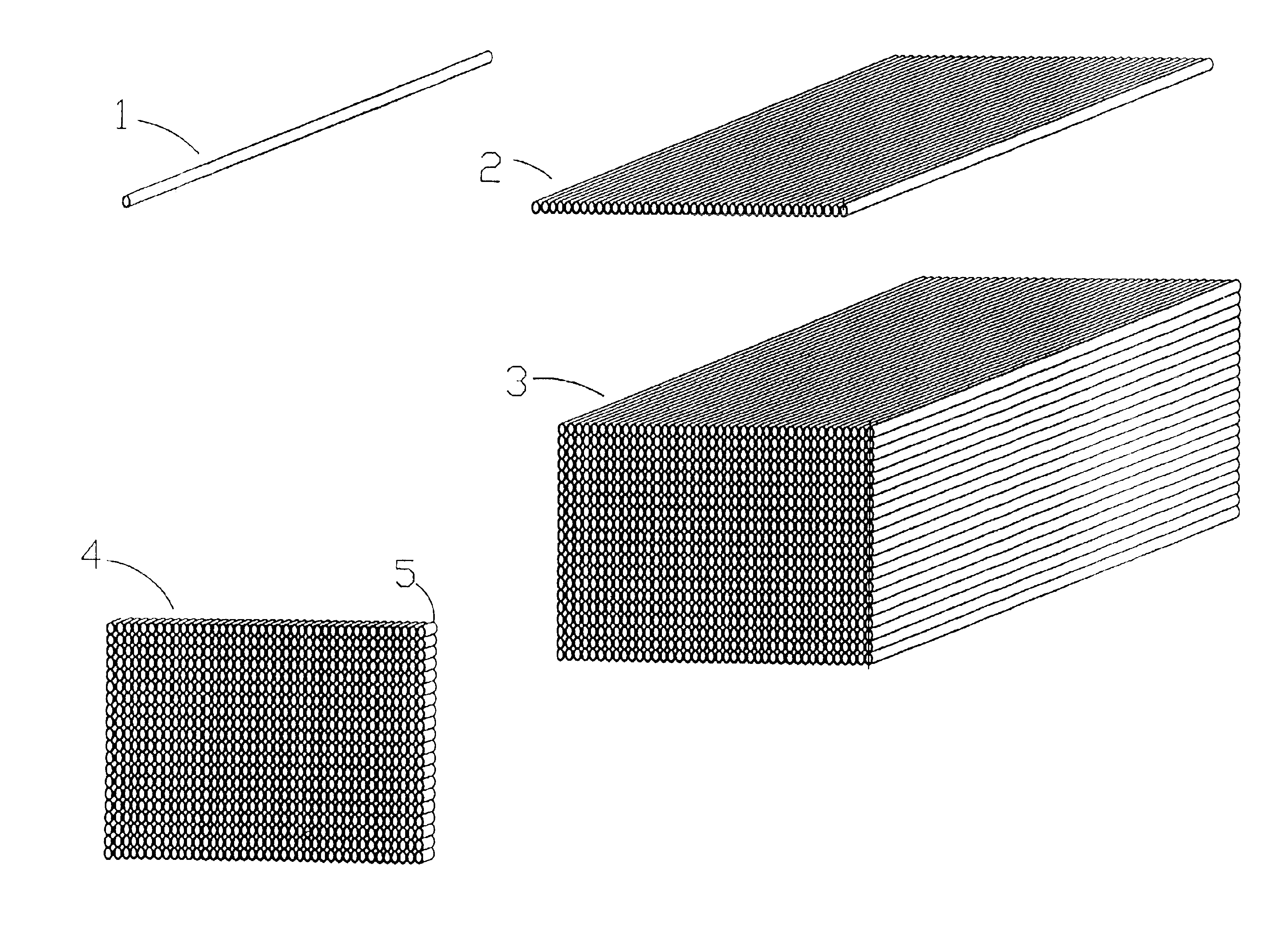
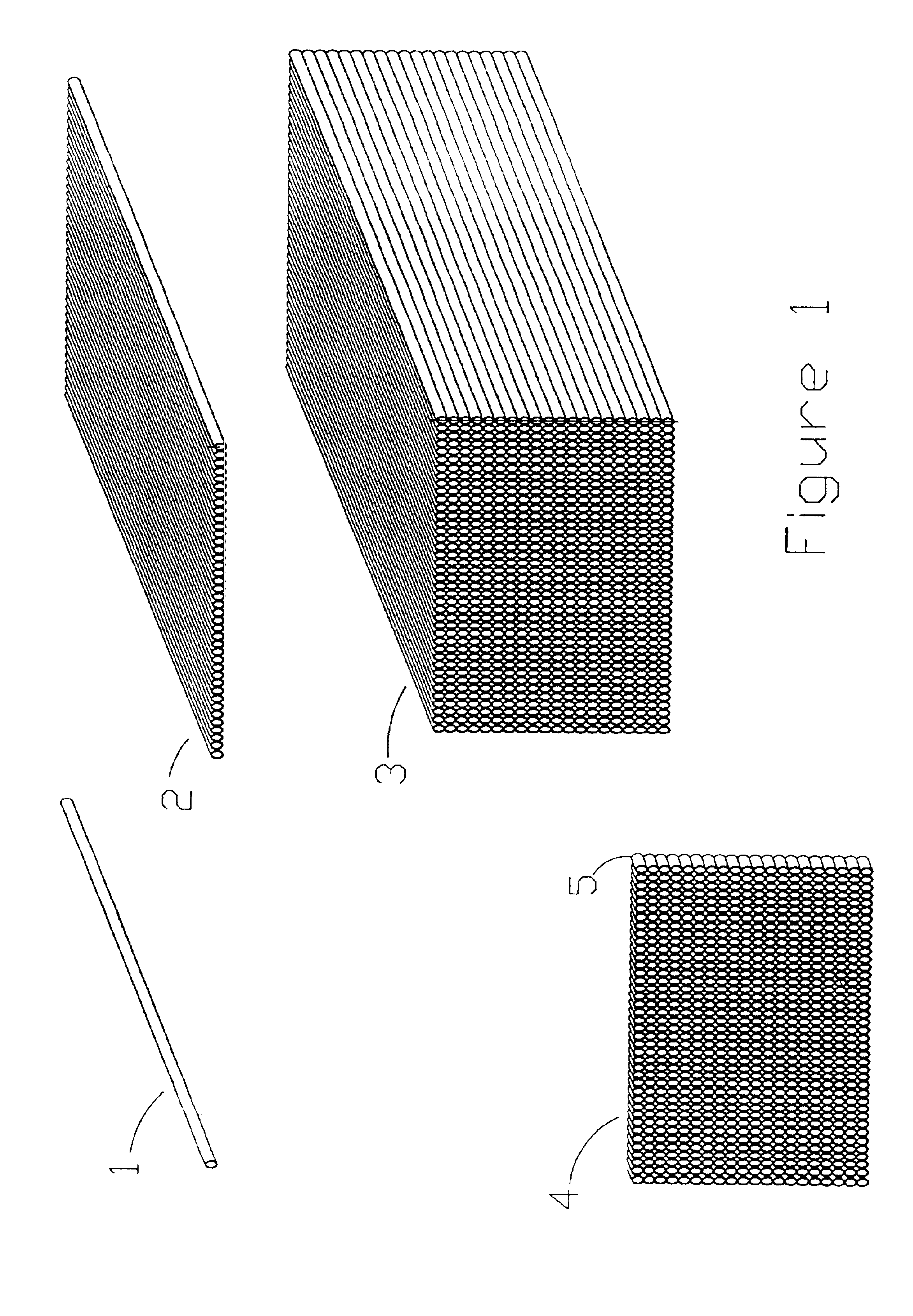
Formation and Analysis of a Microarray

[0180]Antibodies were prepared by affinity purification by reversible binding to the respective immobilized antigens and subsequently immobilized on particulate supports (Poros G, made by PE Biosystems). An Integral 100Q biochromatography workstation.

[0181]Each antibody support was made by trapping the antibody on a column of Poros G (commercially available Poros particles pre-coated with protein G, a bacterial protein capable of binding many immunoglobulins by their Fc domain) and subsequently cross-linking the antibody and the protein G with dimethylpimelimidate (following the PE Biosystems protocol) to covalently immobilize the antibody on the Poros particles. Such antibody columns can be reused (with an acid elution of bound antigen) more than 100 times in a subtractive mode, and are therefore extremely stable. Each antibody support was characterized to demonstrate specificity for a single antigen.

[0182]Antibodies directed against human seru...

example 2

Manufacture and Use of Diagnostic Array Detecting Autoantibodies to Mitochondrial or Lysosomal Proteins

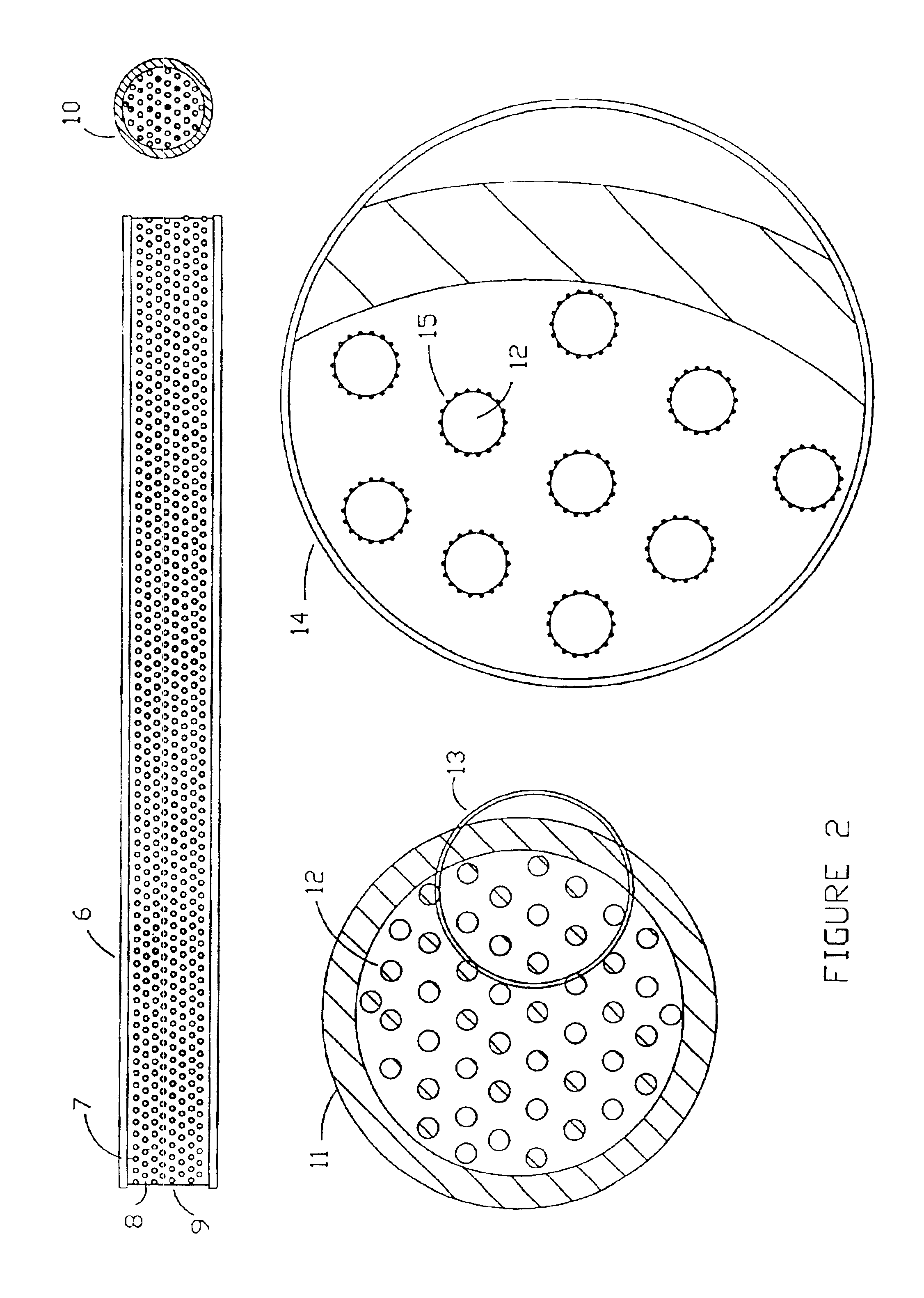
[0193]Suspensions of whole isolated rat and mouse liver mitochondria, lysosomes, and expressed proteins are suspended or dissolved in an aqueous buffer, at 10 mg / ml concentration, and optionally fixed with glutaraldehyde (1%). 1 ml of each preparation is mixed according to the kit instructions with 20 ml of JB-4 (Polysciences) catalyzed infiltration resin prepared by mixing 20 ml of monomer A containing 0.17 g of catalyst. After complete mixing, 40 mL of monomer B containing 0.17 g catalyst is added with stirring. When completely dissolved, 0.8 g of Accelerator is added, the mixture placed in a syringe and injected into 0.0625 inch internal diameter Teflon tubing under anaerobic conditions. Polymerization occurs at room temperature in approximately 50 minutes. The ends of the tubes are then heat sealed and stored cold until used, or are immediately extruded for use in preparing a f...

example 3

Manufacture and Use of a Diagnostic Array Using Histological Embedding Support

[0195]Arrays are prepared which incorporate fixed infectious particles to be used to detect convalescent antibodies appearing late in the history of an infection. These are important in following sentinel populations to determine what infectious are occurring.

[0196]Immuno-Bed GMA water-miscible embedding medium is made up as directed (Polysciences Inc.), and small batches are mixed with different suspensions of fixed selected viruses (average titer 109 / ml) or fixed bacterial cells (average 107 particles / ml). The suspension is placed in a syringe and forced under pressure into Teflon® tubing of {fraction (1 / 16)}-inch internal diameter, and allowed to polymerize at room temperature. The tubing is pre-treated with metallic sodium in an organic medium to provide a surface, which will adhere to epoxy resins. The polymerized fiber is stored in the coiled Teflon® tubing in the cold.

[0197]The arrays are assembled...


PUM

PropertyMeasurementUnit
thickaaaaaaaaaa
thickaaaaaaaaaa
lengthaaaaaaaaaa

Abstract

The microarrays of the present invention are prepared by using a separate fiber for each compound being used in the microarray. The fibers are bundled and sectioned to form a thin microarray that is glued to a backing.

Description

CROSS REFERENCE TO RELATED APPLICATIONS[0001]This is a division of application Ser. No. 09 / 482,460, filed Jan. 13, 2000, and is a continuation-in-part of Provisional Patent Application Ser. No. 60 / 146,653 filed Jul. 30, 1999, the content of which is incorporated herein in entirety. The content of Disclosure Document Nos. 286,465 filed Jul. 11, 1991; 418,726 filed May 9, 1997; 444,028 filed Aug. 31, 1998; 445,769 filed Oct. 5, 1998; and 458852 filed 14 Jul. 1999 are hereby incorporated herein by reference.FIELD OF THE INVENTION[0002]The present invention relates to microarrays containing bioreactive molecules, their uses and methods for their manufacture. The arrays are constructed by sectioning bundles of tubules or rods containing unique reactants to produce large numbers of identical arrays.BACKGROUND OF THE INVENTION[0003]Microarrays are known in the art and are commercially available from a number of sources. Microarrays have been used for a number of analytical purposes, typica...

Claims


Application Information

Patent Timeline
Patent Type & Authority Patents(United States)
IPC IPC(8): B01L3/00B01J19/00C12Q1/68G01N33/543G01N31/22C12M1/00C12M1/34C12N15/09C12Q1/00C12Q1/02C40B40/06C40B40/12C40B70/00G01N27/447G01N33/15G01N33/50G01N33/53G01N33/566G01N37/00
CPCB01J19/0046B01L3/50857C12Q1/6837G01N33/5436G01N33/54366Y10S436/805B01J2219/0052B01J2219/00524B01J2219/00547B01J2219/00585B01J2219/00596B01J2219/00605B01J2219/0061B01J2219/00612B01J2219/00617B01J2219/00619B01J2219/00621B01J2219/00626B01J2219/0063B01J2219/00637B01J2219/00644B01J2219/00659B01J2219/00664B01J2219/00673B01J2219/00707B01J2219/0072B01J2219/00722B01J2219/00731C40B40/06C40B40/12C40B70/00G01N2500/00Y10S435/808Y10S436/809B01J2219/00515Y10T442/20G01N33/543
Inventor ANDERSON, NORMAN G.ANDERSON, N. LEIGH
Owner LARGE SCALE PROTEOMICS
Why Eureka
Try Eureka