[0026]The present invention provides an improved apparatus and method for the rapid, non-intrusive determination of the concentration of blood analytes. In one embodiment, it provides a portable tabletop unit for measurement of blood analyte concentrations where the subject may walk up to the device for measurement from a body part, such as a finger. However, there are many situations where blood analyte measurement must be done outside of a domestic or laboratory environment. Thus, another embodiment of the present invention provides a portable
system which may be positioned on
body tissue and transported on the user's person. Features such as small size, a
wireless sensor, battery operation, portability, and downloadability demonstrably increase the utility and range of the analyte measurement apparatus of the present invention beyond the hospital or laboratory setting.
[0027]The present invention also provides a method and apparatus with increased sensitivity and accuracy. A problem encountered in the area of blood analyte measurement via IR
spectroscopy is accuracy and drift. In general, other analytes and various other substances present interfere with the IR measurement of the desired analyte. These analytes vary in concentration and thus vary the IR spectrum in the regions being used to determine specific analyte concentration. The present invention corrects for all other analytes with concentrations sufficient to interfere in the determination of the concentration of the analyte or analytes of interest. Measuring the entire visible and IR spectrum provides enough data to simultaneously determine all of the analytes and thereby compensate for any accuracy or drift problems their concentration may cause in measuring the concentration of the analyte(s) of interest.
Data processing using orthogonal functions is used to accomplishing this task. Other properties of blood may also effect the IR measurement of the desired analyte. For example,
turbidity of the blood, as may be caused by elevated
white cell count or high
blood lipids, may affect the measurement. These factors appear in the spectra and are compensated for by the present invention. The analyte measurement apparatus of the present invention is sufficiently sensitive to detect blood glucose or lactate with accuracy within, approximately, 10% of the level actually present, and may do so in a short period of time (e.g. 5 seconds or less). Due to the non-intrusive nature of the measurement and its relative
rapidity, it is also possible to monitor blood analyte levels essentially continuously.
[0028]The blood analyte measurement apparatus of the present invention includes a radiation source for generating and transmitting a spectrum of radiation onto a portion of tissue (for transmission therethrough or reflection therefrom), one or more sensors for detecting the radiation either transmitted through or reflected from the tissue over a
broad spectrum and generating an output in response to the detected radiation, and a processor for receiving output from the sensors to determine the concentration of blood analytes in the portion of tissue. In a preferred embodiment, the apparatus also makes use of a mounting device to position the radiation source and the sensors relative to a portion of tissue so the one or more sensors may receive a substantial portion of the radiation produced by the radiation source and transmitted through or reflected by the portion of tissue. In a further preferred embodiment, the information regarding absorption of the radiation is then algorithmically processed to clarify the
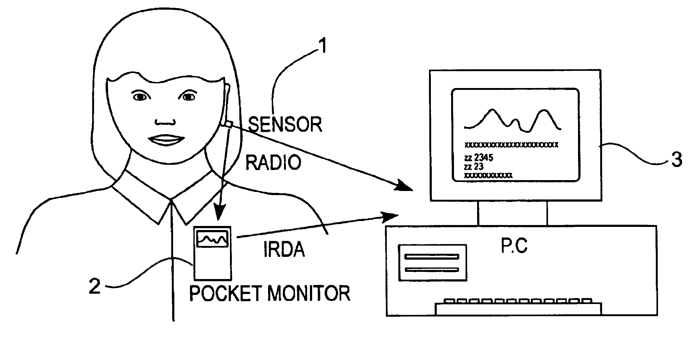
signal(s) of the desired blood analytes. Thus, the invention, in a typical configuration, includes a sensor module which is preferably attached to an
earlobe, a pocket monitor for immediate readout and data
logging, and a
data link to a PC for long term storage and compilation of data. Thus, blood analyte levels may be continuously monitored without the constraints of attachment wires or bulky apparatus.
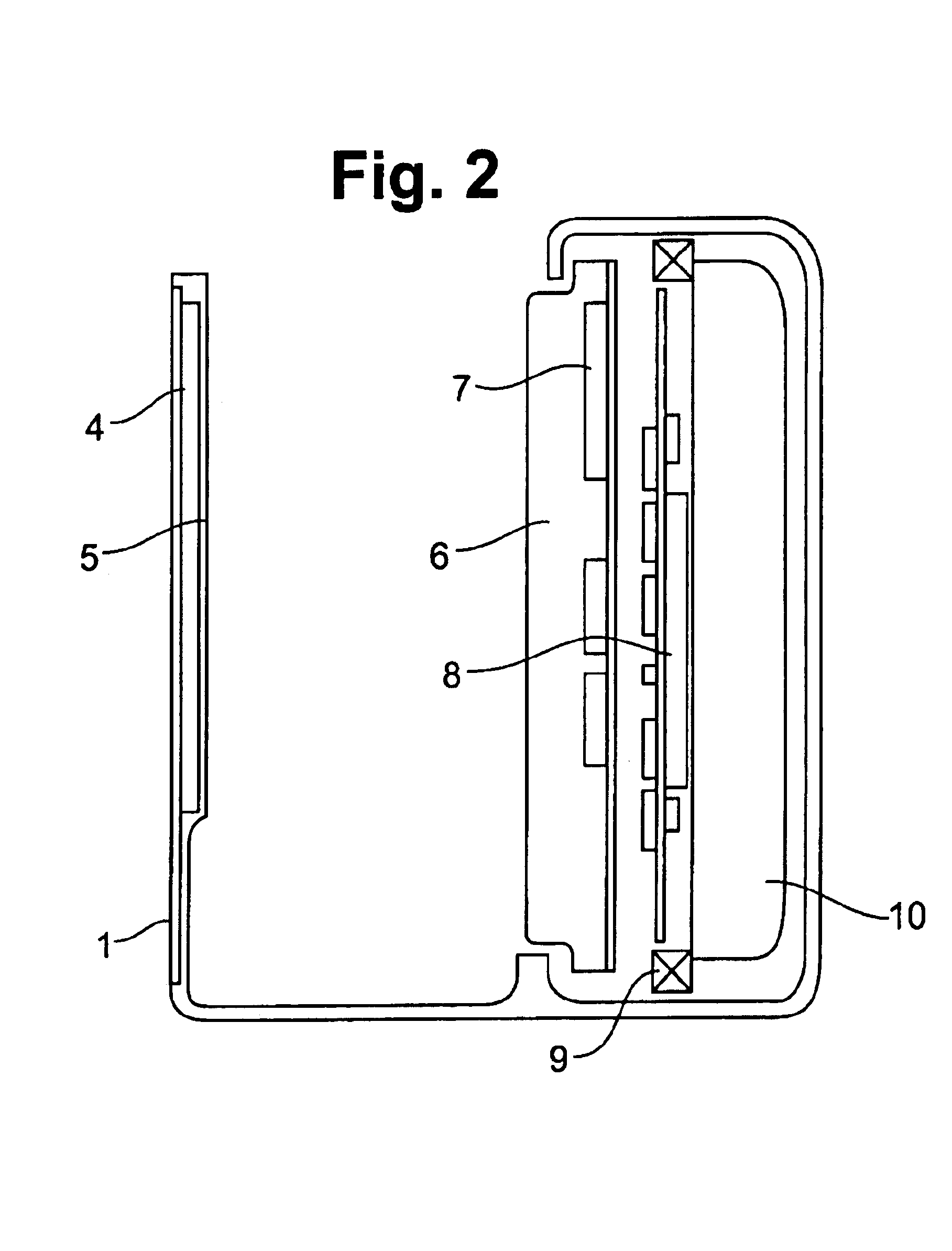
[0029]The blood analyte sensor module is integrated as much as possible to reduce the size and weight. In one embodiment, the sensor module is completely self-contained. The sensor module illuminates the
measurement site with a built-in radiation source tailored to the spectral
region of interest. The radiation source and the sensors are each positioned on a
chip. The radiation source may be integrated onto a custom
chip in transmission mode, or onto the same
chip as the sensors in reflection mode. That is, when it is desired to receive and interpret radiation transmitted through the tissue, the apparatus is working in transmission mode and the radiation source is positioned on a chip separate from the chip on which the sensors are positioned. In contrast, when it is desired to receive and interpret radiation that is reflected from the tissue, the apparatus is working in reflection mode and the radiation source may be positioned on the same chip as the chip on which the sensors are positioned. Preferably, the radiation source is a thermal radiator made up of
tungsten or
tantalum positioned over a reflective
heat shield.
[0033]A processor is provided for
processing the output from the sensors. If desired, an RF
transmitter or other device may be provided for wirelessly transmitting the signals from the sensors to the processor. This processor is preferably a
CMOS microprocessor, which uses a Boolean
algorithm to process the output from the sensors. Various
processing algorithms are used to enhance the value of the data obtained from the sensors. The blood analyte measurement apparatus may also include a display, typically a
liquid crystal display, for the immediate display of data to the user. The data may be downloaded to a computer or other device via an I / O port, typically an RS-232 port.
[0036]An important aspect of the present invention is the interpreting of the signals communicated to the processor by an
algorithm. One type of
algorithm used to interpret this data is
linear regression. A more preferred algorithm makes use of orthogonal functions. The concept is to use the reference spectrum for each blood analyte as basis functions and determine
a weighting function or functions that create an orthogonal set. This permits easy separation algorithms for mixed spectra. The use of algorithms is very helpful for self-calibrating to eliminate data artifacts caused by individual variation in tissue character.


