Multimodal imaging sources

a multi-modal imaging and source technology, applied in the field of medical and molecular imaging, can solve the problems of inconvenient use of markers, difficult registration and alignment of images obtained from two machines, and great difficulty in remaining motionless, and achieve the effect of avoiding higher energy emissions

Inactive Publication Date: 2005-08-09
ISO SCI LAB
View PDF8 Cites 42 Cited by
  • Summary
  • Abstract
  • Description
  • Claims
  • Application Information

AI Technical Summary

Benefits of technology

[0013]The radionuclide can best be selected from known calibrators for the detector system with which the source is to be used, or can be selected from possible radionuclides that have radiation energies similar to radionuclides used with the detector systems to be used. These radionuclides include, but are not limited to Ag-110m, Am-241, Au-195, Ba-133, C-14, Cd-109, Ce-139, Co-57, Co-60, Cs-137, Eu-152, Gd-151, Gd-153, Ge-68, Hg-203, Ir-192, I-125, I-129, I-131, Lu-173, Lu-177m, Mn-54, Na-22, Ra-226, Rh-101, Ru-103, Ru-106, Sb-125, Se-75, Sn-113, Sr-90, Ta-182, Te-123m, Tl-204, Th-228, Th-229, Th-230, Y-88, Zn-65, and Zr-95, with Ba-133, Co-57, Ge-68, Na-22, Gd-153, Cs-137 and Se-75 being particularly good nuclides to provide for PET and SPECT visibility. The particular radionuclides to be used are preferably chosen based upon factors such as their half-life (which are typically much longer than the half lives of the radionuclides used in imaging humans and animals), their energy peak correlations with radionuclides used in imaging humans and other animals, and an absence of higher energy emissions.

Problems solved by technology

However, since these combination machines are costly, many hospitals, physicians and researchers continue to rely on scanning patients and animals consecutively with a CT scanner and then a PET or SPECT scanner.
Since patients often experience great difficulty in remaining motionless not only while being scanned on one machine, but being moved from machine to machine, it can be problematic to register and align the images obtained from the two machines.
These markers are inconvenient to use since they require filling with the radioactive solution by the end user immediately before use.
Although the encapsulation portion of known sealed radioactive sources may be formed of materials that show up on a CT scan, these sealed radioactive sources are not designed or intended for use in the calibration, registration or alignment of images taken on different scanners.

Method used


Image

  • Multimodal imaging sources
  • Multimodal imaging sources
  • Multimodal imaging sources

Examples


Embodiment Construction

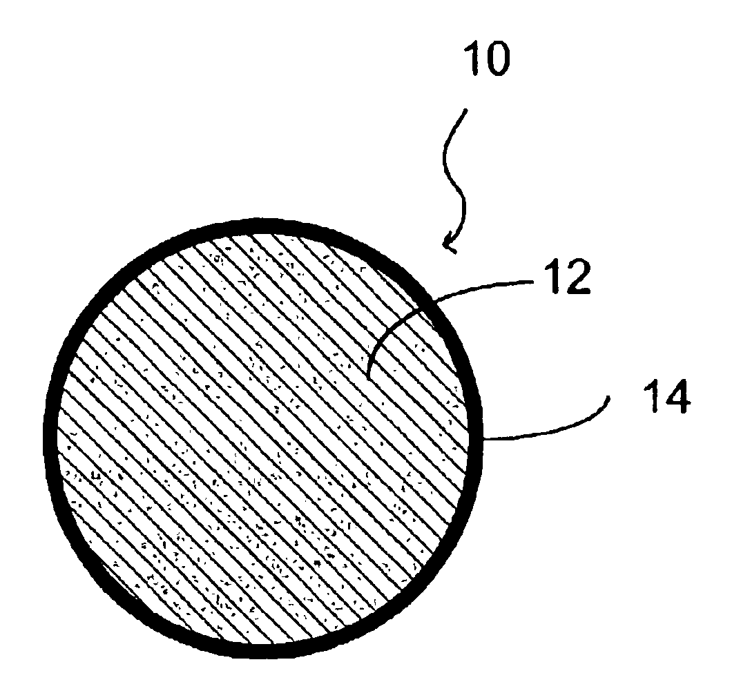
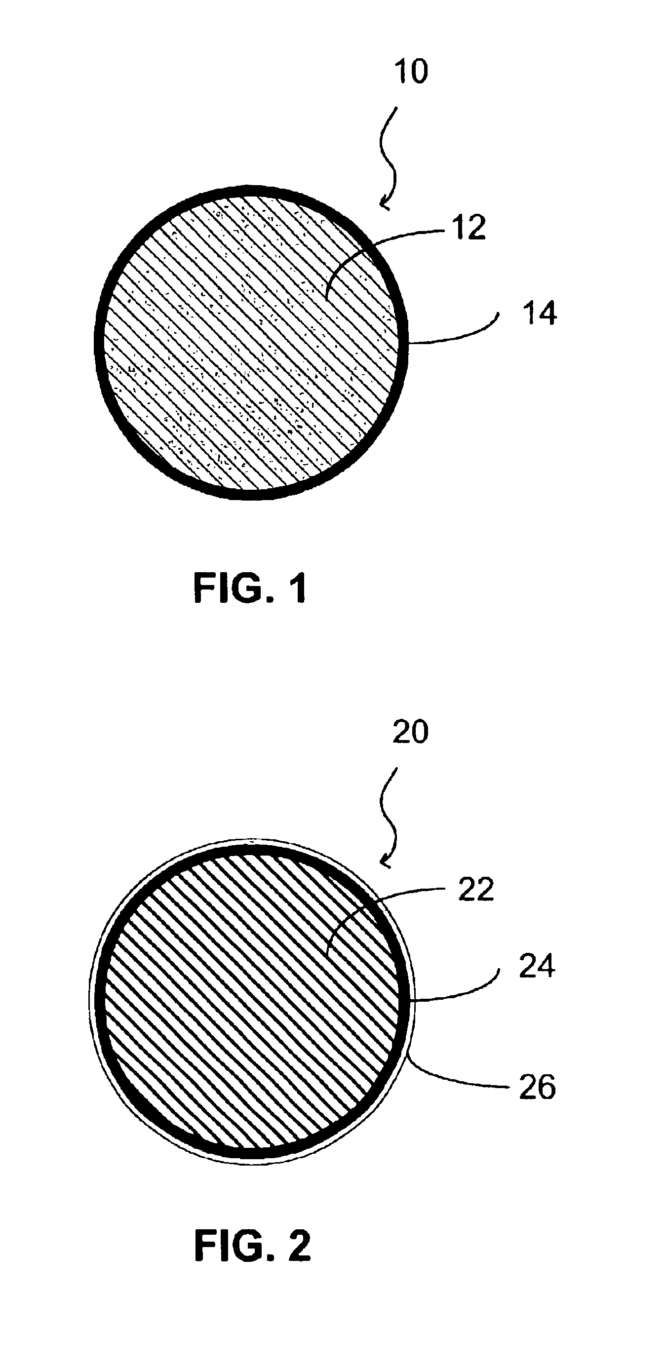
[0028]The multimodal source of the invention can be provided in a wide variety of configurations. FIG. 1 shows a cross sectional first embodiment of the source 10 of the invention comprising a point source in the form of a spherical source with both radioactive and CT / MRI / optical target materials. The point source can be conveniently sized at approximately 5 mm in diameter or smaller. In this embodiment, a core 12 of the source can be formed of a matrix (such as a polymeric resin, a cement, a silicone, a ceramic, a polymer gel, etc.) with one or more radionuclides such as Ag-110m, Am-241, Au-195, Ba-133, C-14, Cd-109, Ce-139, Co-57, Co-60, Cs-137, Eu-152, Gd-151, Gd-153, Ge-68, Hg-203, Ir-192, I-125, I-129, I-131, Lu-173, Lu-177m, Mn-54, Na-22, Ra-226, Rh-101, Ru-103, Ru-106, Sb-125, Se-75, Sn-113, Sr-90, Ta-182, Te-123m, Tl-204, Th-228, Th-229, Th-230, Y-88, Zn-65, and Zr-95, (with Ba-133, Co-57, Ge-68, Na-22, Gd-153, Cs-137 and Se-75 being particularly good nuclides) mixed in, and...


PUM


Abstract

A multimodal source for imaging with at least one of a gamma camera, a positron emission tomography (PET) scanner and a single-photon-emission computed tomography (SPECT) scanner, and at least one of a computed tomography (CT) scanner, magnetic resonance imaging (MRI) scanner and optical scanner. The multimodal source has radioactive material permanently incorporated into a matrix of material, at least one of a material that is a target for CT, MRI and optical scanning, and a container which holds the radioactive material and the CT, MRI and/or optical target material. The source can be formed into a variety of different shapes such as points, cylinders, rings, squares, sheets and anthropomorphic shapes. The material that is a target for gamma cameras, PET scanners and SPECT scanners and/or CT, MRI and/or optical scanners can be formed into shapes that mimic biological structures.

Description

BACKGROUND OF THE INVENTION[0001]The invention is related to medical and molecular imaging of patients and animals with various scanning machines and methods, and more particularly to sealed calibration and reference sources for such scanning machines.[0002]Scientists and physicians have found that in imaging patients and animals, there are advantages in using combinations of two or more of computed tomography (CT) scanning, magnetic resonance imaging (MRI) scanning, gamma camera scanning, positron emission tomography (PET) scanning, single-photon-emission computed tomography (SPECT) scanning, and optical scanning. Recently, scanners that combine CT and PET functionality have gained favor among physicians. However, since these combination machines are costly, many hospitals, physicians and researchers continue to rely on scanning patients and animals consecutively with a CT scanner and then a PET or SPECT scanner. Since patients often experience great difficulty in remaining motionl...

Claims


Application Information

Patent Timeline
Patent Type & Authority Patents(United States)
IPC IPC(8): A61B6/00
CPCA61B6/583A61B19/54A61B2019/542A61B2019/5437A61B6/4258A61B2019/5495A61B6/508A61B6/037A61B2019/5454A61B90/39A61B2090/392A61B2090/3937A61B2090/3954A61B2090/3995
Inventor ZYROMSKI, KRISTIANA E.
Owner ISO SCI LAB
Why Eureka
Try Eureka