Methods and reagents for quantitation of cell-surface molecule expression on peripheral blood cells

a cell surface molecule and reagent technology, applied in the field of methods and reagents for quantitation of cell surface molecule expression on peripheral blood cells, can solve the problems of achieve the effects of high intensity, high uniformity and clustering, and ineffective stabilization of hla-dr expression

Inactive Publication Date: 2005-10-04
BECTON DICKINSON & CO
View PDF24 Cites 12 Cited by
  • Summary
  • Abstract
  • Description
  • Claims
  • Application Information

AI Technical Summary

Benefits of technology

[0022]The invention is based, in part, upon the novel finding that including chloroquine during staining of peripheral blood with anti-HLA antibody stabilizes the HLA-DR-specific signal obtained from CD14+ monocytes. This is surprising: chloroquine has been reported to be unable to block the cellular recycling of MHC glycoproteins, and other agents that affect protein processing, such as Brefeldin A and monensin, have been shown ineffective in stabilizing HLA-DR expression. Equally surprising is the novel finding that including chloroquine during staining of unfractionated peripheral blood with anti-CD11b antibody stabilizes the CD11b-specific signal.
[0023]The invention is also based in part upon the finding that conjugation of an anti-CD14 antibody to a fluorescence energy resonance transfer tandem fluorophore, PerCP / CY5.5, provides a high intensity, highly uniform and clustered signal from peripheral blood monocytes, permitting the ready discrimination of monocytes from granulocytes. Incorporating these improvements provides an assay well-suited for clinical use, permitting the rapid and reliable quantitative measurement of peripheral monocytic HLA-DR levels.

Problems solved by technology

This is surprising: chloroquine has been reported to be unable to block the cellular recycling of MHC glycoproteins, and other agents that affect protein processing, such as Brefeldin A and monensin, have been shown ineffective in stabilizing HLA-DR expression.

Method used


Image

  • Methods and reagents for quantitation of cell-surface molecule expression on peripheral blood cells
  • Methods and reagents for quantitation of cell-surface molecule expression on peripheral blood cells
  • Methods and reagents for quantitation of cell-surface molecule expression on peripheral blood cells

Examples


example 1

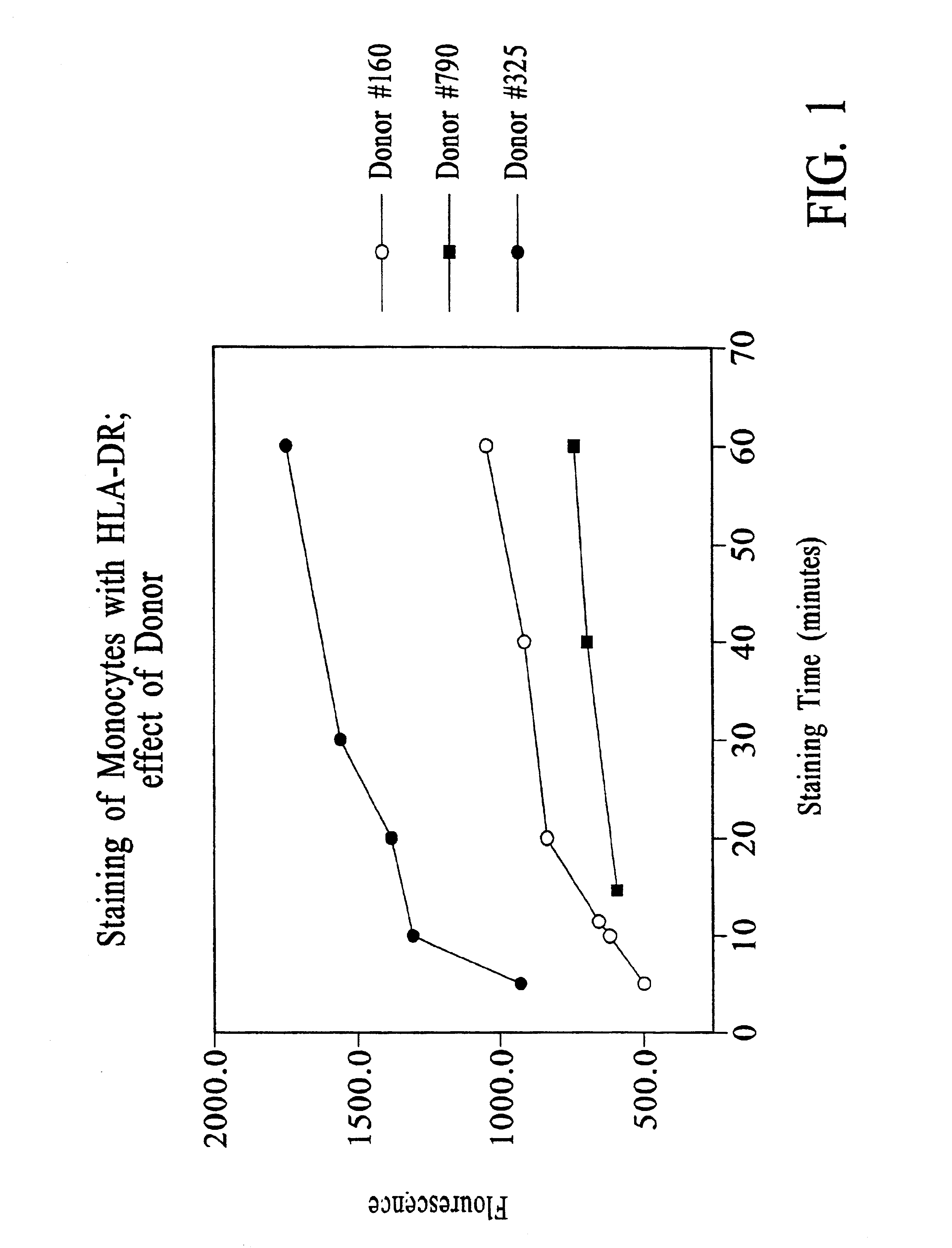
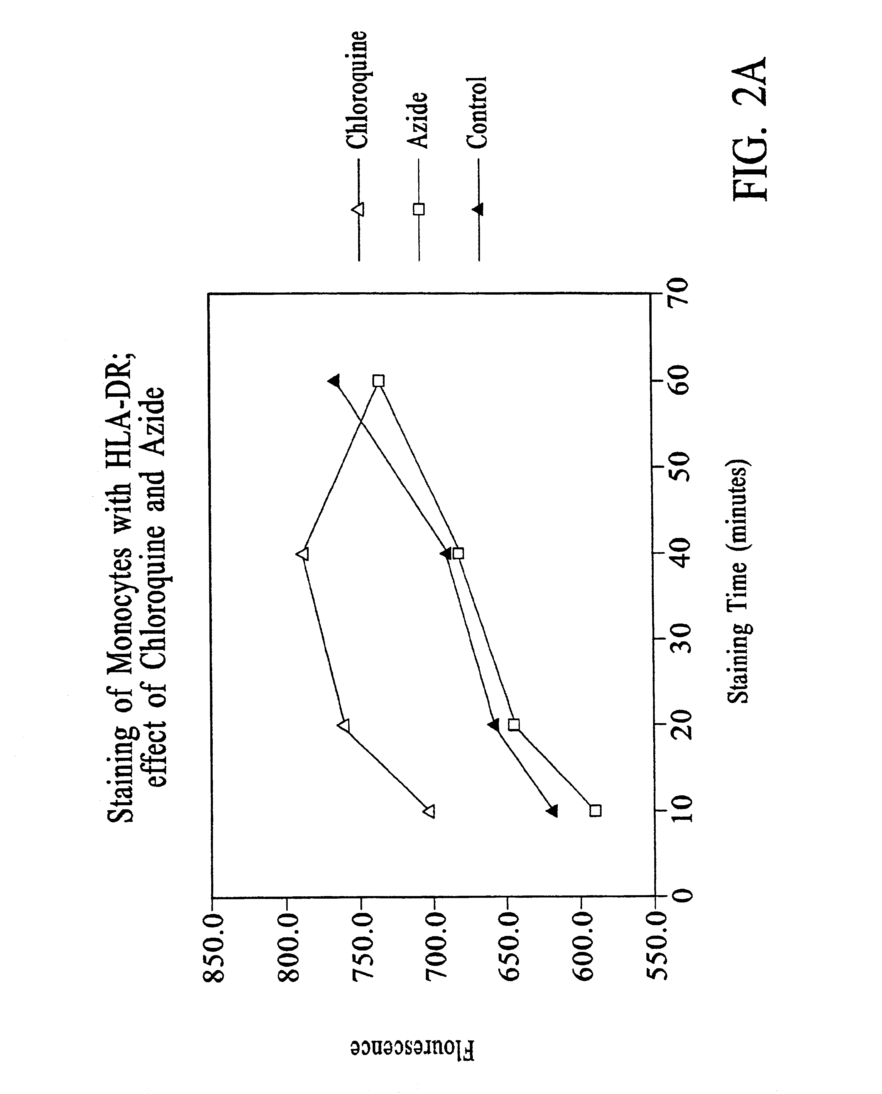
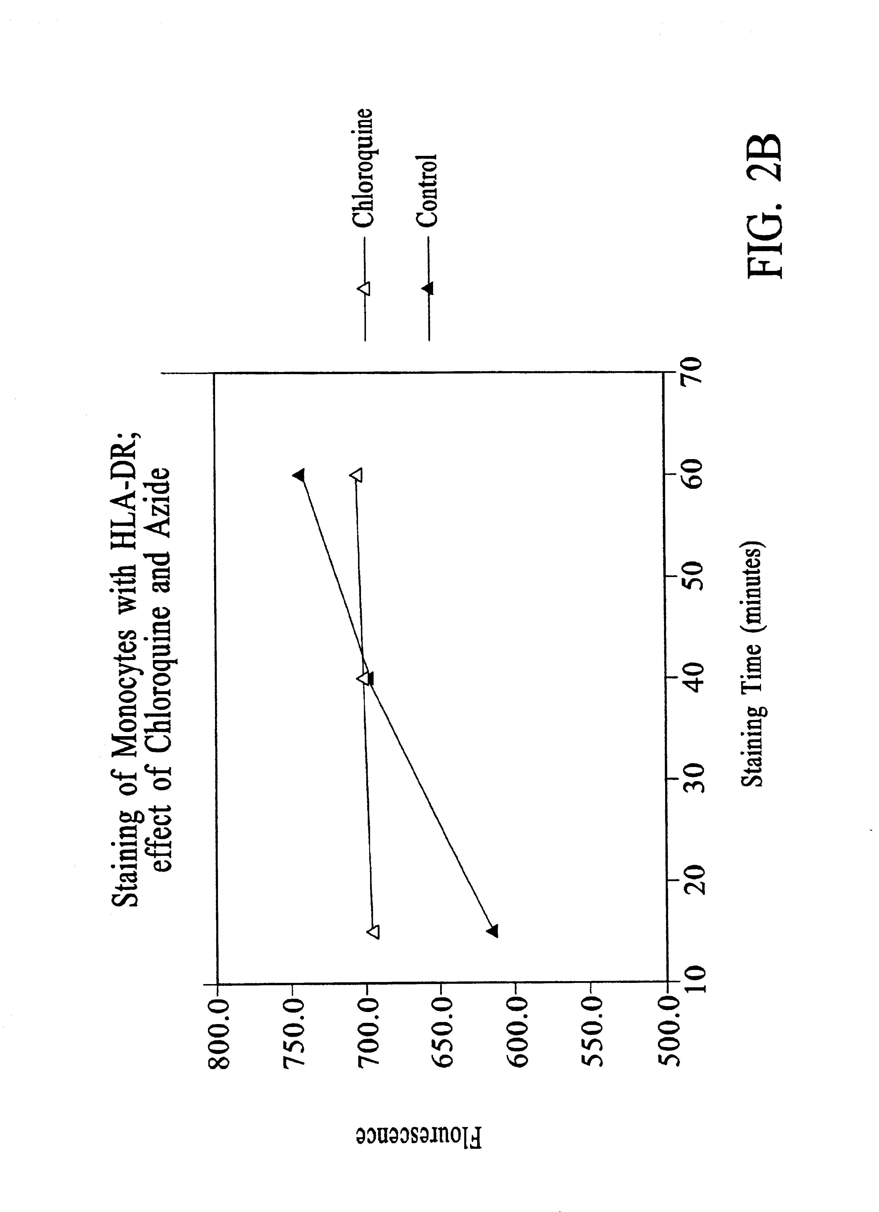
Chloroquine Inhibition of Time-Dependent Variation in HLA-DR-Specific Fluorescent Antibody Staining

[0114]Peripheral blood samples were drawn from normal volunteers.

[0115]The time-dependence of the HLA-DR-specific signal detectable by flow cytometry of antibody-stained peripheral blood monocytes was assessed by varying the duration of antibody staining.

[0116]For the experiments reported in FIG. 1, to 50 μl EDTA-anticoagulated whole blood was added 10 μl of staining reagent containing either (a) CD14PerCP / CY5.5 (63 ng) and HLA-DR-PE (250 ng) (donors #160 and #325) or (b) CD14-PerCP / CY5.5 (63 ng) and HLA-DR-PE (500 ng) (donor #790).

[0117]Samples were incubated with staining reagent at room temperature for the times indicated, after which 1 ml of 1× FACS™ Lysing Solution (10× stock from BDIS, catalog no. 349202) was added, and incubation continued for 10 minutes.

[0118]For donor #160, the samples were then centrifuged at 250×g for 5 minutes, the supernatant removed, the pellet resuspende...

example 2

Measurement of HLA-DR on Patient Peripheral Blood Monocytes

[0133]Peripheral blood samples were obtained by venipuncture from patients admitted to ICU. One aliquot of each sample was assayed using the following protocol.

[0134]To 50 μl of whole blood was added 10 μl of staining reagent containing 50g / ml HLA-DR-PE (clone L243); 6.3 μg / ml CD14-PerCP / CY5.5; and 18 mM chloroquine diphosphate. HLA-DR-PE (BDIS cat. no. 347367, repurified to provide a 1:1 antibody:PE conjugate) is a mouse IgG2a / κ antibody that reacts with a nonpolymorphic HLA-DR epitope and does not cross-react with HLA-DQ or HLA-DP molecules. CD14-PerCP / CY5.5 (BDIS custom conjugate of antibody LeuM3, available unconjugated as BDIS catalogue number 347490) is a mouse IgG2b / K antibody specific for CD14.

[0135]The aliquot was incubated for 25-35 minutes in the dark at room temperature, and then 1 ml 1× FACS™ Lysing Solution (obtained as 10× stock, BDIS catalog No. 349202) was added and incubation continued for 8-10 minutes. The...

example 3

Stabilization of CD11b-Specific Fluorescent Signal by Chloroquine

[0139]Blood from three normal adult donors was collected in standard EDTA Vacutainer® tubes and placed directly on ice. Fifty (50) μl of whole blood was transferred to a tube containing 20 μl of a cocktail of CD11b PE and CD45 PerCP antibodies with or without chloroquine, with the chloroquine-containing tubes having a final chloroquine concentration of 3 mM. Blood was stained for the indicated time at room temperature before addition of 1.0 ml FACS lysing reagent (1×). Flow cytometric analysis was performed essentially as described in Examples 1 and 2. Results are shown in FIG. 7.


PUM

PropertyMeasurementUnit
pHaaaaaaaaaa
fluorescentaaaaaaaaaa
molar ratioaaaaaaaaaa

Abstract

Improved methods, reagents, and kits for quantitation of HLA-DR and / or CD11b expression on peripheral blood cells are presented. Inclusion of a lysosomotropic amine, such as chloroquine, during staining stabilizes HLA-DR and CD11b expression. Use of a novel anti-CD14 conjugate, anti-CD14-PerCP / CY5.5, permits the ready discrimination of monocytes. The improved methods, reagents, and kits may be used to assess immune competence, and to direct and monitor immunostimulatory therapies in septic patients exhibiting monocyte deactivation.

Description

CROSS REFERENCE TO RELATED APPLICATIONS[0001]This application is a continuation U.S. patent application Ser. No. 09 / 406,013, filed Sep. 24, 1999, which issued as U.S. Pat. No. 6,423,505, which is a continuation-in-part of U.S. patent application Ser. No. 09 / 204,860, filed Dec. 3, 1998, which issued as U.S. Pat. No. 6,200,766, the disclosures of both of which are incorporated herein by reference in their entirety.FIELD OF THE INVENTION[0002]The present invention relates to improved methods, reagents and kits for quantitating the expression of HLA-DR and / or CD11b on the surface of human peripheral blood cells, particularly on the surface of peripheral blood monocytes. The invention further relates to methods for assessing immune competence and for directing and monitoring immunostimulatory therapies for sepsis based upon the levels of monocyte HLA-DR expression so measured.BACKGROUND OF THE INVENTION[0003]Sepsis is one of the most common causes of death in developed countries. Inciden...

Claims


Application Information

Patent Timeline
Patent Type & Authority Patents(United States)
IPC IPC(8): C07K16/18C07K16/28G01N33/569G01N33/53G01N33/531G01N33/533G01N33/536
CPCC07K16/2896G01N33/56972G01N33/56977G01N2333/70539G01N2333/70553Y10S435/975Y10S435/962Y10S436/80Y10S436/808Y10S435/961Y10S436/826
Inventor DAVIS, KENNETH A.
Owner BECTON DICKINSON & CO
Why Eureka
Try Eureka