Cryotherapy method for detecting and treating vulnerable plaque

a technology of vulnerable plaque and cryotherapy, which is applied in the field of cryotherapy for detecting and treating vulnerable plaque, can solve problems such as sudden cardiac death, and achieve the effect of maintaining the patency of the body lumen

Inactive Publication Date: 2005-10-18
BOSTON SCI SCIMED INC
View PDF36 Cites 162 Cited by
  • Summary
  • Abstract
  • Description
  • Claims
  • Application Information

AI Technical Summary

Benefits of technology

[0014]The present invention provides detection and treatment of vulnerable plaque within a blood vessel of a patient. The blood vessel may be any blood vessel in the patient's vasculature, including veins, arteries, and particularly coronary arteries. The vessel will typically be partially stenosed, at least in part from vulnerable plaque. In particular, the present invention may inhibit release of retained fluid within the vulnerable plaque so as to inhibit acute coronary syndrome and to help maintain the patency of a body lumen. The present invention may also provide for the treatment of vulnerable plaque in carotid arteries for stroke prevention. Where the patient's vasculature has both the vulnerable plaque and standard stenotic plaque, the treatment techniques described herein may be selectively directed to the vulnerable plaque, optionally without substantial cooling of the standard stenotic plaque. In other embodiments, both types of plaque may be treated.
[0017]Differentiation of vulnerable plaque from standard stenotic plaque may be enhanced by the use of a balloon catheter inflated with a gas. The differentiation may be effected using a temperature sensor for sensing a temperature of a tissue adjacent the balloon. Surprisingly, plaque differentiation benefits from gas temperatures within the balloon of about 20° C. or more, preferably being 30° C. or more. Hence, the balloon may be inflated so that an initial temperature of the gas within the balloon is about 30° C. Inflation with such a warm gas can amplify the measured temperature differences (for example) sensed by temperature sensors distributed along a balloon wall and in thermal engagement with the vulnerable plaque (at a first sensor) and a standard stenotic plaque or healthy luminal wall (at a second sensor).
[0019]Usually, the temperature at the surface of the blood vessel lumen is in the range from about 10° C. to −40° C., preferably from about 10° C. to −20° C., more preferably from about 5° C. to −10° C. The tissue is typically maintained at the desired temperature for a time period in the range from about 15 seconds to 120 seconds, preferably from 30 seconds to 60 seconds, optionally being in a range from about 20 seconds to about 60 seconds. Vulnerable plaque stabilization may be enhanced by repeating cooling in cycles, typically with from about 1 to 3 cycles, with the cycles being repeated at a rate of about one cycle every 120 seconds.
[0020]Surprisingly, cooling temperatures above 0° C. can effect a transition of the vulnerable plaque's lipid core from a disordered cystalline state fluid to a ordered crystalline state solid or gel. Thus, vulnerable plaque can be stabilized by cooling the lipid-rich liquid sufficiently to change a state of the lipid-rich liquid, typically to a highly ordered hexagonal lattice at transition temperatures generally in the range from about 10° C. to −10° C. The stabilization may, at least in part, remain in effect after subsequent return of the tissue to a normal body temperature. The stabilization may, at least in part, be transitory, with the stabilization effect diminishing and / or disappearing after cooling is terminated. The vulnerable plaque may be treated while it remains at least partially stabilized.
[0021]Advantageously, the cooling may be accurately controlled to tailor a desired tissue response. Cooling may be performed so as to cause apoptosis and / or necrosis in the tissues comprising or adjacent to the vulnerable plaque. Alternatively, the cooling may also be performed in a manner that avoids causing apoptosis and / or necrosis. Cooling may stabilize the vulnerable plaque while inhibiting necrosis and / or apoptosis of tissue adjacent the lipid-rich liquid, particularly of the tissues defining a cap of cells between the lipid-rich liquid and the lumen of the blood vessel. Cooling may also inhibit inflammation and deterioration of the vulnerable plaque. The cooling treatment may further inhibit rupture of the cap of cells of the vulnerable plaque.
[0026]The inflating step may be performed so that the gas within the balloon has a temperature of about 20° C. or more, the balloon preferably being inflated so that the gas has an initial temperature in the balloon of 30° C. or more. Surprisingly, temperature sensors along the balloon wall can detect a temperature differential of greater than about 1° C. between a vulnerable plaque and an adjacent portion of the luminal wall with such a system. Additionally, the size of the “hot” region disposed along the vessel wall may increase when the balloon is inflated with the gas. Hence, the use of a warm gas within a balloon catheter can act to amplify the sensitivity of the system for detection of vulnerable plaques.

Problems solved by technology

For example, releasing of the retained fluid from a vulnerable plaque in some cardiac arteries may result in sudden cardiac death.
Releasing of the retained fluid from a vulnerable plaque in the carotid arteries may result in stroke.

Method used


Image

  • Cryotherapy method for detecting and treating vulnerable plaque
  • Cryotherapy method for detecting and treating vulnerable plaque
  • Cryotherapy method for detecting and treating vulnerable plaque

Examples


Embodiment Construction

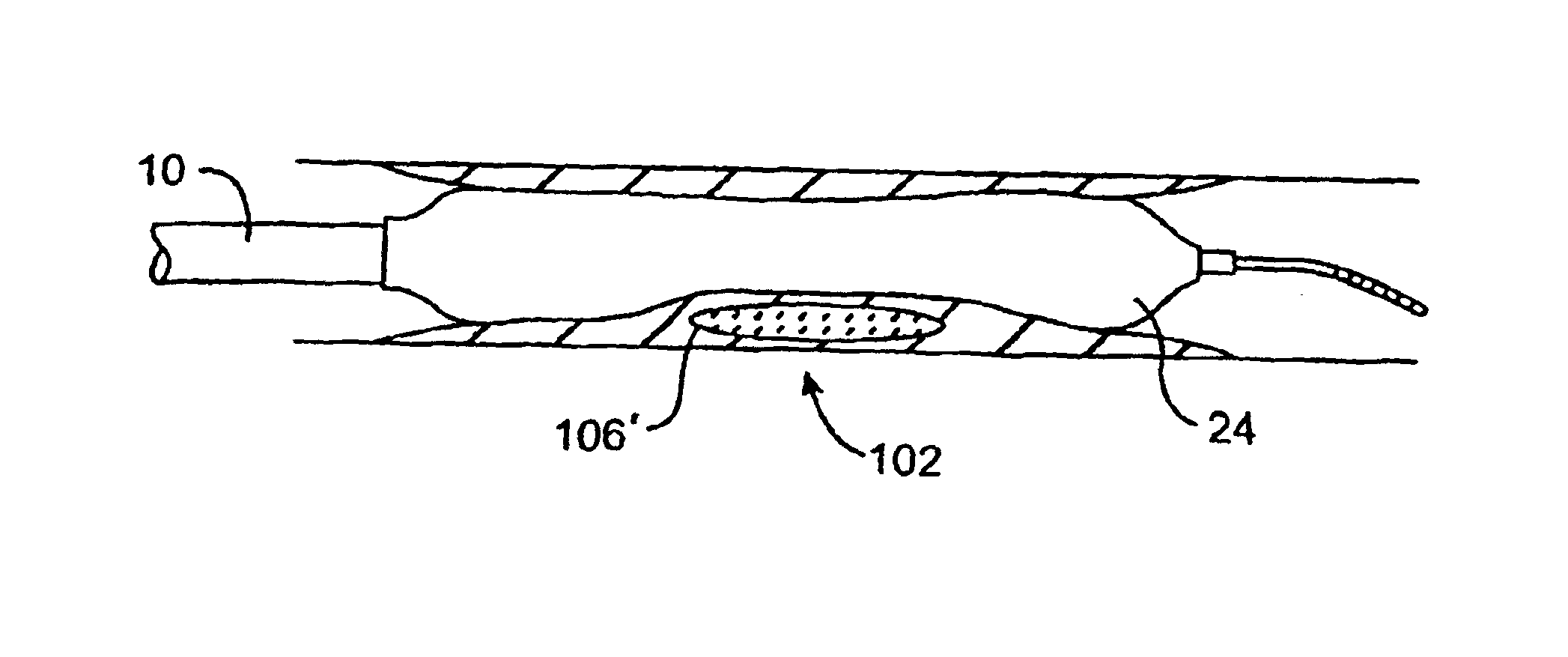
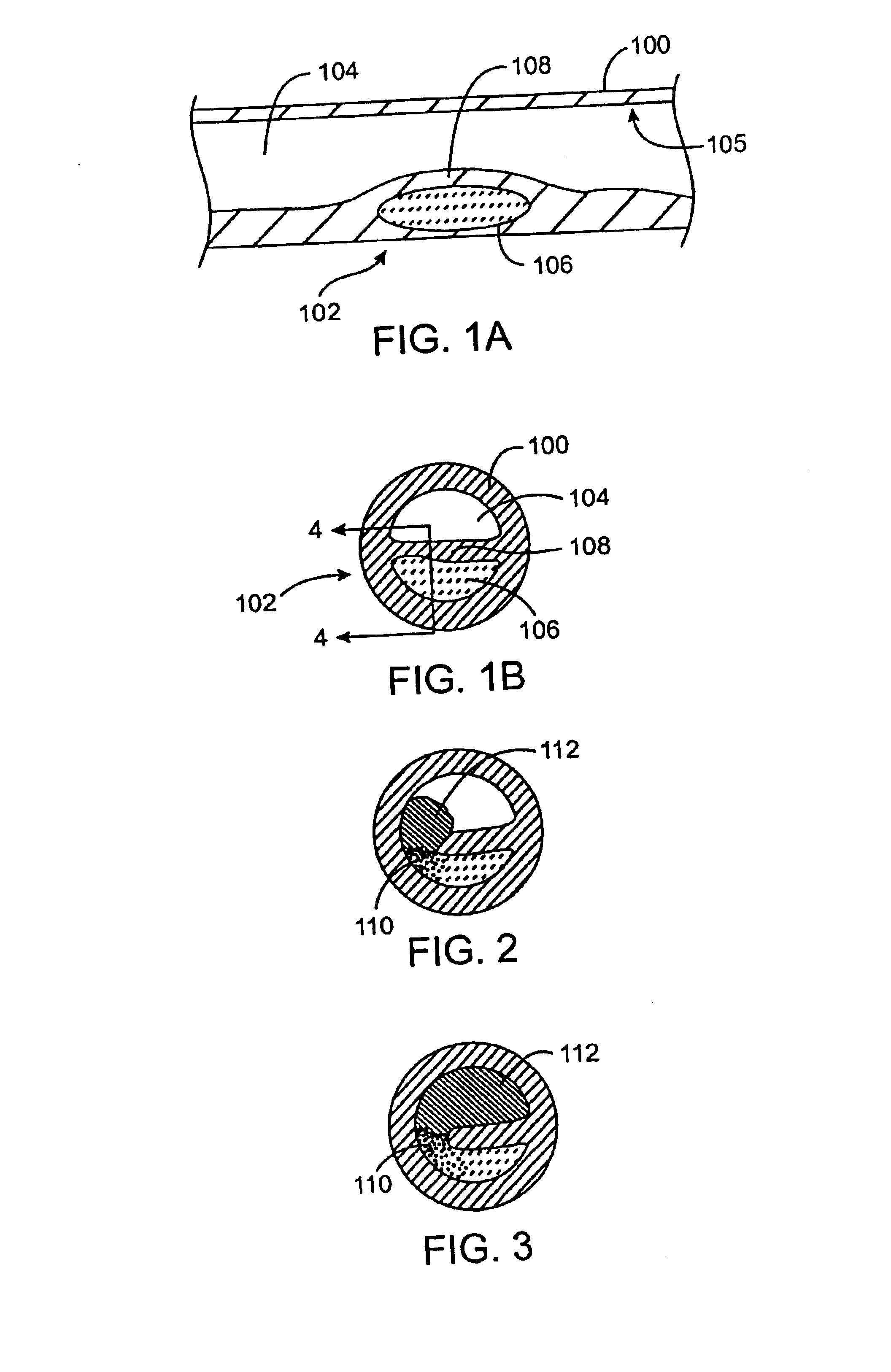
[0051]As used herein, the terms “vulnerable plaque” and “hot plaque” refer to atherosclerotic plaque that is thrombosis-prone. FIGS. 1A and 1B illustrate cross-sectional views of a blood vessel 100 containing a mature vulnerable plaque 102 within a lumen 104 of the vessel. The vulnerable plaque 102 generally comprises a necrotic core 106 of soft, lipid-rich, atheromatous gruel and a fibrous, sclerotic cap 108 of a collagen matrix of smooth muscle cells that covers the core 106. The gruel generally comprises a liquid of esterified cholesterol and low density lipoproteins which is releasably retained by the vulnerable plaque 102. Disruption or fissuring of the cap 108 may cause plaque hemorrhage 110 (release of the highly thrombogenic lipid-rich liquid 106 through the ruptured plaque), as seen in FIG. 2. As a result of plaque hemorrhage 110, the highly thrombogenic lipid-rich liquid 106 is exposed to flowing blood of the vessel lumen 104. As illustrated in FIG. 3, release of the throm...


PUM


Abstract

Methods, apparatus, and kits detect and / or treat vulnerable plaque of a blood vessel. A temperature differential can be sensed along a lumen surface with temperature sensors on a balloon filled with warm gas. Treatment methods include controlled and safe cryogenic cooling of vulnerable plaque to inhibit release of retained fluid within the vulnerable plaque so as to inhibit acute coronary syndrome and to help maintain patency of a body lumen.

Description

CROSS-REFERENCE TO RELATED APPLICATION[0001]This is a continuation-in-part patent application which claims priority from U.S. patent application Ser. No. 09 / 641,462 filed now U.S. Pat. No. 6,602,246, Aug. 18, 2000, the full disclosure of which is incorporated herein by reference.BACKGROUND OF THE INVENTION[0002]1. Field of the Invention[0003]The present invention relates generally to methods, apparatus, and kits for treating blood vessels. More particularly, the present invention provides methods, apparatus, and kits for identifying and / or treating a lesion. In one exemplary embodiment, the invention provides devices which are particularly useful for identification and treatment of vulnerable atherosclerotic plaque within a patient's vasculature to inhibit harmful releases within the vasculature, such as those which may be responsible for strokes or acute coronary syndromes of unstable angina, myocardial infarction, and sudden cardiac death.[0004]Atherosclerotic plaque is present to...

Claims


Application Information

Patent Timeline
Patent Type & Authority Patents(United States)
IPC IPC(8): A61B18/02A61B18/00A61B17/22A61B17/00A61BA61B1/12A61B5/00A61B18/18
CPCA61B1/00082A61B1/128A61B5/01A61B5/015A61B5/6853A61B18/02A61B2017/22002A61B2017/22051A61B2018/0022A61B2018/0212A61B2018/0262A61B1/12A61B1/3137A61B2017/00084A61B2017/00101A61B2017/22001
Inventor JOYE, JAMESTATSUTANI, KRISTINEWILLIAMS, JOSEPH J.
Owner BOSTON SCI SCIMED INC
Why Eureka
Try Eureka