Method for performing automated microfracture

a micro-fracture and automated technology, applied in the field of articular cartilage repair, can solve the problems of limited ability to repair or heal itself, low or insignificant blood flow to the articular cartilage, frequent injuries and defects, etc., and achieve the effect of reducing or eliminating the risk of penetrating too deeply into the bone plate and quick creation of micro-fractures

Inactive Publication Date: 2005-11-01
ZIMMER INC
View PDF9 Cites 139 Cited by
  • Summary
  • Abstract
  • Description
  • Claims
  • Application Information

AI Technical Summary

Benefits of technology

[0017]The present invention is directed to a method for repairing defects in articular cartilage and, more particularly, to a new method for performing automated microfracture on subchondral bone to repair full thickness chondral defects. The important advantage of the present invention is that the microfractured holes on the surface of the subchondral bone plate are formed with an automated process. In particular, this automated process of the present invention uses a pneumatic, hydraulic, electric powered, or otherwise orthopedic gun or instrument to create the microfractures. The microfracture inserter generally comprises a body portion with a microfracture pin assembly removeably connected to the end of the body portion. The microfracture pin assembly generally includes a guide tube, a connector, a biasing member, and a fracture pin. The inserter forceably moves the fracture pin through the end of the guide tube until a sharp end of the fracture pin punctures or penetrates the subchondral bone plate and creates a microfracture or hole in the bone. The biasing member retracts the pin back into guide tube and positions the pin to complete another cycle.
[0018]Broadly described, the method is directed toward performing microfracture surgery on subchondral bone, and this method includes the steps of establishing arthroscopic access to the knee; identifying the chondral defect in the knee; debriding the subchondral bone at the chondral defect and creating a pool to receive the formation of a super-clot; providing an automated microfracture instrument; creating multiple holes in the subchondral bone with the automated microfracture instrument; penetrating the bone to sufficient depth to induce the formation of fat droplets; removing all instruments from the knee; and closing the arthroscopic access to the knee.
[0019]One important advantage of the present invention is that the microfractures are not manually made. Instead, the microfractures are formed with an automated orthopedic gun or instrument, such as an orthopedic microfracture inserter or instrument. The use of an automated instrument has many advantages over the prior method of manually forming the microfractures. The holes in the subchondral bone plate can be formed to have a consistent depth. Further, the depth of the holes can be controlled. Thus, the risk of penetrating too deeply into the bone plate is reduced or eliminated.
[0020]As another advantage, the microfractures are quickly created with the automated orthopedic instrument. A surgeon merely pulls a trigger, pushes a button, or steps on a control switch to activate movement of the fracture pin and create a microfracture in the bone plate. Successive microfractures are created with repeated activation of the instrument.
[0021]As yet another advantage, the holes can easily be spaced 3 to 4 millimeters apart from one another. The microfracture pin assembly also includes a mechanism to guide the placement of the holes.

Problems solved by technology

Injuries and defects to articular cartilage are frequent.
Unfortunately, articular cartilage is generally thin with an extremely low or insignificant blood flow and, as such, has a very limited ability to repair or heal itself.
Partial-thickness chondral defects, for example, cannot spontaneously heal.
If these defects are left untreated, they often degenerate at the articular surface and progress to osteoarthritis.
Even in spite of the formation of fibrocartilage, clinical evidence shows that full-thickness defects continue to degenerate and progress to osteoarthritis if these defects are left untreated.
Although this arthroscopic technique is common, relief for the patient can be incomplete and temporary.
This procedure is inherently limited to the amount or availability of healthy autologous osteochondral grafts in the patient.
The process of growing cells outside the patient can be expensive.
Further, this procedure can require a relatively larger incision to place the cartilage cells.
As one disadvantage, the microfractures or holes are made when the surgeon manually strikes or otherwise forces the awl into the subchondral bone plate.
On the other hand, if the holes are too deep, then the subchondral bone plate can be damaged and lead to unwanted consequences and complications.
As another disadvantage, many microfractures may be placed in a single surgery.
Each hole must be manually placed, and the creation of the many holes can take a lot of time during the surgery.
As yet another disadvantage, the microfractures should be placed 3 to 4 millimeters apart from each other on the bone plate.

Method used


Image

  • Method for performing automated microfracture
  • Method for performing automated microfracture
  • Method for performing automated microfracture

Examples


Embodiment Construction

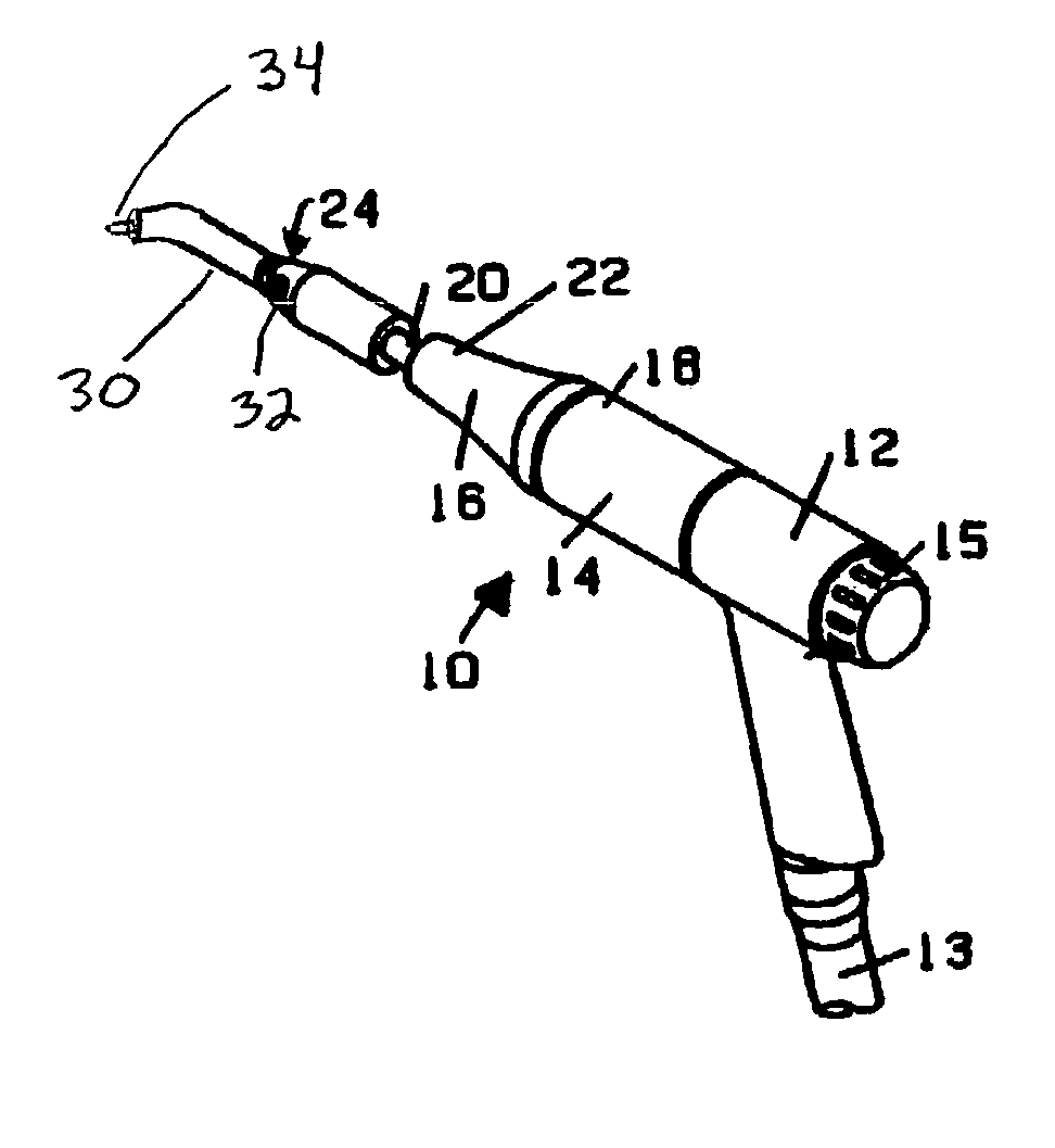
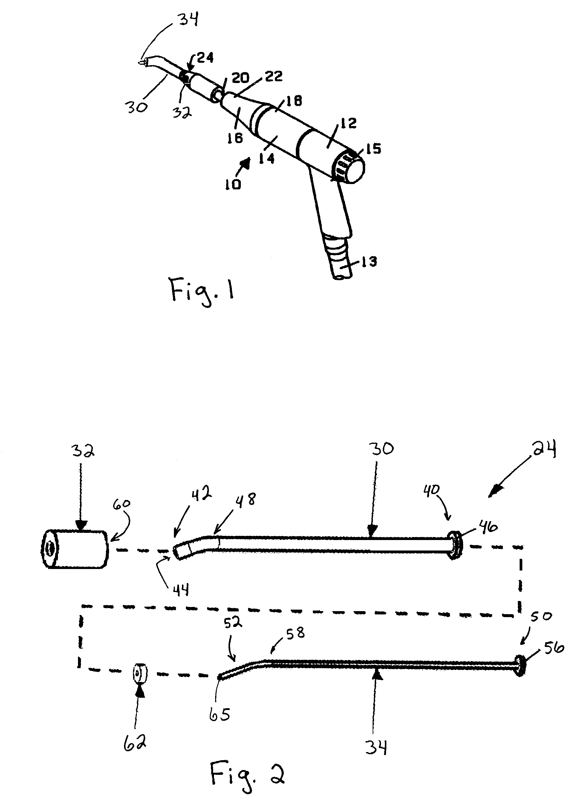
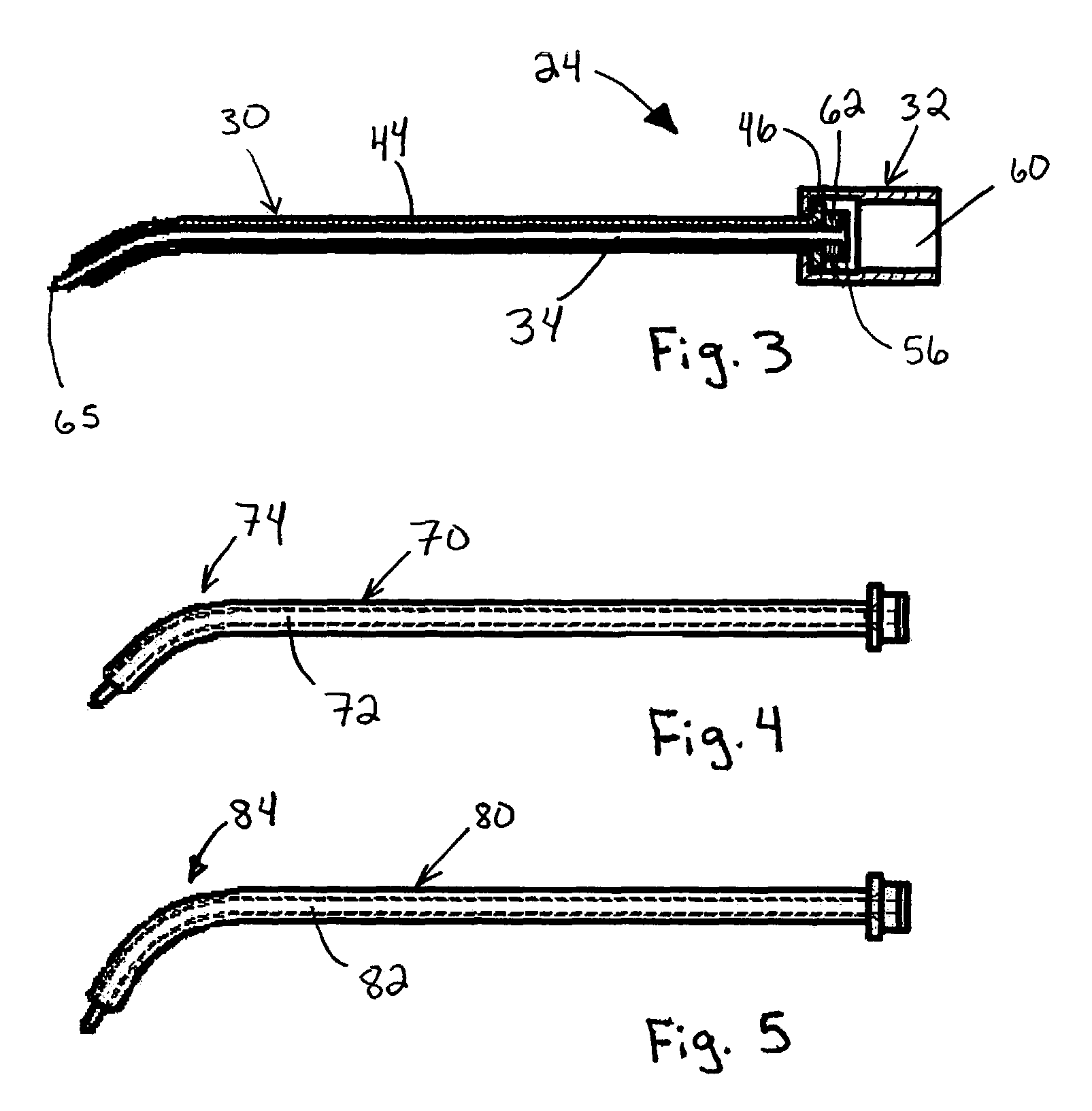
[0027]The instruments, method, and steps of the present invention are now described in more detail. The method describes the steps to perform an automated surgical microfracture procedure on subchondral bone to repair or regenerate articular cartilage at a full-thickness defect. Some of these steps described in the method are known to those skilled in the art and will not be discussed in great detail. Further, one skilled in the art will appreciate that certain steps may be altered or omitted while other steps may be added without departing from the scope of the invention.

[0028]Further, the novel microfracture method of the present invention will be described in connection with arthroscopic knee surgery; though one skilled in the art will appreciate that the microfracture method may be done as an “open” procedure as well. Specifically, the method will address a patient having unstable cartilage covering the underlying bone or a full-thickness defect (i.e., loss of articular cartilag...


PUM


Abstract

The present invention is directed to a method for repairing defects in articular cartilage and, more particularly, to a new method for performing automated microfracture on subchondral bone to repair articular cartilage. The microfractured holes on the surface of the subchondral bone plate are formed with an automated process using a pneumatically driven orthopedic microfracture instrument. The instrument moves a fracture pin through the end of a guide tube until a sharp end of the fracture pin punctures or penetrates the subchondral bone plate and creates a microfracture or hole in the bone.

Description

CROSS-REFERENCE TO RELATED APPLICATIONS[0001]This application claims benefit of priority of U.S. Provisional Application Ser. No. 60 / 418,545 filed Oct. 15, 2002.FIELD OF THE INVENTION[0002]The disclosure herein generally relates to a method for repairing defects in articular cartilage and, more particularly, to a new method for performing automated microfracture on subchondral bone to repair articular cartilage.BACKGROUND OF THE INVENTION[0003]Articular cartilage is a highly organized avascular tissue composed of chondrocytes formed in an extracellular matrix. This tissue is extremely important to the normal, healthy function and articulation of joints. Articular cartilage enables joint motion surfaces to articulate smoothly with a very low coefficient of friction. It also acts as a cushion to absorb compressive, tensile, and shearing forces and, thus, helps protect the ends of bone and surrounding tissue.[0004]Injuries and defects to articular cartilage are frequent. Traumatic chon...

Claims


Application Information

Patent Timeline
Patent Type & Authority Patents(United States)
IPC IPC(8): A61B17/16
CPCA61B17/1604
Inventor BURKINSHAW, BRIAN
Owner ZIMMER INC
Why Eureka
Try Eureka