X-ray diagnostic apparatus

a diagnostic apparatus and x-ray technology, applied in the field of x-ray diagnostic apparatus, can solve the problems of medium starting to diffuse, limited field of view of imaging system, and deterioration of the ability to extract blood vessels

Inactive Publication Date: 2005-11-01
TOSHIBA MEDICAL SYST CORP
View PDF5 Cites 9 Cited by
  • Summary
  • Abstract
  • Description
  • Claims
  • Application Information

AI Technical Summary

Benefits of technology

[0015]It is therefore an object of the invention to provide a d

Problems solved by technology

The field of view of the imaging system is limited to the field of view of the imaging device.
A diluted contrast medium gives rise to deterioration in the ability to extract blood vessels.
The contrast medium starts to diffuse little by little with time

Method used


Image

  • X-ray diagnostic apparatus
  • X-ray diagnostic apparatus
  • X-ray diagnostic apparatus

Examples


Embodiment Construction

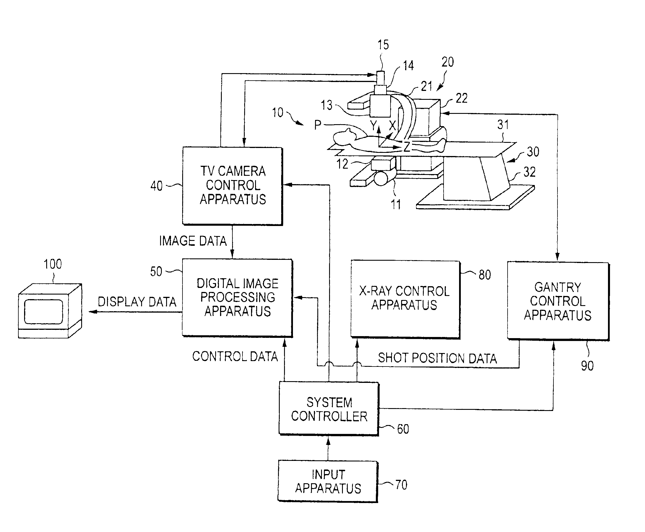
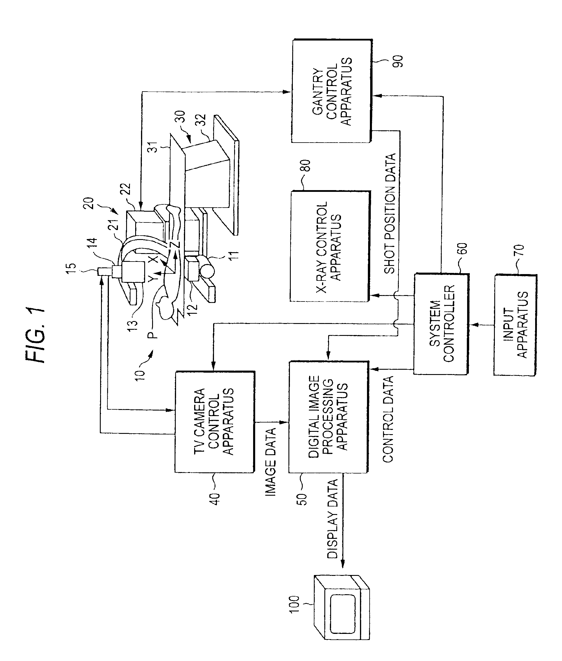
[0033]The following description will describe a preferred embodiment of an X-ray diagnostic apparatus of the invention with reference to the drawings.

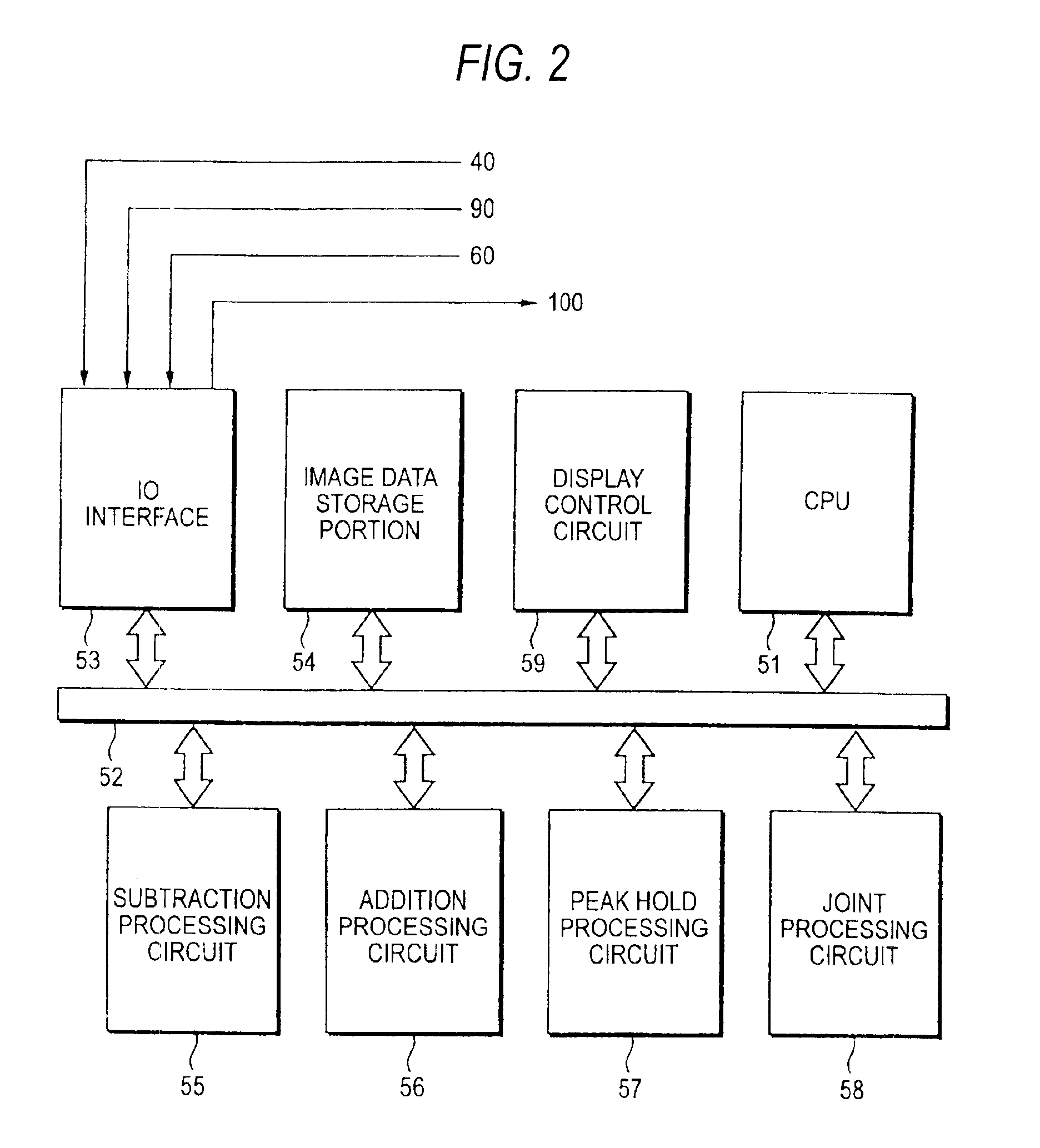
[0034]FIG. 1 is a view depicting an arrangement of an X-ray diagnostic apparatus according to one embodiment of the invention. FIG. 2 is a block diagram of a digital image processing apparatus of FIG. 1. An imaging system 10 generates image data sets from shots by subjecting a patient to X-ray exposure. The imaging system 10 includes an X-ray tube 11 and an imaging device. An X-ray collimator 12 is attached to the X-ray tube 11. The imaging device comprises an image intensifier 13, an optical system 14, and a TV camera 15. The imaging device may comprise a flat panel detector adopting a solid-state image sensor. The solid-state image sensor comprises, for example, a semiconductor layer made of selenium or the like, a voltage applying electrode formed on the surface of the semiconductor layer, and a signal electrode formed on the back s...


PUM


Abstract

A diagnostic X-ray system includes an imaging system that generates image data sets from shots by subjecting a patient to X-ray exposure, a supporting mechanism that supports the imaging system movably with respect to the patient, a system controller that controls the imaging system and the supporting mechanism in such a manner that shots are repeated at each of a plurality of shot positions set discretely along the body axis of the patient, and an image processing apparatus that generates another image data set covering a range wider than the field of view of the imaging system from the image data sets.

Description

CROSS-REFERENCE TO RELATED APPLICATIONS[0001]This is a Continuation Application of PCT Application No. PCT / JP02 / 08417, filed Aug. 21, 2002, which was not published under PCT Article 21(2) in English.[0002]This application is based upon and claims the benefit of priority from the prior Japanese Patent Applications No. 2001-282256, filed Sep. 17, 2002, the entire contents of which are incorporated herein by reference.BACKGROUND OF THE INVENTION[0003]1. Field of the Invention[0004]The present invention relates to an X-ray diagnostic apparatus that takes a shot by subjecting a patient to X-ray exposure.[0005]2. Description of the Related Art[0006]An imaging system includes an X-ray tube and an imaging device. Most of today's imaging devices include an image intensifier and a TV camera. An imaging device adopting a flat panel detector equipped with a solid-state image sensor is expected to come into widespread use in the near feature. The field of view of the imaging system is limited to...

Claims


Application Information

Patent Timeline
IPC IPC(8): A61B6/02A61B6/00G06T7/00
CPCA61B6/4225A61B6/4441A61B6/481A61B6/504A61B6/5229A61B6/5235A61B6/5241G06T7/0024G06T2200/32G06T2207/30101G06T7/30
Inventor HAYASHI, TAKASHI
Owner TOSHIBA MEDICAL SYST CORP
Why Eureka
Try Eureka