Automated verification and inspection device for sequentially inspecting microscopic crystals
a technology of inspection device and microscopic crystal, which is applied in the direction of crystal growth process, chemistry apparatus and processes, instruments, etc., can solve the problems of difficult to grow successful crystals, limited x-ray crystallography, and often larger crystallographic results
- Summary
- Abstract
- Description
- Claims
- Application Information
AI Technical Summary
Benefits of technology
Problems solved by technology
Method used
Image
Examples
second preferred embodiment
[0080]In a second preferred embodiment, the depth of view of camera 135 is approximately 50 to 100 micrometers. The crystal in the drop of liquid may be larger than the depth of view or there may be crystals growing at various levels within the hanging drop of liquid, as shown in FIG. 29. Therefore, in the second preferred embodiment, lens 145 is focused at multiple different levels 721–724 and a set of images are recorded at the different levels so that the entire crystal may be analyzed.
Specimen Auto-Focus
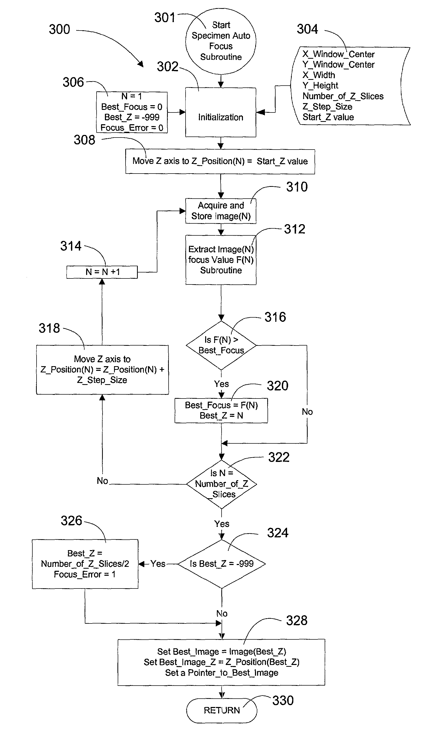
[0081]The third preferred embodiment of the present invention utilizes a specimen auto-focus subroutine 300 (FIG. 31). Subroutine 300 ensures that the specimen within the micro-well is in-focus at the desired zoom (or magnification ratio) of image lens 145. Utilizing the auto-focus feature, the present invention causes camera 135 to take a number of images defined by a Number—of—Z—Slices. Typically, there are between 5 and 10 slices separated in the Z-axis from one another by a Z...
fourth preferred embodiment
[0083]In the first preferred embodiment, it was disclosed how an operator could manually score each drop of liquid as either “CRYSTAL” or “NO CRYSTAL”. In the fourth preferred embodiment, the operator is given a greater variety of options in deciding on how to score each drop. Table 1 shows listing of the operator's scoring options, including number, text description, and the corresponding color code. Once a micro-well drop has been scored a 9, the operator can further classify the crystals in a scoring shown in Table 2.
[0084]
TABLE 1SCOREDESCRIPTIONDISPLAY COLOR0clearWhite1light precipitationRed2heavy precipitationYellow3ugly precipitationBlue4phase separationOrange5unknownViolet6SpherolitesBlack7Grainy precipitationGray8MicrocrystalsBrown9CrystalGreen
[0085]
TABLE 2SCOREDESCRIPTION9.0crystal (no comments)9.1needles, intergrown9.2needles, single9.3plates, intergrown9.4plates, single9.5chunks, 9.6chunks, 9.7chunks, >50 microns, intergrown9.8chunks, >50 microns, single9.9gorgeous >50 mi...
fifth preferred embodiment
[0086]In the forth preferred embodiment, it was disclosed how an operator can manually score each drop of liquid into one of 10 categories with corresponding color coding, and how the operator score category 9 into further subcategories of 9.0 through 9.9. In the fifth preferred embodiment, the inspection device automatically scores and classifies each drop specimen by executing computer software subroutines as shown in FIGS. 33, 34a, 34b, 34c, 34d, and 35a and 35b under control of the program flow shown in FIG. 36. The automatic classification can occur at three levels of detail, the first level, Type—of—Classification=1, simply discriminates between a drop that is clear or not-clear (unknown), the second level, Type—of—Classification=2, scores and classifies the drop into classes 0 through 9 as described in Table 1 above, and the third level, Type—of—Classification=3, performs second level scoring and classification, plus adds an additional 10 subcategories to the CLASS 9, crystal...
PUM
| Property | Measurement | Unit |
|---|---|---|
| thick | aaaaa | aaaaa |
| thick | aaaaa | aaaaa |
| depth of view | aaaaa | aaaaa |
Abstract
Description
Claims
Application Information
 - R&D
- Intellectual Property
- Life Sciences
- Materials
- Tech Scout
- Unparalleled Data Quality
- Higher Quality Content
- 60% Fewer Hallucinations
Browse by: Latest US Patents, China's latest patents, Technical Efficacy Thesaurus, Application Domain, Technology Topic, Popular Technical Reports.
© 2025 PatSnap. All rights reserved.Legal|Privacy policy|Modern Slavery Act Transparency Statement|Sitemap|About US| Contact US: help@patsnap.com


