X-ray computed tomography apparatus

a computed tomography and x-ray technology, applied in the field of multitube type xray computed tomography apparatus, can solve the problems of deterioration in positioning precision, large installation area, and enormous cost, and achieve the effect of improving positioning precision in radiotherapy

Inactive Publication Date: 2006-01-24
TOSHIBA MEDICAL SYST CORP
View PDF15 Cites 205 Cited by
  • Summary
  • Abstract
  • Description
  • Claims
  • Application Information

AI Technical Summary

Benefits of technology

[0007]It is an object of the present invention to provide an X-ray computed...

Problems solved by technology

This requires a large installation area and an enormous...

Method used


Image

  • X-ray computed tomography apparatus
  • X-ray computed tomography apparatus
  • X-ray computed tomography apparatus

Examples


Embodiment Construction

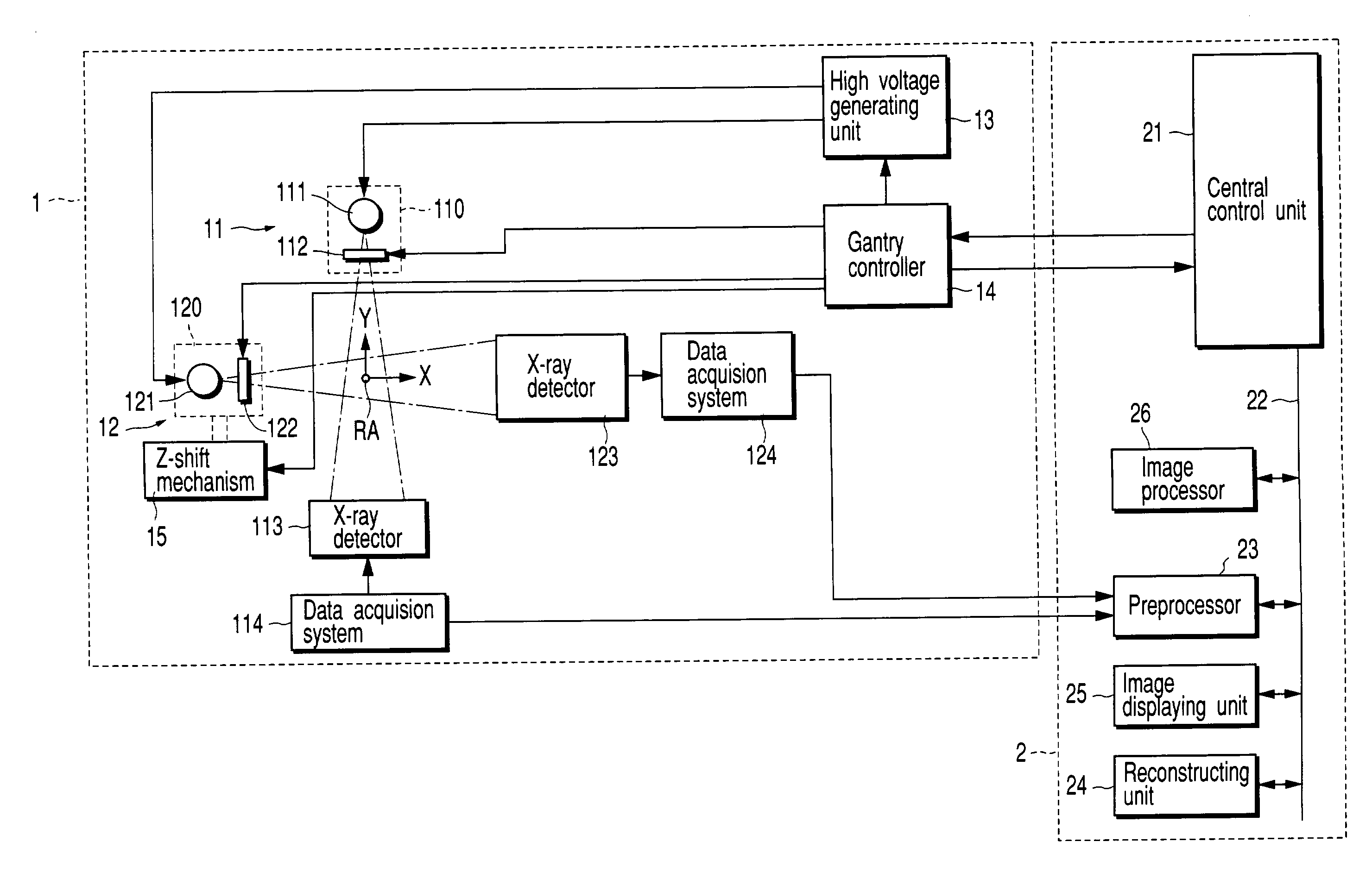
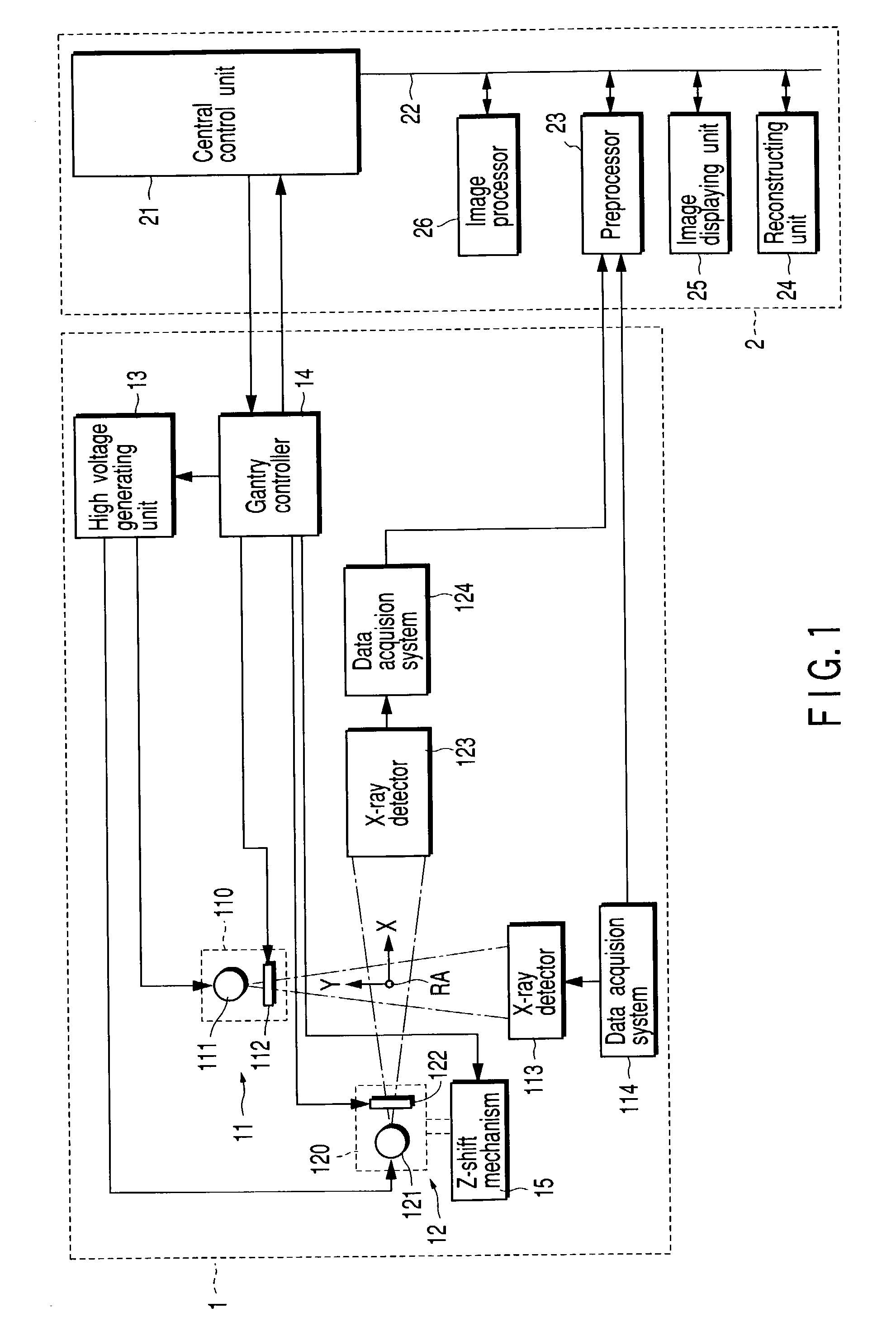
[0023]An X-ray computed tomography apparatus (X-ray CT apparatus) according to a preferred embodiment of the present invention will be described below with reference to the views of the accompanying drawing. Note that the scanning scheme of X-ray computed tomography apparatuses include various types, e.g., a rotate / rotate type in which an X-ray tube and X-ray detector rotate together around a subject to be examined, and a stationary / rotate type in which many detection elements are arrayed in the form of a ring, and only an X-ray tube rotates around a subject to be examined. The present invention can be applied to either type. In this case, the rotate / rotate type will be exemplified. In order to reconstruct one-slice tomographic image data, 360° projection data corresponding to one rotation around a subject to be examined is required, or (180°+ fan angle) projection data is required in the half scan method. The present invention can be applied to either of these reconstruction scheme...


PUM


Abstract

An X-ray computed tomography apparatus includes a first X-ray tube configured to generate X-rays with which a subject to be examined is irradiated, a first X-ray detector configured to detect X-rays transmitted through the subject, a second X-ray tube which generates X-rays with which a treatment target of the subject is irradiated, a rotating mechanism which rotates the first X-ray tube, the first X-ray detector, and the second X-ray tube around the subject, a reconstructing unit configured to reconstruct an image on the basis of data detected by the first X-ray detector, and a support mechanism which supports the second X-ray tube. The central axis of X-rays from the second X-ray tube tilts with respect to a body axis of the subject. This makes it possible to reduce the dose on a portion other than a treatment target.

Description

CROSS-REFERENCE TO RELATED APPLICATIONS[0001]This application is based upon and claims the benefit of priority from the prior Japanese Patent Application No. 2001-320927, filed Oct. 18, 2001, the entire contents of which are incorporated herein by reference.BACKGROUND OF THE INVENTION[0002]1. Field of the Invention[0003]The present invention relates to a multi-tube type X-ray computed tomography apparatus.[0004]2. Description of the Related Art[0005]Conventionally, X-ray computed tomography apparatuses which generate image data from the data (projection data) of X-rays transmitted through a subject to be examined have been known. A doctor refers to the generated image to obtain medical information such as the position, size, and degree of morbidity of a treatment target, and plans a radiotherapy treatment operation based on the information. That is, the doctor sets an irradiation range, irradiation position, dose, and the like. One of the biggest purposes of the treatment plan is to...

Claims


Application Information

Patent Timeline
IPC IPC(8): A61N5/10H05G1/70A61B6/00A61B6/03A61B6/06
CPCA61B6/032A61B6/488A61N5/1049A61B6/06A61N2005/1061A61B6/4429
Inventor NAKASHIMA, SHIGEYUKIYANAGIDA, YUJISHINNO, TOSHIYUKI
Owner TOSHIBA MEDICAL SYST CORP
Why Eureka
Try Eureka