Meniscal and tibial implants

a technology of meniscus and implants, applied in the field of surgical implants, can solve the problems of inability to reproduce the mechanical properties of a normal meniscus, inability to function and move, and the lack of functional features of the normal meniscus in the typical existing knee replacement, etc., and achieve the effect of high mobility and stable join

Inactive Publication Date: 2006-02-07
HOWMEDICA OSTEONICS CORP
View PDF26 Cites 128 Cited by
  • Summary
  • Abstract
  • Description
  • Claims
  • Application Information

AI Technical Summary

Benefits of technology

[0023]The anatomical meniscal-bearing knee implant of the present invention has one or more compliant prosthetic menisci that are attached to the joint capsule and meniscal horns in a similar fashion to the way a natural meniscus is attached to a natural knee. Like a natural meniscus, the meniscal implant will be able to pivot and glide on the prosthetic tibial plateau. Arthrokinematic constraint will come from the meniscal implant's attachments, which will gently guide movements, providing a highly mobile but stable joint. Also through its attachments, the Anatomical Meniscal-Bearing Knee will provide proprioceptive input, giving the central nervous system feedback for refined motor control. Like a natural knee with intact menisci, the outer border of the menisci implants will be mechanically linked to the tibial plateau via the coronary ligament, such as for example by the implant being attached such as by its being sutured directly to the joint capsule / coronary ligament or indirectly by attaching it to the remaining meniscal rim which is in turn attached to the coronary ligament. Tendon slips from the quadriceps, attached to both medial and lateral meniscus, will pull the meniscal replacements forward during active extension and likewise, the semimembranosus (medial meniscus) and / or popliteus tendons (lateral meniscus) will pull the meniscal replacements posteriorly during active flexion.

Problems solved by technology

In many prosthetic knee implants, function and mobility are impaired because rigid structures are used to replace the natural soft tissues.
Typical existing knee replacements lack the functional features normally provided by the menisci and the common polyethylene on metal such as cobalt chrome (CoCr) bearing interface lacks the wear-reducing fluid film bearing surface.
However, these implants do not reproduce the mechanical properties of a normal meniscus.
However, because all four of the menisci inside a person's knees are in high-stress locations, and are subjected to frequently-repeated combinations of compression and tension (and sometimes abrasion as well, especially in people suffering from arthritis or other forms of cartilage damage), meniscal damage often occurs in the knees of humans, and occasionally other large animals.
However, because of the complex structures and anchoring involved, and because of the need to create and sustain extremely smooth and constantly wet surfaces on the inner portions of each meniscal wedge, prior methods of replacing or repairing damaged meniscal are not entirely adequate.
Many meniscal implants for the knee address the need for attachment to the surrounding soft tissue but they do not address the need to resurface the femoral and / or the tibial articulating surfaces.
Because this implant is rigid and because it is disconnected from the soft tissues it lacks the ability to shock absorb and / or provide proprioceptive input.
In fact, because it is approximately 10–20 times more rigid than bone it may actually cause concentrated loading, increased contacts stresses, and therefore accelerate degenerative joint changes.
Because the polyethylene meniscus is semi-rigid it has a limited capacity to absorb shock or conform to the femoral component.
Because of its materials, the Oxford knee also lacks a wear-reducing fluid film bearing surface.

Method used


Image

  • Meniscal and tibial implants
  • Meniscal and tibial implants
  • Meniscal and tibial implants

Examples


Embodiment Construction

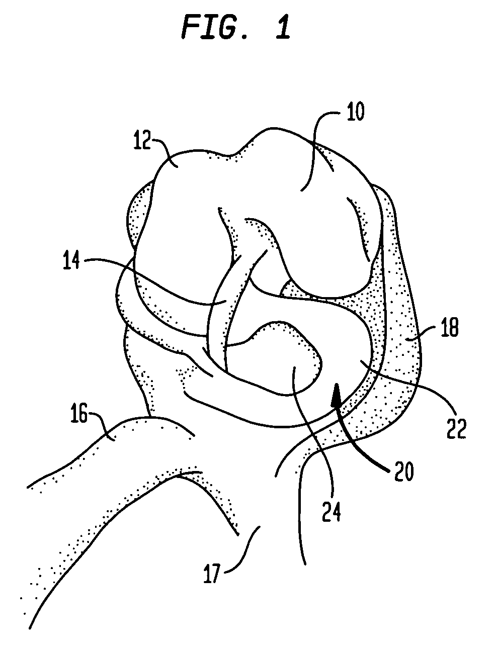
[0052]Referring to FIG. 1 there is shown, for purposes of reference, an open knee joint capsule including a lateral femoral condylar surface 10 and a medial femoral condylar surface 12. The anterior cruciate ligament 14 is shown running through the joint. The quadriceps 16 is shown coupled to the tibia 17 and the lateral collateral ligament 18 is shown connecting the tibia and the femur. The lateral meniscus 20 which includes a rim area 22 is located above the tibial plateau 24.

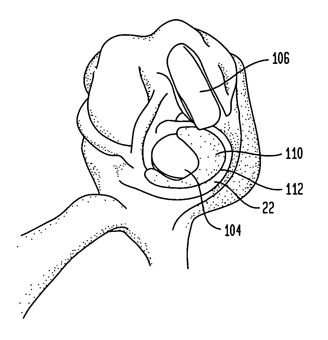
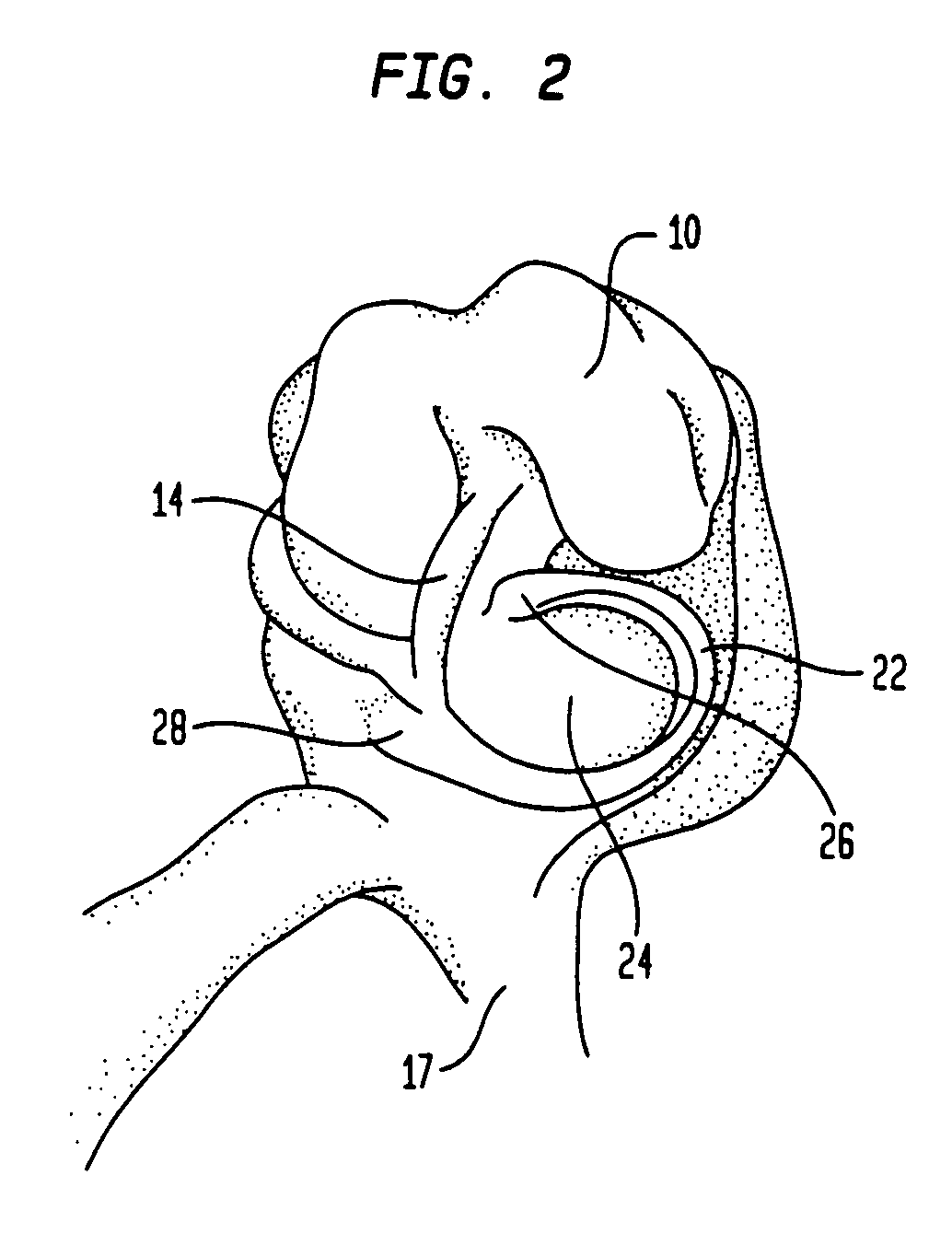
[0053]Referring to FIG. 2, there is shown the joint capsule of FIG. 1 with the inner portion of the meniscus 20 removed leaving meniscal rim 22. In the preferred method, which will be discussed below, the incision / resection of the meniscus 20 is made within or at the border of what is known as the red zone of the meniscus, i.e., the vascularized region of the meniscus. The resection of the inner part of meniscus 20 leaves meniscal horns 26, 28 in place. Since the meniscal rim 22 remains, all the attachment po...


PUM

PropertyMeasurementUnit
depthaaaaaaaaaa
depthaaaaaaaaaa
depthaaaaaaaaaa

Abstract

Instrumentation and a method for resurfacing a joint capsule having cartilage and meniscal surfaces such as a knee joint includes resecting a central portion of the joint cartilage on one joint member such as the tibia while leaving a meniscal rim attached to the peripheral joint capsule. A cavity is then formed in the bone underlying the central portion of the joint surface such as the lateral tibial surface. A resurfacing implant is then coupled, by cementing for example, to the cavity. A soft prosthetic meniscal implant is then coupled to the remaining meniscal ring such as by suturing.

Description

BACKGROUND OF THE INVENTION[0001]This invention relates to surgical implants that are designed to replace meniscal tissue and cartilage in a mammalian joint, such as a knee joint and methods to implant the same. While a knee is the primary joint of concern, the invention applies to other body joints as the hip, shoulder, elbow, temporomandibular, sternoclavicular, zygapophyseal, and wrist.[0002]Compared to the hip the knee has a much greater dependence on passive soft tissues (menisci, ligaments, and the joint capsule) for stability and function. Although the mechanics of the two joints are different, known hip and knee implants are very similar in design, both consisting of a semi-rigid on rigid (polyethylene on CoCr) bearing surface. In many prosthetic knee implants, function and mobility are impaired because rigid structures are used to replace the natural soft tissues.[0003]Normal anatomical knees have two pliable, mobile menisci that function to absorb shock, distribute stress,...

Claims


Application Information

Patent Timeline
Patent Type & Authority Patents(United States)
IPC IPC(8): A61F2/38A61B17/00A61B17/06A61B17/15A61B17/16A61B17/17A61B19/00A61F2/00A61F2/30A61F2/46
CPCA61B17/155A61B17/1675A61B17/1764A61F2/30756A61F2/3868A61F2/3872A61B17/00234A61F2310/00976A61B2017/0046A61B2017/1602A61B2019/304A61F2/30767A61F2/3859A61F2/389A61F2002/30016A61F2002/30064A61F2002/30166A61F2002/30187A61F2002/30616A61F2002/30884A61F2002/30909A61F2002/30957A61F2002/3895A61F2002/4631A61F2230/0028A61F2230/0034A61F2250/0019A61F2310/00023A61F2310/00029A61F2310/00167A61F2310/00179A61F2310/00203A61F2310/00239A61B17/06166A61B2090/034
Inventor POSNER, ELLIOT
Owner HOWMEDICA OSTEONICS CORP
Why Eureka
Try Eureka