System for endoscopic suturing

a technology of endoscope and suture, applied in the field of systems, can solve the problems of multiple passes of endoscope, long patient recovery period, and insufficient suture size or size to be able to apply sutures in tissue, and achieve the effect of limiting the functionality of endoscop

Inactive Publication Date: 2006-02-14
LSI SOLUTIONS
View PDF31 Cites 2115 Cited by
  • Summary
  • Abstract
  • Description
  • Claims
  • Application Information

AI Technical Summary

Benefits of technology

[0010]It is another object of the present invention to provide an improved system for endoscopic suturing which allows single insertion of an endoscope and accessory tube assembly in the gastrointestinal tract of a patient through which multiple instruments for suturing and securing sutures can be used without removal of the endoscope between suturing and suture securing operations.
[0012]Yet a further object of the present invention is to provide an improved system for endoscopic suturing in which a suturing instrument separate from an endoscope can utilize suction to capture tissue to be sutured.
[0013]A still further object of the present invention is to provide an improved system for remote suturing in which can readily adapted to different types of flexible endoscopes to provide an external accessory tube through which medical or surgical instruments can pass.
[0015]A further object of the present invention is to provide an improved system for endoscopic suturing using instruments having shafts with enhanced flexibility.
[0017]Yet a further object of the present invention is to provide an improved system for endoscopic suturing in which remote viewing with an endoscope of suture and suture securing operations is provided in the stomach without hindering endoscope functionality.
[0018]Briefly described, a preferred embodiment of this system embodying the invention includes an endoscope, such as a gastroscope, having a distal end locatable in the body of a patient, such as in the gastrointestinal or gastroesophageal tract, and a flexible shaft extending to the distal end, a flexible accessory tube coupled to the endoscope to be movable relative to the endoscope's shaft, and an attachment tip coupled to the shaft of the endoscope having an opening through which one end of the accessory tube is received. The accessory tube is coupled to the shaft of the endoscope with multiple tube guides enabling the accessory tube to slide through the tube guides in response to bending of the endoscope's shaft. The accessory tube has a cannula through which an instrument may pass in the accessory tube. The system includes a tissue suturing instrument having a partially flexible shaft locatable through the accessory tube, and a tissue engaging end coupled to the shaft which is viewable by the endoscope at its distal end when the instrument is fully inserted through the accessory tube. The tissue engaging end has a vacuum sleeve enabling suction to be selectably applied at the tissue engaging end to capture tissue in a gap of a sew tip through an opening in the vacuum sleeve. Suction is applied via a vacuum connection assembly to a channel which extends down the shaft to the sew tip. A valve is provided to close one end of the suture carrying channel to enable such suction at the sew tip. Two needles are provided which extends through the shaft of the suturing instrument. Each needle is separately actuated into the gap of the sew tip through suctioned tissue to capture a ferrule having one end of a loop of suture. The system further includes a suture securing instrument having a partially flexible shaft locatable through the accessory tube, and a distal end coupled to the shaft which is viewable by the endoscope at its distal end when the instrument is fully inserted through the accessory tube. After removal of the suturing instrument from the accessory tube, a loop of suture extends through the tissue through the accessory tube, the suture securing instrument receives the free ends of the loop of suture at its distal end through a sleeve member, and the suture securing instrument is then inserted through the accessory tube to the location of the suture in the tissue. The suture securing instrument crimps the sleeve member and cuts the free ends of the suture to retain the suture closed. The endoscope enable an operator, such as a surgeon, gastroenterologist, or other skilled physician, to view the engaging end of the suturing instrument for selecting placement of the suture through tissue, and of the distal end of the suture securing instrument to secure the suture closed.

Problems solved by technology

Such procedures are invasive, as laparoscopy requires small access incision(s) made in the body of the patient through which a laparoscope and other surgical enabling tools are provided, while open surgical techniques are traditionally invasive and can have complications and long patient recovery periods.
Although gastroscopes often have a working channel to a port at the distal end of the gastroscope through which a biopsy tool may be provided to obtain tissue samples, they are not currently designed or typically large enough to be capable of applying sutures in tissue.
Since the sewing device of U.S. Pat. No. 5,792,153 does not allow normal use of its biopsy channel of the endoscope upon which the sewing device is mounted, a second endoscope must be used to secure and cut the suture through its biopsy channel using the device described in U.S. Pat. No. 5,755,730.
This results in multiple passes of endoscopes back and forth through the gastroesophageal tract, especially if single sutures are each applied and secured at multiple locations in tissue.
However, the overtube may be uncomfortable to patients, and can cause complications, such as mucosal tears in the esophagus.
Moreover, mounting on an endoscope limits the use of the endoscope for full visualization of tissue, as the sewing device partially obstructs the viewing area at the distal end of the endoscope.
Further the use of the biopsy or working channel of an endoscope for needle placement does not allow use of the channel for other purposes, such as obtaining a biopsy.

Method used


Image

  • System for endoscopic suturing
  • System for endoscopic suturing
  • System for endoscopic suturing

Examples


Embodiment Construction

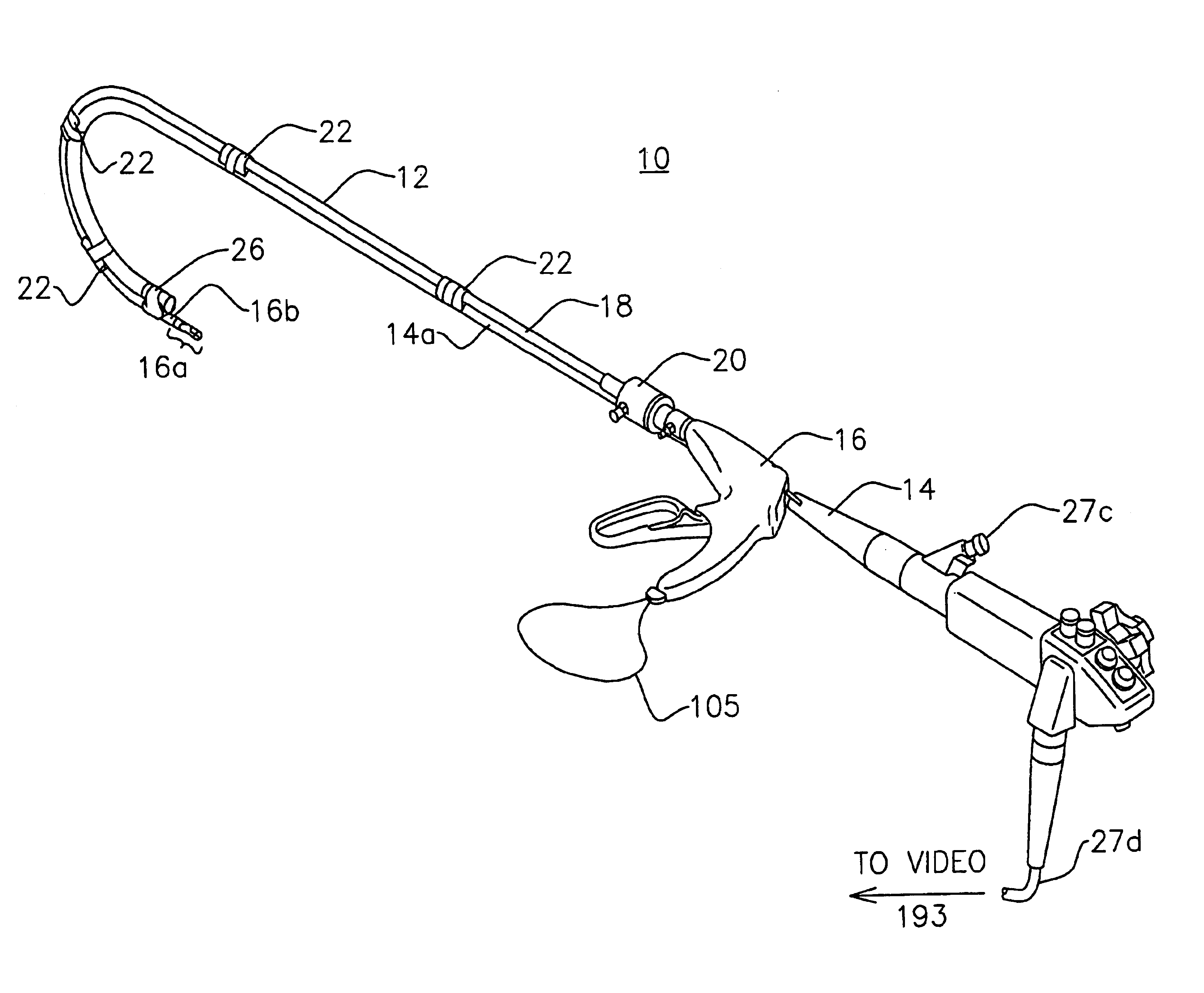
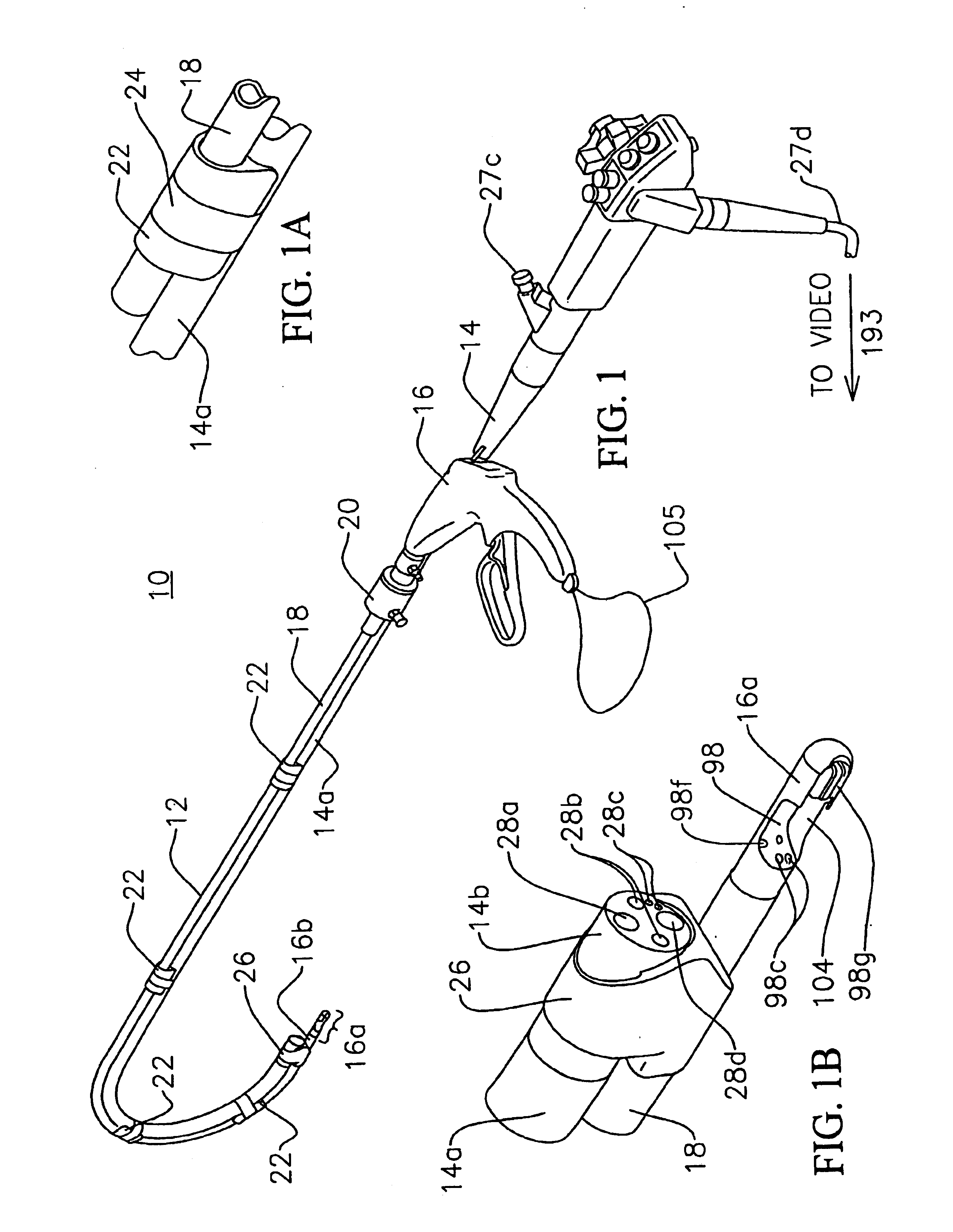
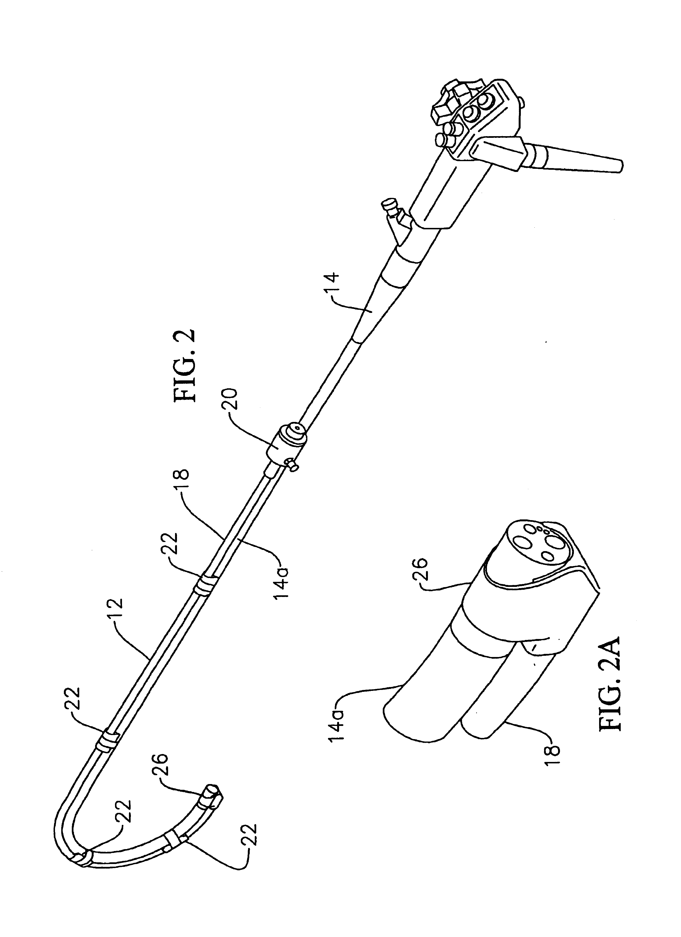
[0102]Referring to FIG. 1, a system 10 for suturing is shown including an accessory tube 12 and an endoscope 14, referred to herein after as a gastroscope, or other type of flexible endoscope having a shaft 14a coupled to the accessory tube, and a suturing instrument 16. The suturing instrument 16 may be inserted in the accessory tube 12 as shown in FIG. 1, and is removable from the accessory tube 12 as shown in FIG. 2. The accessory tube 12 has access tubing 18 which is sufficiently flexible to be movable with the flexible shaft 14a of the gastroscope. Tubing 18 is braid reinforced with a braid of stainless steel, nylon, or Kevlar, to maintain the integrity of the tubing's circular cross-sectional shape and avoid kinking as the shaft 14a of the gastroscope bends when placed through the mouth into the gastrointestinal tract of a patient. The braiding may be located between two layers of tubing 18, which are integrated with the braiding during their extrusion forming tubing 18. For e...


PUM


Abstract

A system for endoscopic suturing is provided having an endoscope, such as a gastroscope, with a distal end locatable in the body of a patient and a flexible shaft extending to the distal end, a flexible accessory tube coupled to the endoscope to be movable relative to the endoscope's shaft, and a tip coupled to the shaft of the endoscope having an opening through which one end of the accessory tube is received. The system further includes a tissue suturing instrument and a suture securing instrument. Each of these instruments has a distal end coupled to a partially flexible shaft locatable through the accessory tube.

Description

FIELD OF THE INVENTION[0001]The present invention relates to a system (and method) for endoscopic suturing, and in particular to a system for suturing through an accessory tube coupled to a flexible endoscope, which may be placed in the stomach through mouth and the esophagus of a patient utilizing a tissue suturing instrument and a suture securing instrument. The invention is suitable, for example, for applying at least one suture in the soft tissue lining of the stomach for different procedures such as gastroplasty, fundoplication, anterior gastropexy, or other procedures requiring suturing in the stomach, without the need for laparotomy or laparoscopy.BACKGROUND OF THE INVENTION[0002]Application of sutures in the gastrointestinal tract is required for several different types of medical procedures, for example, transoral endoscopic valvuloplasty for gastroesophageal reflux disease, gastroplasty, fundoplication, anterior gastropexy, suturing esophageal perforations, or closure of e...

Claims


Application Information

Patent Timeline
Patent Type & Authority Patents(United States)
IPC IPC(8): A61B17/10A61B17/04A61B17/30
CPCA61B1/0014A61B17/0469A61B1/018A61B17/0467A61B2017/306A61B17/0482A61B2017/00296A61B2017/0496
Inventor SAUER, JUDE S.FITZSIMMONS, MICHAEL W.BOVARD, MARK A.
Owner LSI SOLUTIONS
Why Eureka
Try Eureka