Method and apparatus for processing an image of a tissue sample microarray

a tissue sample and microarray technology, applied in the field of methods and apparatus for processing an image of a tissue sample microarray, can solve the problem that tissue dots may even be missing, and achieve the effect of reducing the number of controls

Inactive Publication Date: 2006-04-18
OLYMPUS AMERICA
View PDF77 Cites 22 Cited by
  • Summary
  • Abstract
  • Description
  • Claims
  • Application Information

AI Technical Summary

Benefits of technology

[0016]A method and apparatus for processing an image of a tissue sample microarray include placing a plurality of tissue samples in an array or a rectangular grid on a conventional glass microscope slide. The tissue samples, which have been sectioned and form the array, may then be treated simultaneously as by staining the entire slide with a suitable biological stain for providing contrast to image various structures within the tissues or cells making up the multiple samples of the microarray. The advantage of doing this is that it allows multiple samples taking from multiple patients to be treated in substantially the same way and reduces the number of controls which must be put in place during the experiment.

Problems solved by technology

Some tissue dots even may be missing.

Method used


Image

  • Method and apparatus for processing an image of a tissue sample microarray
  • Method and apparatus for processing an image of a tissue sample microarray
  • Method and apparatus for processing an image of a tissue sample microarray

Examples


Embodiment Construction

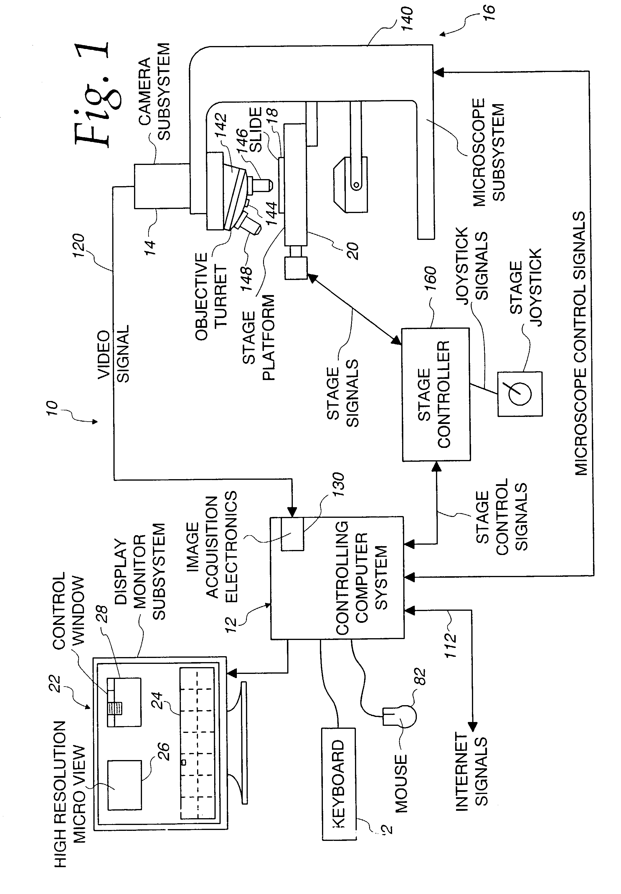
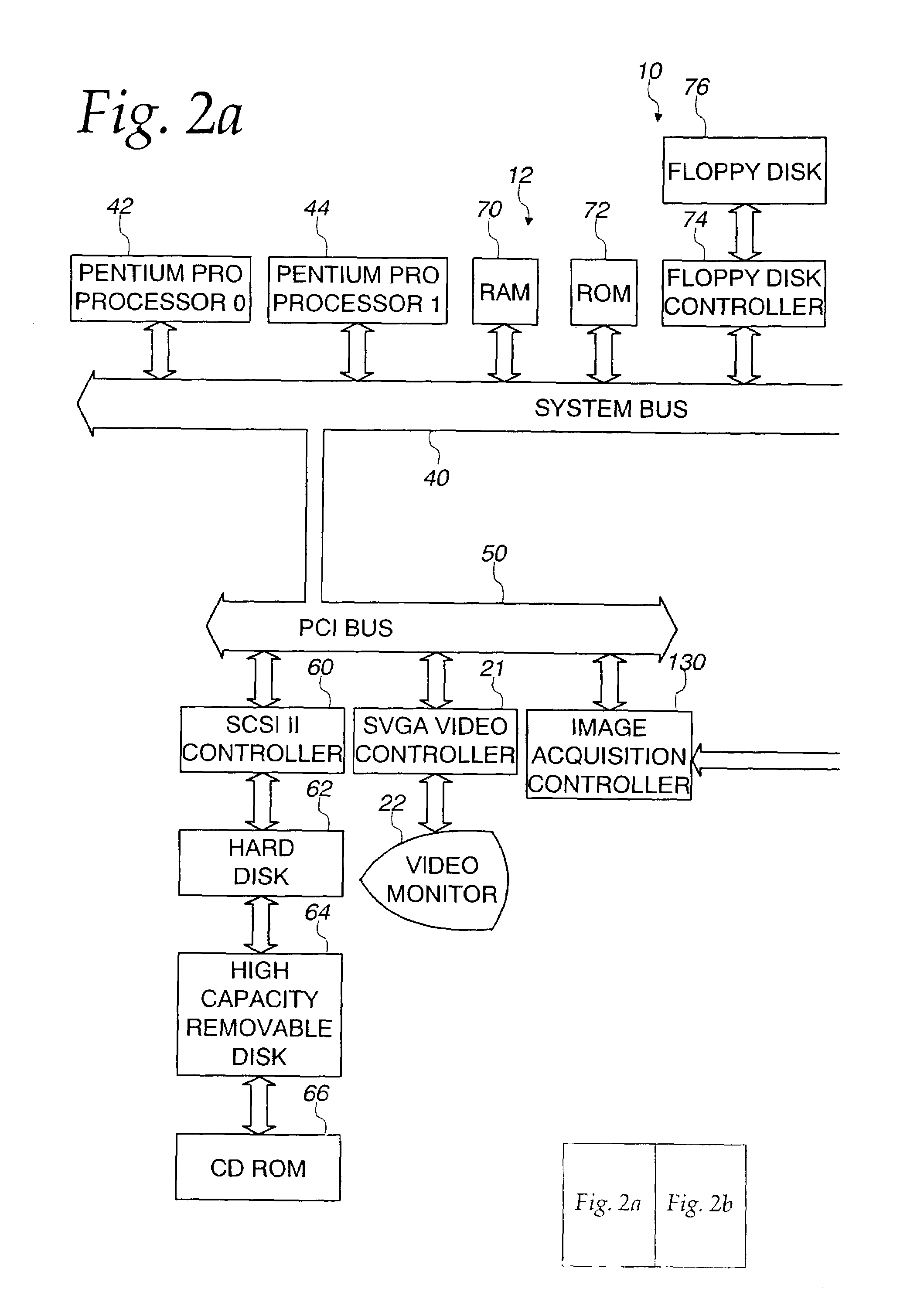
[0041]Referring now to the drawings and especially to FIG. 1, an apparatus embodying the present invention is shown therein and generally identified by reference numeral 10. The apparatus 10 is adapted for synthesizing low magnification and high magnification microscopic images of tissue sample microarrays. The apparatus 10 includes a computer 12 which is a dual microprocessor personal computer in combination with a Hitachi HV-C20 video camera 14 associated with a Zeiss Axioplan 2 microscope 16. The computer system 12 receives signals from the camera 14 which captures light from the microscope 16 having a microscope slide 18 positioned on an LUDL encoded motorized stage 20. The encoded motorized stage 20 includes a MAC 2000 stage controller for controlling the stage in response to the computer 12.

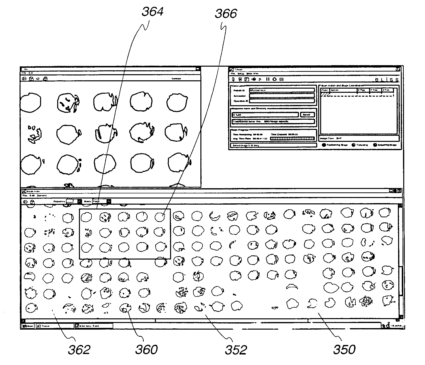
[0042]A microscope slide 18 includes a plurality of tissue sample microarrays 19 each comprising a tissue sample microarray made up of a grid or array of circular tissue sample sections or ...


PUM

PropertyMeasurementUnit
cellular structureaaaaaaaaaa
data structureaaaaaaaaaa
optical microscopeaaaaaaaaaa

Abstract

A method and apparatus for processing an image of a tissue sample microarray include placing a plurality of tissue samples in an array on a microscope slide. The tissue samples are then simultaneously and uniformly treated, as by staining. Images of the tissue making up the microarray are captured and stored together with identifying information related thereto. The images may be displayed from the digital storage medium using a programmed processor which can select various magnifications for display. The images also can be accessed by network and remotely.

Description

FIELD OF THE INVENTION[0001]This is a continuation of prior application Ser. No. 09 / 740,711, filed Dec. 19, 2000, U.S. Pat. No. 6,466,690 which is hereby incorporated herein by reference in its entiretyBACKGROUND OF THE INVENTION[0002]The invention relates in general to a method and apparatus for processing images of a tissue sample microarray made up of a plurality of tissue microarray dots using an optical microscope. More particularly, the invention relates to a method and apparatus for capturing tissue sample images from the tissue sample microarray, indexing such images and manipulating and transmitting them.[0003]It is known that in the treatment and prevention of cancers it is often important periodically to examine persons at risk for cancer. In some instances it may be necessary to biopsy tissue from such persons. As medical care has become available to more people and as the need for such increased vigilance has been recognized, the number of biopsies has increased.[0004]O...

Claims


Application Information

Patent Timeline
Patent Type & Authority Patents(United States)
IPC IPC(8): G06K9/00G01N33/48G16B25/30G01N33/483G06F17/30G06Q50/00G06T1/00G16B40/00G16B45/00
CPCG06F19/20G06K9/00127G06F19/26G06F19/24G16B25/00G16B40/00G16B45/00G16B25/30G06V20/69
Inventor BACUS, JAMES W.BACUS, JAMES V.
Owner OLYMPUS AMERICA
Why Eureka
Try Eureka