Method for characterizing shapes in medical images

a technology of medical images and shapes, applied in the field of medical imaging, can solve the problems of high false positive rate, difficult segmentation or elimination of false positive shapes, and increase detection sensitivity or accuracy, and achieve the effects of reducing false positives, sacrificing detection sensitivity, and high detection specificity

Inactive Publication Date: 2006-05-09
THE BOARD OF TRUSTEES OF THE LELAND STANFORD JUNIOR UNIV
View PDF25 Cites 46 Cited by
  • Summary
  • Abstract
  • Description
  • Claims
  • Application Information

AI Technical Summary

Benefits of technology

[0011]The advantage of the present invention is that the computer-implemented method provides a high detection specificity, i.e. reducing false positives, without sacrificing sensitivity of the detection of a shape of interest. Another advantage is that the present invention provides computer-aided assistance of physicians in the interpretation of medical images, making the process considerably more efficient than current human viewing interpretation and enabling a cost-effective medical diagnosis to be widely deployed for screening purposes. Yet another advantage is that the present invention is deterministic and thus, does not require any training, is very fast, and easy to implement.
[0012]The objectives and advantages of the present invention will be understood by reading the following detailed description in conjunction with the drawings, in which:
[0013]FIG. 1 shows an overview of the method of characterizing a shape in a medical image according to the present invention including the step of locating and detecting a shape in a medical image.
[0014]FIG. 2 shows a method of locating and detecting a shape in a medical image that could be used in the present invention;
[0015]FIG. 3 shows a method of locating and detecting a shape in a medical image including the step of scaling normal vectors that could be used in the present invention;
[0016]FIGS. 4–7 show several embodiments related to a 2-D representation of the methods shown in FIGS. 2–3;

Problems solved by technology

However, one of the limitations of detection methods focusing on increased sensitivity or accuracy of detection is that they could easily lead to a high false positive rate due to structures in the colon or lung with convex surfaces, such as haustral folds or pulmonary blood vessels.
Furthermore, a detection due to false positive structures is usually based on the shape of the structure, which is often adjacent to other anatomical structures, thus making segmentation or elimination of false positive shapes difficult.
However, automatically determining which portions of the image correspond to the shape of interest and which correspond to adjacent but distinct anatomical structures is very difficult.

Method used


Image

  • Method for characterizing shapes in medical images
  • Method for characterizing shapes in medical images
  • Method for characterizing shapes in medical images

Examples


Embodiment Construction

[0026]Although the following detailed description contains many specifics for the purposes of illustration, anyone of ordinary skill in the art will readily appreciate that many variations and alterations to the following exemplary details are within the scope of the invention. Accordingly, the following preferred embodiment of the invention is set forth without any loss of generality to, and without imposing limitations upon, the claimed invention.

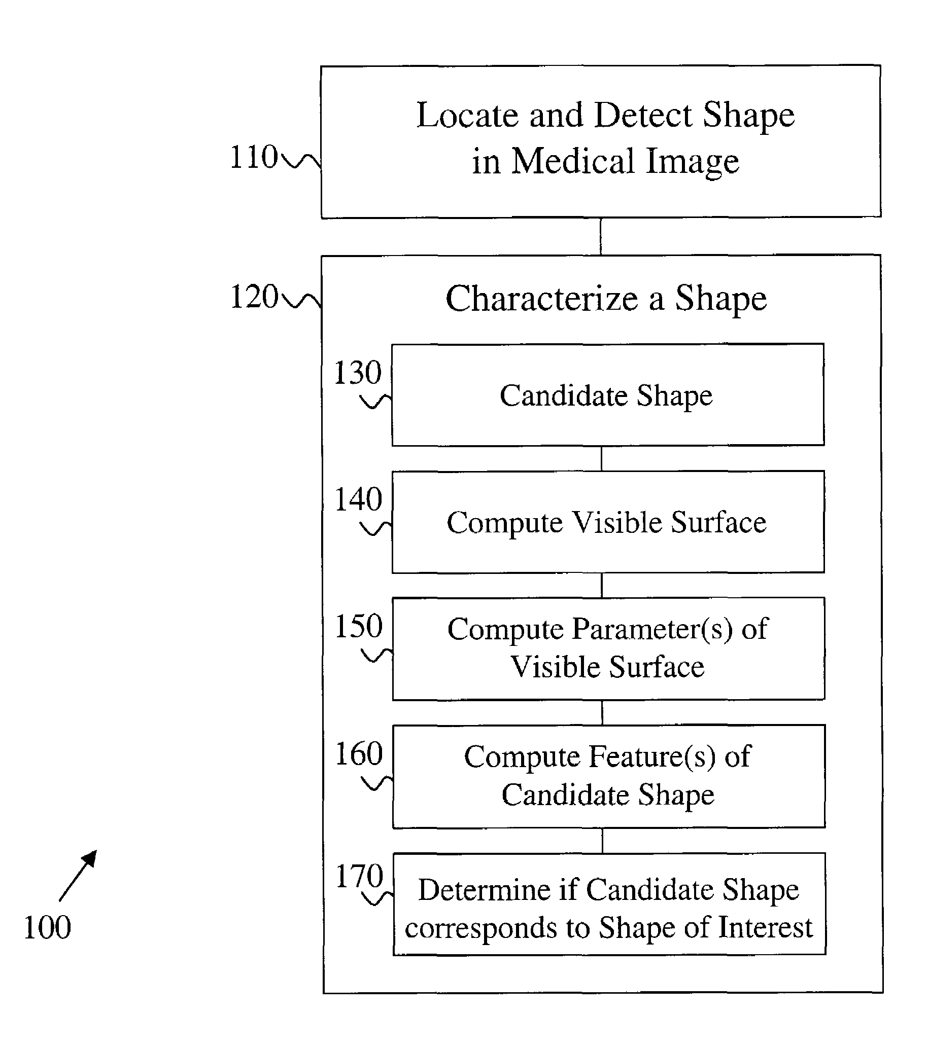
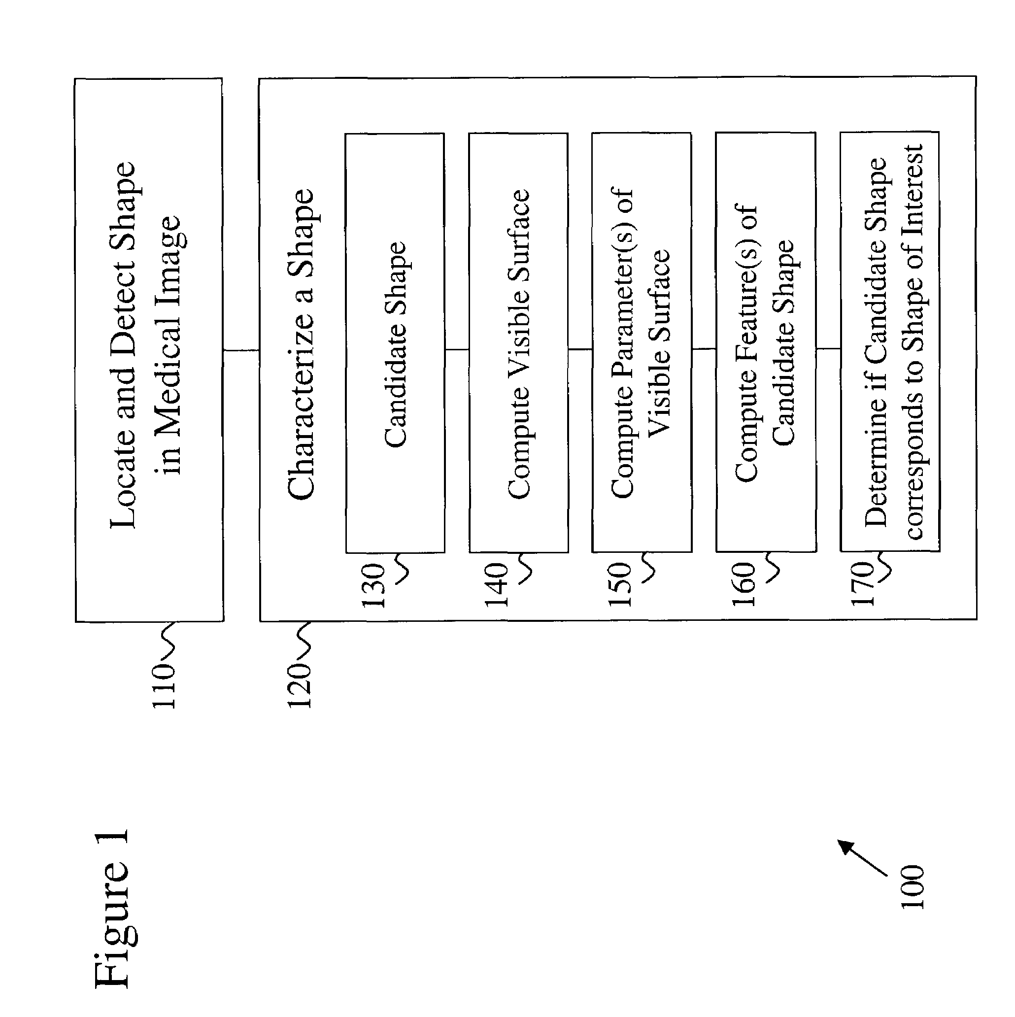
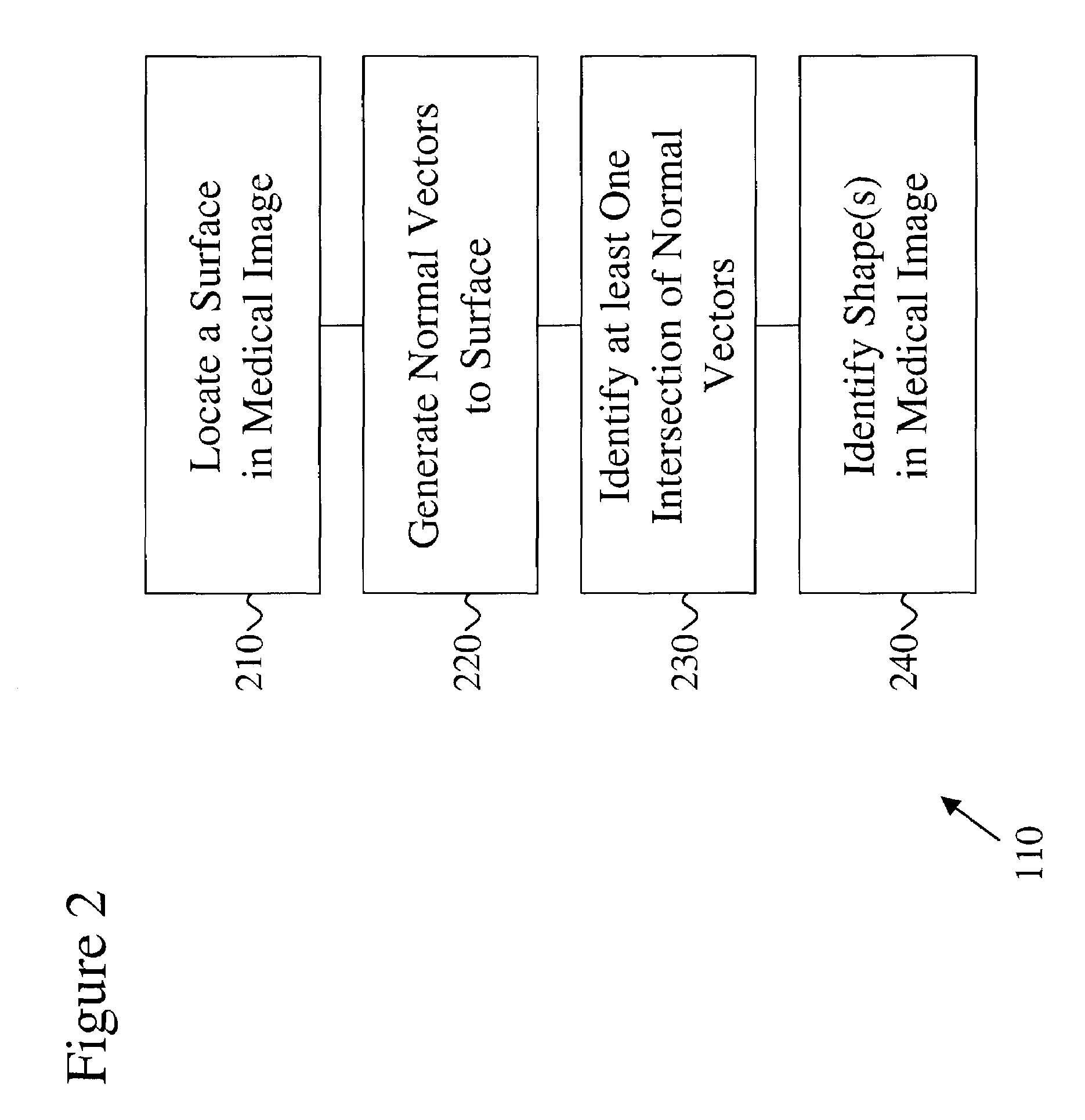
[0027]The present invention provides a computer-implemented method for characterizing one or more shapes in a medical image to provide accurate and early detection of pre-cancerous or cancerous growths and more particularly to eliminate false positives from such a detection. The present invention enables a user, such as, but not limited to, a radiologist, to determine which portions of the medical image corresponds to a shape of interest and which portions of the medical image correspond to distinct anatomical features. The medical images...


PUM


Abstract

A computer-implemented method for determining and characterizing, which portions or shapes of a medical image correspond to a shape of interest is provided. A candidate shape is obtained after which a visible surface is computed adjacent to this candidate shape. A visible surface includes one or more portions of the medical image that are visible by the candidate shape. Once the visible surface is determined, parameters of the visible surface are computed. Then the method further includes the step of determining whether the candidate shape corresponds to a shape of interest. The method further includes the step of computing features of the candidate shape and / or classifying the candidate shape. The advantage of the computer-implemented method is that it provides a high detection specificity, i.e. reducing false positives, without sacrificing sensitivity of the detection of a shape of interest.

Description

CROSS-REFERENCE TO RELATED APPLICATIONS[0001]The present application is cross-referenced to and claims priority from U.S. Provisional Applications 60 / 288,674 filed May 4, 2001 and 60 / 288,621 filed May 4, 2001, which are both hereby incorporated by reference. This application is also cross-referenced to co-pending U.S. Patent Application entitled “Method for Detecting Shapes in Medical Images” filed with the USPTO on May 3, 2002, which is hereby incorporated by reference.FIELD OF THE INVENTION[0002]This invention relates generally to medical imaging. More particularly, the present invention relates to a method for characterizing shapes in medical images according to a shape of interest.BACKGROUND[0003]In the United States, lung cancer and colon cancer are the first and second leading cancer killers, respectively. Early detection of colonic polyps and lung nodules, the precursors to these diseases, has been shown to improve survival. Therefore, early detection of precancerous growths ...

Claims


Application Information

Patent Timeline
Patent Type & Authority Patents(United States)
IPC IPC(8): G06K9/00G06T7/00G06V10/46
CPCG06K9/48G06T7/0012Y10S128/922G06K2209/053G06V10/46G06V2201/032
Inventor PAIK, DAVID S.NAPEL, SANDY A.RUBIN, GEOFFREY D.BEAULIEU, CHRISTOPHER F.
Owner THE BOARD OF TRUSTEES OF THE LELAND STANFORD JUNIOR UNIV
Why Eureka
Try Eureka