MR-compatible methods and systems for cardiac monitoring and gating

a technology of cardiac monitoring and gating, which is applied in the field of magnetic resonance imaging, can solve the problems of ecg signal corruption, erroneous cardiac gating, and none of these conventional methods have been shown to provide reliable monitoring and gating ability, and achieve high spatial and temporal resolution

Inactive Publication Date: 2006-05-30
DUKE UNIV
View PDF8 Cites 10 Cited by
  • Summary
  • Abstract
  • Description
  • Claims
  • Application Information

AI Technical Summary

Benefits of technology

[0003]A fundamental problem associated with using a conventional electrocardiograph (ECG) to monitor a patient's cardiac activity during MR imaging is the corruption of the ECG signal due to adverse electromagnetic effects. This effect is particularly pronounced in MR microscopy of small animals (e.g., laboratory rodents), where strong, rapidly-switching, magnetic field gradients are needed to obtain high spatial and temporal resolution, and the animal's ECG signal is less than a millivolt in amplitude. The spurious signals often resemble the QRS spike and can lead to erroneous cardiac gating. Furthermore, the artifacts often do not disappear until tens of milliseconds after the gradients turn off.

Problems solved by technology

A fundamental problem associated with using a conventional electrocardiograph (ECG) to monitor a patient's cardiac activity during MR imaging is the corruption of the ECG signal due to adverse electromagnetic effects.
The spurious signals often resemble the QRS spike and can lead to erroneous cardiac gating.
However, none of these conventional methods has been shown to provide reliable monitoring and gating ability in small rodents during cardiac MR microscopy.

Method used


Image

  • MR-compatible methods and systems for cardiac monitoring and gating
  • MR-compatible methods and systems for cardiac monitoring and gating
  • MR-compatible methods and systems for cardiac monitoring and gating

Examples


example

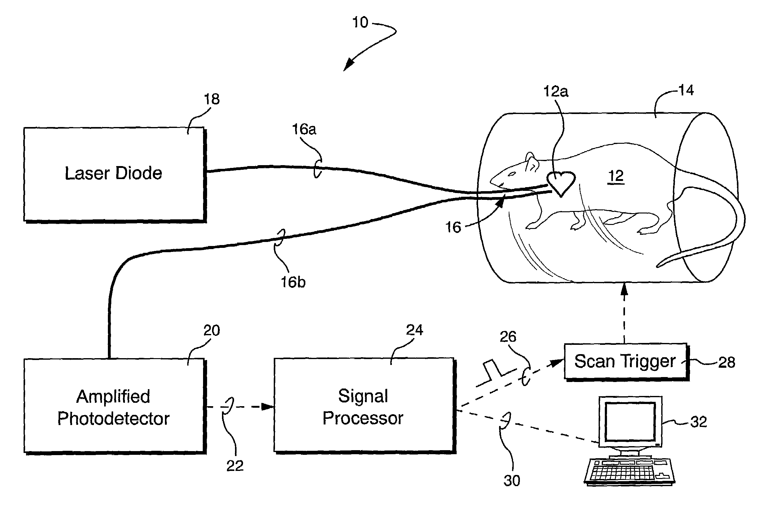
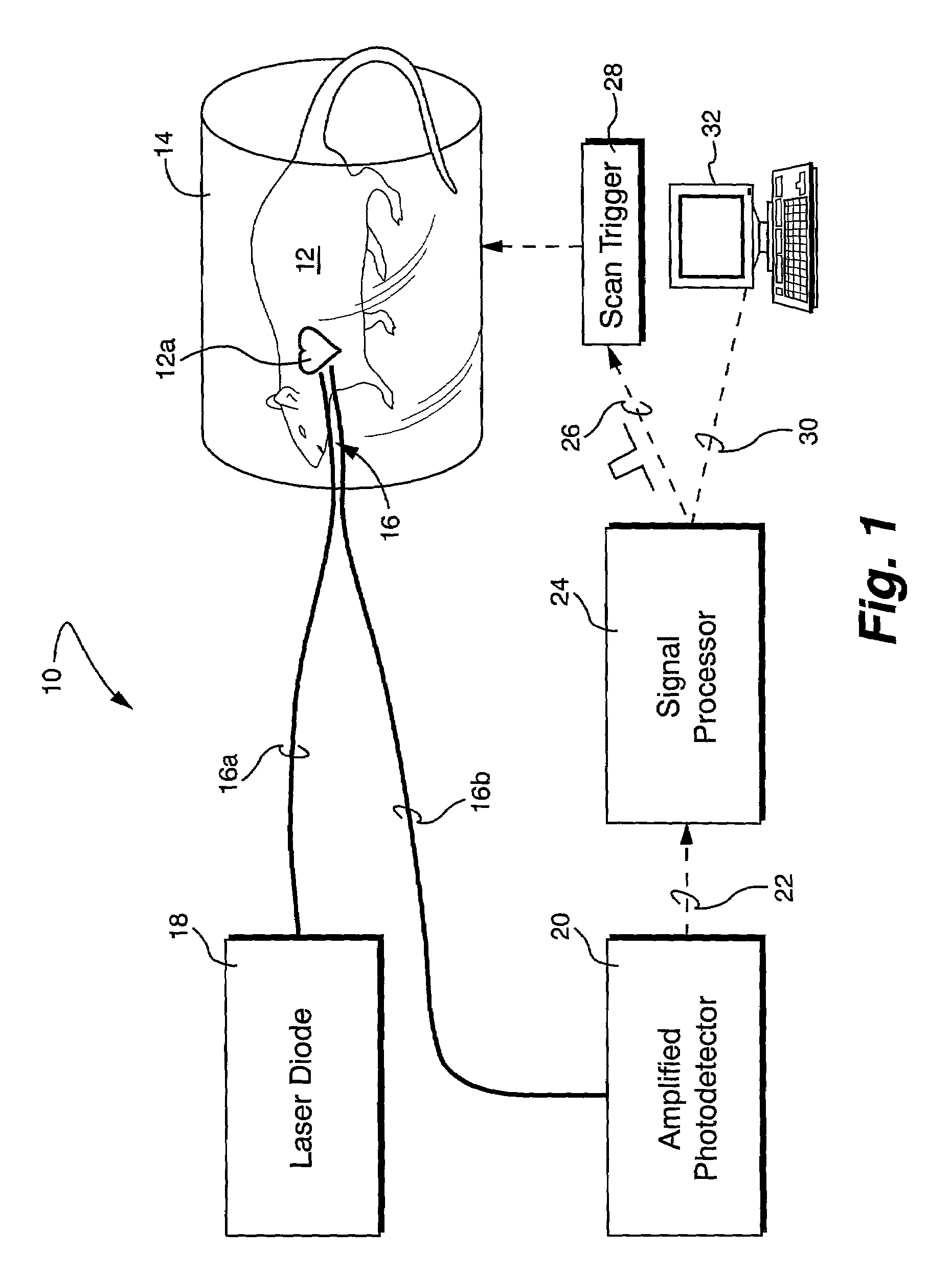
[0016]A system 10 as shown in FIG. 1 was employed. Specifically, two 5-m step-index multimode optical fibers (Thorlabs, Newton, N.J.) were used as the transmit and receive optical fibers 16a, 16b, respectively. The last 10 cm of each fiber was stripped of buffer, and the bare fibers were bundled together for total diameter of 250 microns. The fiber tips were cleaved at appropriate angles to maximize light detection. Light from a collimated 40 mW, 650 nm laser diode (Thorlabs), selected for its minimal tissue absorption, was focused into the transmit fiber 16a using an optical lens.

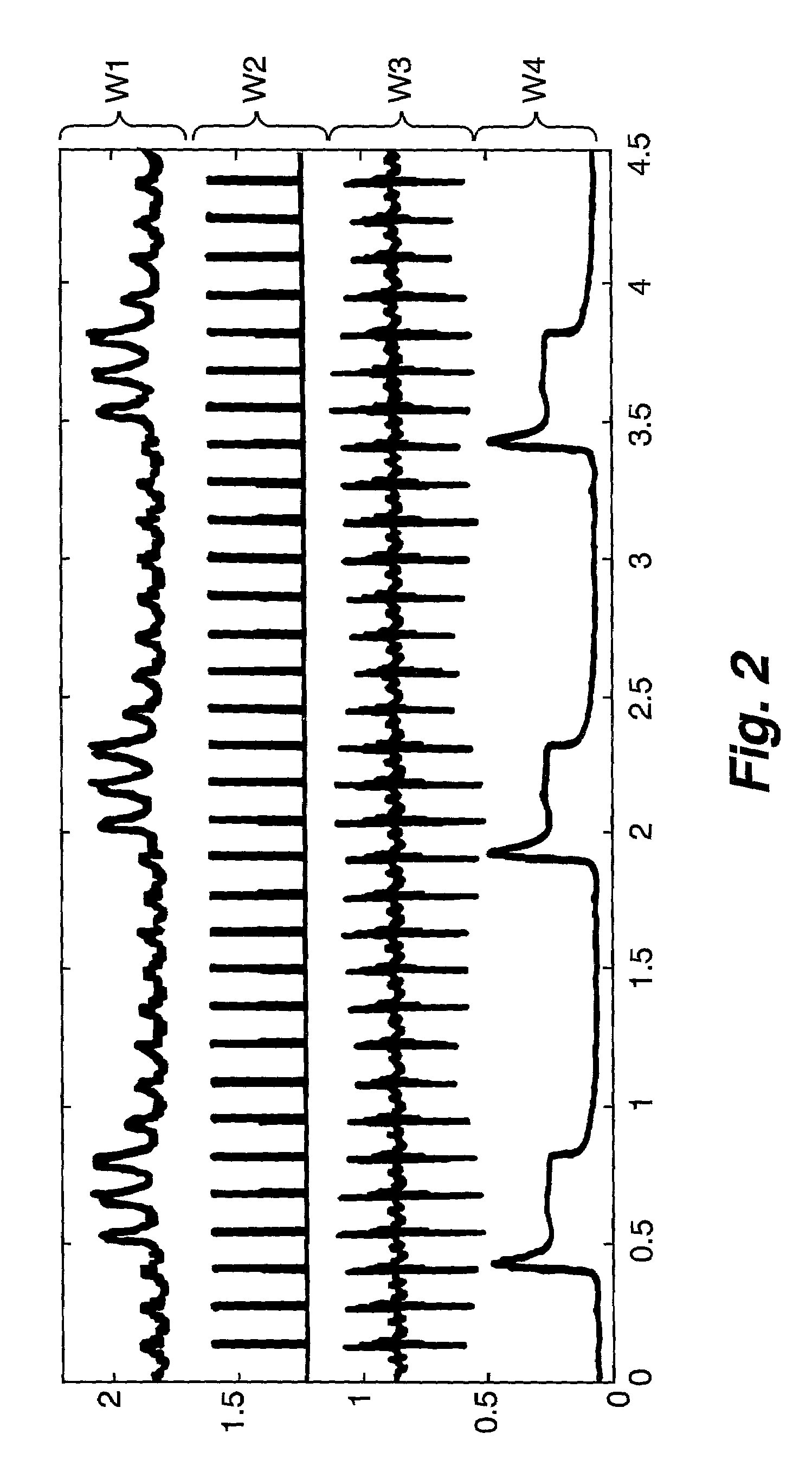
[0017]Twenty-eight rats (150 g–250 g) and one C57 mouse (40 g) were intubated and anesthetized with isoflurane delivered b ventilator as described more fully in Hedlund et al, Magn. Res. Img., 18, 753–759 (2000), the entire content of which is expressly incorporated hereinto by reference. Pediatric electrodes were taped to the animal's footpads to acquire a reference ECG signal. Average heart rates were 30...


PUM


Abstract

Noninvasive, MR-compatible methods and systems optically detect mechanical cardiac activity by anatomic (e.g., esophageal) movements. Most preferably, esophageal motion is detected optically and is indicative rhythmic cardiac activities. This esophageal motion may then be detected and used to provide a signal indicative of periods of cardiac activity and inactivity. The signal may be further processed so as to generate a trigger signal that may be input to a MR scanner. In such a manner, MR microscopy may be accomplished to acquire information at a specific phase of the cardiac cycle, for example, in synchrony with periods of cardiac inactivity. Moreover, since mechanical cardiac activity is detected and employed, instead of electrical activity as is employed in conventional techniques, the present invention is immune to electromagnetic interference during MR microscopy. As a result, robust cardiac signals may be monitored and gated during 2-dimensional and 3-dimensional in vivo microscopy. The present invention is therefore especially well suited for MR microscopy of small animals, such as laboratory mice and rats.

Description

GOVERNMENT RIGHTS STATEMENT[0001]This invention was made with Government support under Grant No. NIH NCRR #P41 RR05959 awarded by the National Institutes of Health. The Government has certain rights to the invention.FIELD OF THE INVENTION[0002]The present invention relates generally to the field of magnetic resonance (MR) imaging. More specifically, the present invention relates to systems and methods for reliably monitoring and gating cardiac activity in patients during MR microscopy.BACKGROUND AND SUMMARY OF THE INVENTION[0003]A fundamental problem associated with using a conventional electrocardiograph (ECG) to monitor a patient's cardiac activity during MR imaging is the corruption of the ECG signal due to adverse electromagnetic effects. This effect is particularly pronounced in MR microscopy of small animals (e.g., laboratory rodents), where strong, rapidly-switching, magnetic field gradients are needed to obtain high spatial and temporal resolution, and the animal's ECG signa...

Claims


Application Information

Patent Timeline
Patent Type & Authority Patents(United States)
IPC IPC(8): A61B5/05G01R33/56G01R33/567
CPCG01R33/5673G01R33/5604
Inventor HEDLUND, LAURENCE W.BRAU, ANJA C. S.WHEELER, CHARLES T.JOHNSON, G. ALLAN
Owner DUKE UNIV
Why Eureka
Try Eureka