Hermetically sealed endoscope image pick-up device

a pickup device and endoscope technology, applied in the field of endoscope image pickup devices, can solve the problems of high toxic gases, large time required for aeration, doctors, nurses,

Active Publication Date: 2006-07-11
OLYMPUS CORP
View PDF16 Cites 83 Cited by
  • Summary
  • Abstract
  • Description
  • Claims
  • Application Information

AI Technical Summary

Problems solved by technology

As is well known, however, sterilization gases are highly toxic, and hence complicated procedures must be carried out during the sterilization of endoscopes and their attachments in order to ensure safety.
Another problem is that a large amount of time is required to perform aeration for removing gas that has bonded to an instrument following sterilization of an endoscope and its attachments.
As a result, doctors, nurses, and so on are unable to use endoscopes and their attachments immediately after sterilization.
Furthermore, running costs are high.
If an antiseptic fluid is used, on the other hand, a problem arises in that management of the antiseptic drug solution is complicated.
Moreover, waste processing of the antiseptic fluid involves a great deal of expense.

Method used


Image

  • Hermetically sealed endoscope image pick-up device
  • Hermetically sealed endoscope image pick-up device
  • Hermetically sealed endoscope image pick-up device

Examples


first embodiment

[0031]the present invention will be described in detail below with reference to the drawings.

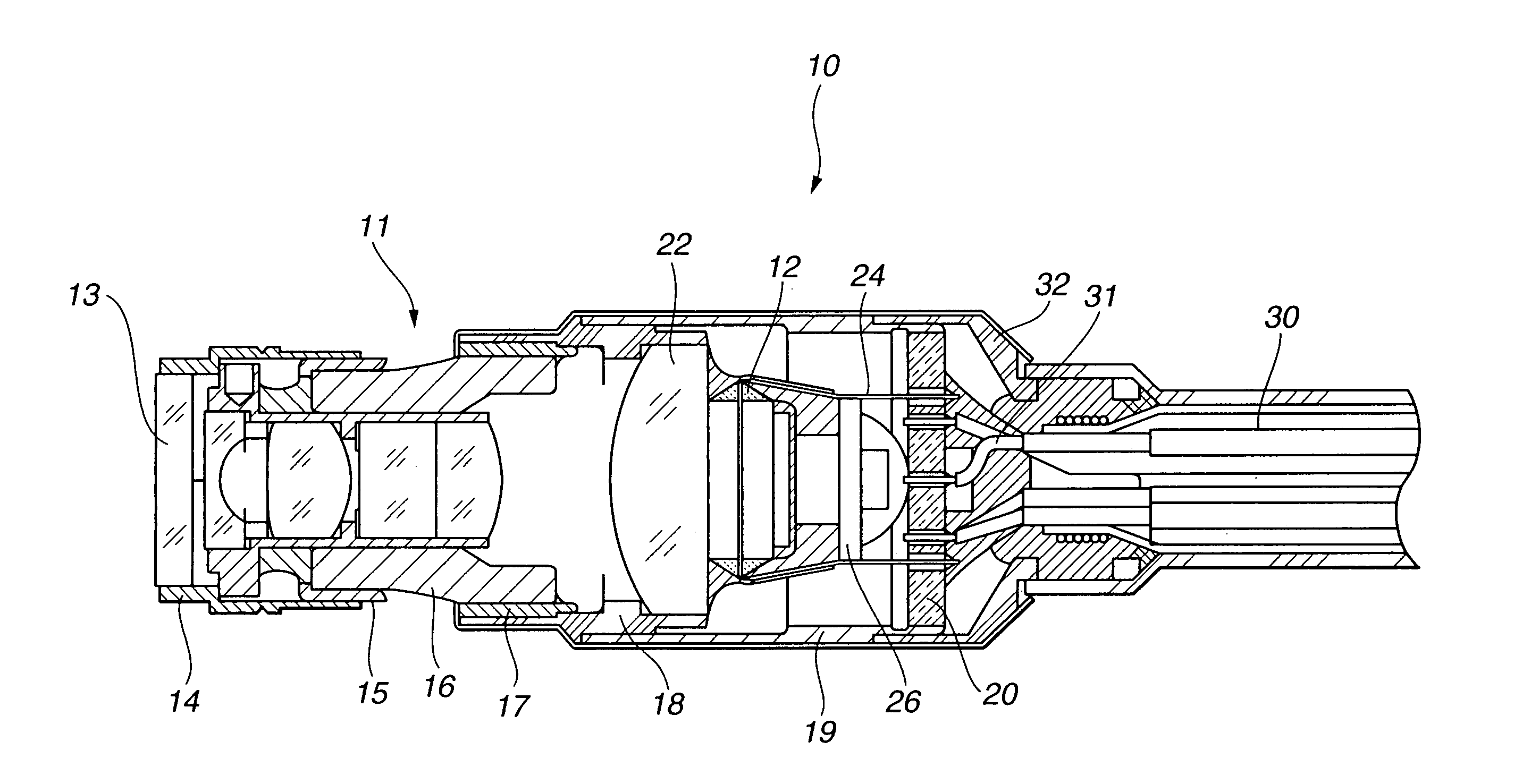
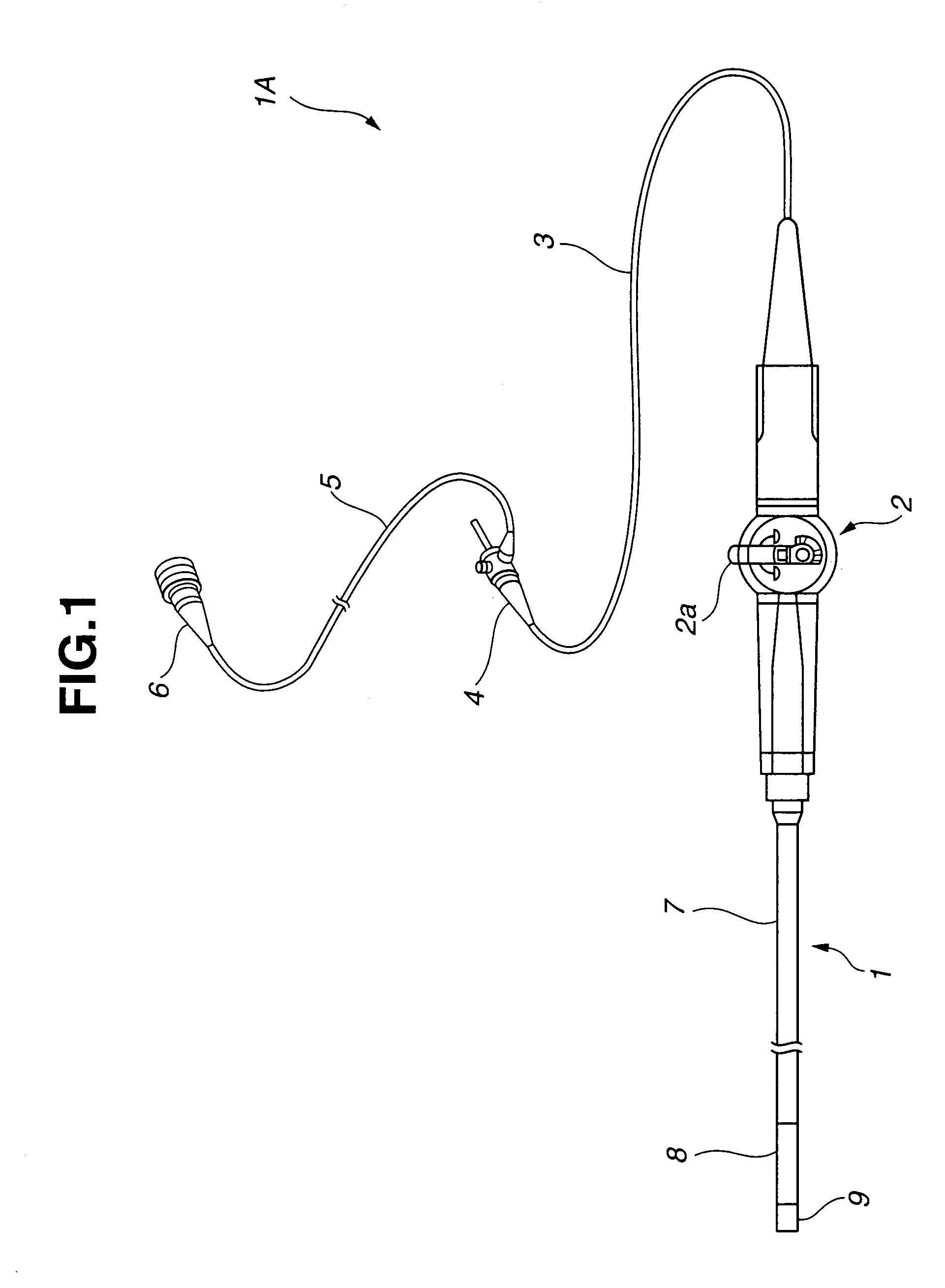
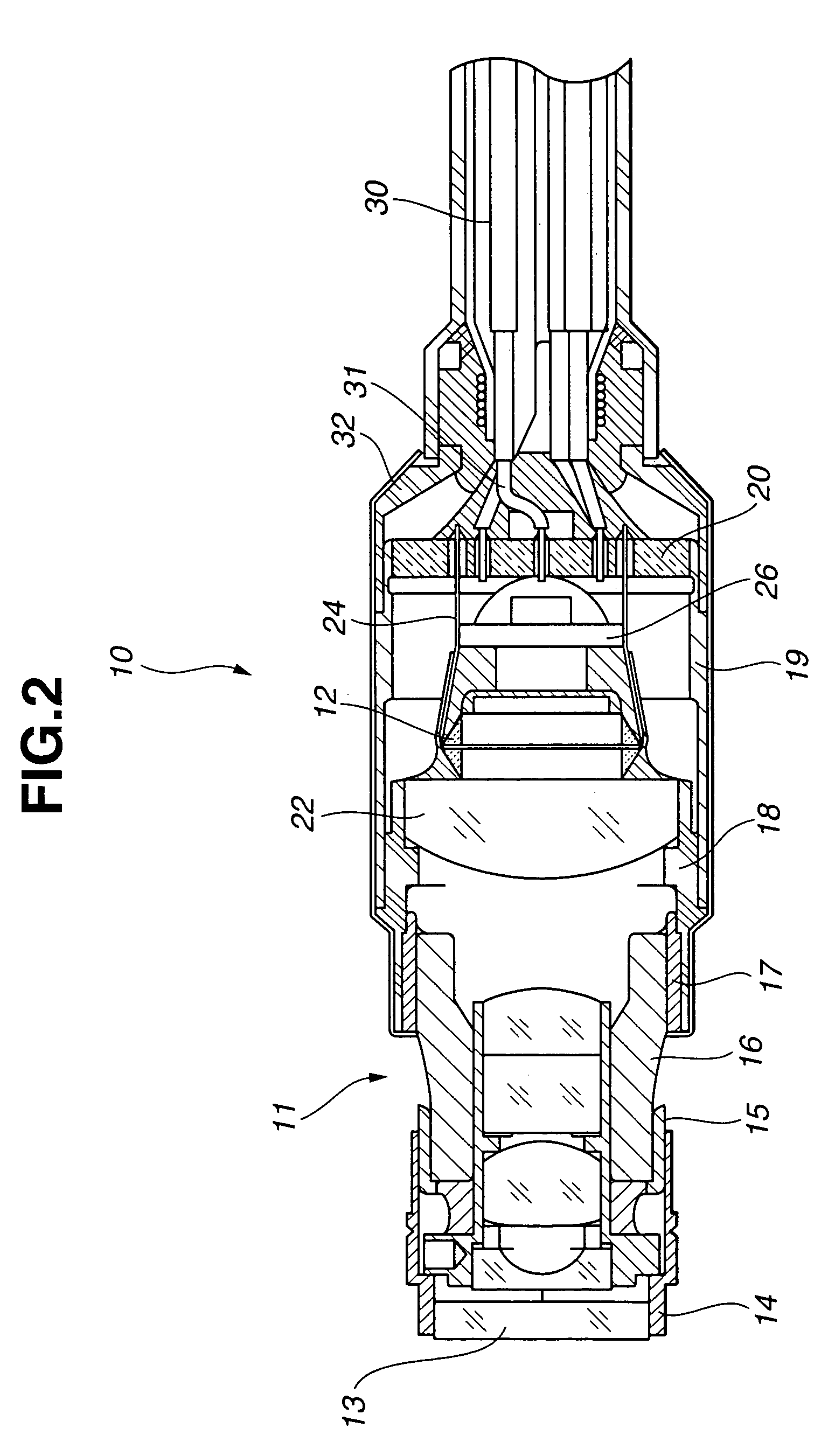
[0032]FIG. 1 is an illustrative diagram showing the constitution of an endoscope using an image pick-up device according to the first embodiment of the present invention. FIG. 2 is a sectional view showing the constitution of the image pick-up device according to the first embodiment of the present invention. FIG. 3 is a plan view showing the constitution of a transparent substrate used in the image pick-up device according to the first embodiment of the present invention. FIGS. 4 through 8 are sectional views illustrating assembly procedures of the image pick-up device according to the first embodiment of the present invention. FIG. 4 is a sectional view showing a condition according to the first embodiment of the present invention in which the transparent substrate is mounted in a substrate frame. FIG. 5 is a sectional view showing a condition according to the first embodiment of the prese...

second embodiment

[0087]In an image pick-up unit 33 of the image pick-up device of the second embodiment, the solid-state image pick-up device 12 is disposed on the rear face of the lens 22 of the lens holder 18. Further, a second transparent substrate 34 pre-installed with the HIC 27 and electronic component 28 is disposed on the rear face of the solid-state image pick-up device 12.

[0088]The second transparent substrate 34 is provided with through holes 36a for attaching, soldering, and connecting the lead wires 24 of the solid-state image pick-up device 12, and through holes 36b into which solderable current-carrying pins 35 are inserted.

[0089]The second transparent substrate 34 is formed of a transparent member formed of sapphire, white board, a glass material, or similar.

[0090]The lead wires 24 of the solid-state image pick-up device 12 are inserted into the through holes 36a of the second transparent substrate 34. The through holes 36a and lead wires 24 are then electrically connected on the opp...

third embodiment

[0097]In an image pick-up unit 37 of the image pick-up device of the third embodiment, the solid-state image pick-up device 12 is disposed on the rear face of the lens 22 of the lens holder 18, and a transparent substrate 38 pre-installed with the HIC 27 and electronic component 28 is disposed on the rear face of the solid-state image pick-up device.

[0098]The transparent substrate 38 is provided with through holes 40a into which the lead wires 24 of the solid-state image pick-up device 12 are inserted and then connected by soldering, and through holes 40b into which solderable terminal pins 39 are inserted.

[0099]The transparent substrate 38 is formed of a similar transparent member to the transparent substrate 20 and second transparent substrate 34 described above.

[0100]The lead wires 24 of the solid-state image pick-up device 12 are inserted into the through holes 40a of the transparent substrate 38. The through holes 40a and lead wires 24 are then electrically connected by solderi...


PUM


Abstract

The present invention provides an image pick-up device and an endoscope in which a lens member and a solid-state image pick-up device are prevented from exposure to water vapor during autoclave sterilization, and in which the diameter of an insertion portion is narrow, a rigid tip end portion is short, and assembly is performed easily. The image pick-up device of the present invention comprises an image pick-up unit for capturing an optical image obtained from incident light entering an internal cavity of a frame, and outputting an image signal of the optical image, wiring extending from the image pick-up unit in an opposite direction to the direction in which the incident light enters, this wiring being capable of transmitting the image signal, and a substrate which is disposed in the internal cavity of a frame, has holes for engaging with the wiring, one surface of which is substantially orthogonal to the axial direction of the frame, and which is formed of a member having a light-transmitting property.

Description

[0001]This application claims benefit of Japanese Application No. 2003-060155 filed on Mar. 6, 2003, the contents of which are incorporated by this reference.BACKGROUND OF THE INVENTION[0002]1. Field of the Invention[0003]The present invention relates to an image pick-up device and an endoscope, and more particularly to an image pick-up device and endoscope which are easily assembled.[0004]2. Description of the Related Art[0005]Conventionally in the medical field, medical endoscopes in which an insertion portion of an elongated endoscope is inserted by an operator or the like into a body cavity have been widely used to observe organs and the like inside the body and perform various curative procedures when necessary using procedural instruments inserted into an instrument channel. In the industrial field, industrial endoscopes in which the elongated insertion portion is inserted into the interior of boilers, turbines, engines, chemical plants, and soon have been widely used to obser...

Claims


Application Information

Patent Timeline
Patent Type & Authority Patents(United States)
IPC IPC(8): A61B1/00A61B1/04A61B1/05G02B23/24H04N5/225
CPCA61B1/05G02B23/2484H04N2005/2255H04N23/555
Inventor FUTATSUGI, YASUYUKI
Owner OLYMPUS CORP
Why Eureka
Try Eureka