Method and system for risk-modulated diagnosis of disease

a risk-modulated diagnosis and disease technology, applied in the field of computer-aided diagnosis, can solve the problems of poor characterization of lesions, high risk of breast cancer in women under 40 years, and complicated clinical acquisition of x-ray mammograms, so as to improve the characterization of lesions and improve the effect of computer-aided diagnosis

Active Publication Date: 2006-10-17
UNIVERSITY OF CHICAGO
View PDF4 Cites 95 Cited by
  • Summary
  • Abstract
  • Description
  • Claims
  • Application Information

AI Technical Summary

Benefits of technology

[0157]Preferred embodiments of the present invention provide a method and system that employ a lesion characterization module and a risk assessment module for characterizing the lesion and the environment local and distal to the lesion in question. A specific example presented here includes a computerized method for the characterization of mammographic lesions combined with a computerized method for the assessment of the breast parenchymal pattern on mammograms, resulting in improved characterization of lesions for specific subpop

Problems solved by technology

Although “subjective” does not necessarily mean “poor judgment,” it does permit sub-optimal and inconsistent decision making.
Although general rules for the differentiation between benign and malignant mammographically identified breast lesions exist, considerable misclassification of lesions occurs with the current methods.
Clinical acquisition of x-ray mammograms is a rather complicated procedure and requires specific techniques in order to obtain high quality images.
Mammographic surveillance for women under age 40 years who are at very high risk of developing breast cancer, however, still remains an issue, since the benefit of screening women in this age group has not been proven.
However, relative risk estimates do not directly approximate the underlying probability of a diagnosis of breast cancer for an individual over time.
Although, the basic mechanism underlying the association between breast cancer and these risk factors is not well understood.
It has been recognized for some time that varying levels of endogenous and exogenous estrogens have been associated with the risk of developing breast cancer.
Increased mammographic density is another factor that has been found to be associated with an increased risk of breast cancer.
47). On the other hand, Gail et al. did not find, in their data, that age at onset was helpful in the prediction of risk once the number of relatives affected was considered
However, the results provoked various criticisms, for example, possible bias in the resu

Method used


Image

  • Method and system for risk-modulated diagnosis of disease
  • Method and system for risk-modulated diagnosis of disease
  • Method and system for risk-modulated diagnosis of disease

Examples


second embodiment

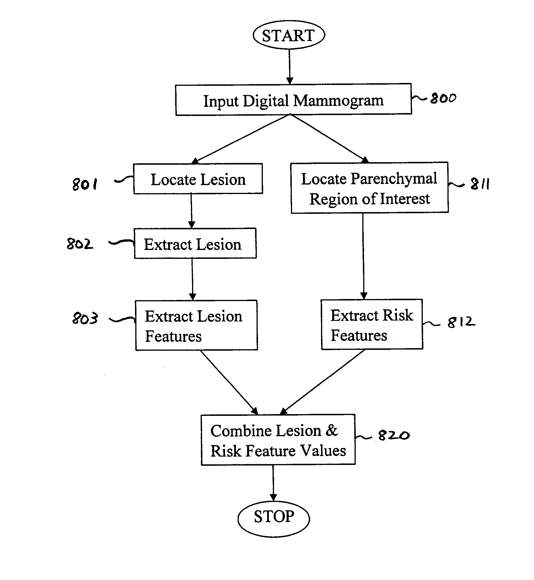
[0248]FIG. 8B illustrates a method for risk-modulated diagnosis according to the present invention. Note that in FIG. 8B, steps 800, 801, 802, 803, 811, and 812 are identical to the similarly identified steps shown in FIG. 8A.

[0249]In step 804, the lesion feature values calculated in step 803 are combined, e.g., using linear discriminant analysis to determine an indication of whether the lesion is benign or malignant. Similarly, in step 813, the risk features values calculated in step 813 are combined to determine whether the individual associated with the digital mammogram is a high or low risk individual. Note that an artificial neural network or other appropriate classifier may be used in step 804 and / or step 813.

[0250]In step 830, the output from each LDA (calculated in steps 804 and 813) is normalized and then multiplied together in step 830 to yield a risk-modulated measure characterizing the lesion. The classifier output that characterizes the lesion is weighted by the output...

third embodiment

[0251]FIG. 8C illustrates a method for risk-modulated diagnosis according to the present invention. Steps 800–803 and 811–813 are the same as described above with reference to FIGS. 8A and 8B. Here the risk classifier (using, e.g., a LDA) is trained with, e.g., four risk parenchymal features, in the task of distinguishing between high- and low-risk individuals.

[0252]In step 814, the output of step 813 is normalized. In step 815, a threshold is then used on the normalized LDA (or classifier) output to separate the individual into the high risk population or the low risk population. For example, based on previous data, one can use the computer-extracted risk features that correspond to a Gail model risk output of less than 20% for the low risk group. Anything higher than 20% can be considered as high risk.

[0253]The lesion classifier is retrained with the (in this case) five lesion features separately, for the high risk group and the low risk group. If the inquiry in step 815 determine...


PUM


Abstract

A method of calculating a disease assessment by analyzing a medical image, comprising (1) extracting at least one lesion feature value from the medical image; (2) extracting at least one risk feature value from the medical image; and (3) determining the disease assessment based on the at least one lesion feature value and the at least one risk feature value. The method employs lesion characterization for characterizing the lesion, and risk assessment based on the lesion's surroundings, i.e., the environment local and distal to the lesion. Computerized methods both characterize mammographic lesions and assess the breast parenchymal pattern on mammograms, resulting in improved characterization of lesions for specific subpopulations, combining the benefits of both techniques.

Description

CROSS-REFERENCE TO RELATED APPLICATION[0001]The present application claims priority to U.S. Provisional Application No. 60 / 354,523, the contents of which are incorporated herein by reference.STATEMENT REGARDING FEDERALLY SPONSORED RESEARCH[0002]The present invention was made in part with U.S. Government support under NIH Grants CA79711 and CA89452. The U.S. Government may have certain rights to this invention.BACKGROUND OF THE INVENTIONField of the Invention[0003]The invention relates generally to the field of computer-aided diagnosis in the detection, characterization, diagnosis, and / or assessment of normal and diseased states.[0004]The present invention also generally relates to computerized techniques for automated analysis of digital images, for example, as disclosed in one or more of U.S. Pat. Nos. 4,839,807; 4,841,555; 4,851,984; 4,875,165; 4,907,156; 4,918,534; 5,072,384; 5,133,020; 5,150,292; 5,224,177; 5,289,374; 5,319,549; 5,343,390; 5,359,513; 5,452,367; 5,463,548; 5,491,...

Claims


Application Information

Patent Timeline
IPC IPC(8): G06K9/00A61B5/00A61B5/05A61B6/00A61B6/04A61B8/00G06FG06F19/00G06T7/00G06V10/25
CPCG06K9/3233G06T7/0012G06F19/3431G06F19/3437G06F19/3443G06F19/345G06T2207/30068G16H50/30G16H50/50G16H50/70G16H50/20G06V10/25
Inventor GIGER, MARYELLEN L.HUO, ZHIMINVYBORNY, CARL J.
Owner UNIVERSITY OF CHICAGO
Why Eureka
Try Eureka