
System and method for performing a three-dimensional virtual segmentation and examination with optical texture mapping

a three-dimensional virtual segmentation and optical texture technology, applied in the field of three-dimensional virtual segmentation and examination with optical texture mapping, can solve the problems of invasive, uncomfortable, painful procedure for patients, and large mortality of colonoscopy, and achieve the effect of reducing the number of computations required

Inactive Publication Date: 2006-12-12
THE RES FOUND OF STATE UNIV OF NEW YORK
View PDF63 Cites 41 Cited by
  • Summary
  • Abstract
  • Description
  • Claims
  • Application Information

AI Technical Summary

Benefits of technology

[0018]It is an object of the invention to use a system and method to perform a relatively painless, inexpensive and non-intrusive in vivo examination of an organ where the actual analysis of the scanned colon can be possibly performed without the patient present. The colon can be scanned and visualized in real-time or the stored data can be visualized at a later time.
[0022]It is still a further object of the invention to use a modified Z buffer technique to minimize the number of computations required for generating the viewing screen.

Problems solved by technology

Colon cancer continues to be a major cause of death throughout the world.
The colonoscopy, although reliable, is both relatively costly in money and time, and is an invasive, uncomfortable painful procedure for the patient.
However, barium enemas may not always provide a view of the entire colon, require extensive pretreatment and patient manipulation, is often operator-dependent when performing the operation, exposes the patient to excessive radiation and can be less sensitive than a colonoscopy.
However, complete control of a camera in a large domain would be tedious and tiring, and an operator might not view all the important features between the start and finishing point of the exploration.
The camera could also easily get “lost” in remote areas or be “crashed” into one of the walls by an inattentive operator or by numerous unexpected obstacles.
However, this second technique does not give the viewer the flexibility to alter the course or investigate an interesting area viewed along the flight path.

Method used


Image

  • System and method for performing a three-dimensional virtual segmentation and examination with optical texture mapping
  • System and method for performing a three-dimensional virtual segmentation and examination with optical texture mapping
  • System and method for performing a three-dimensional virtual segmentation and examination with optical texture mapping

Examples


Embodiment Construction

[0053]While the methods and systems described in this application can be applied to any object to be examined, the preferred embodiment which will be described is the examination of an organ in the human body, specifically the colon. The colon is long and twisted which makes it especially suited for a virtual examination saving the patient both money and the discomfort and danger of a physical probe. Other examples of organs which can be examined include the lungs, stomach and portions of the gastro-intestinal system, the heart and blood vessels.

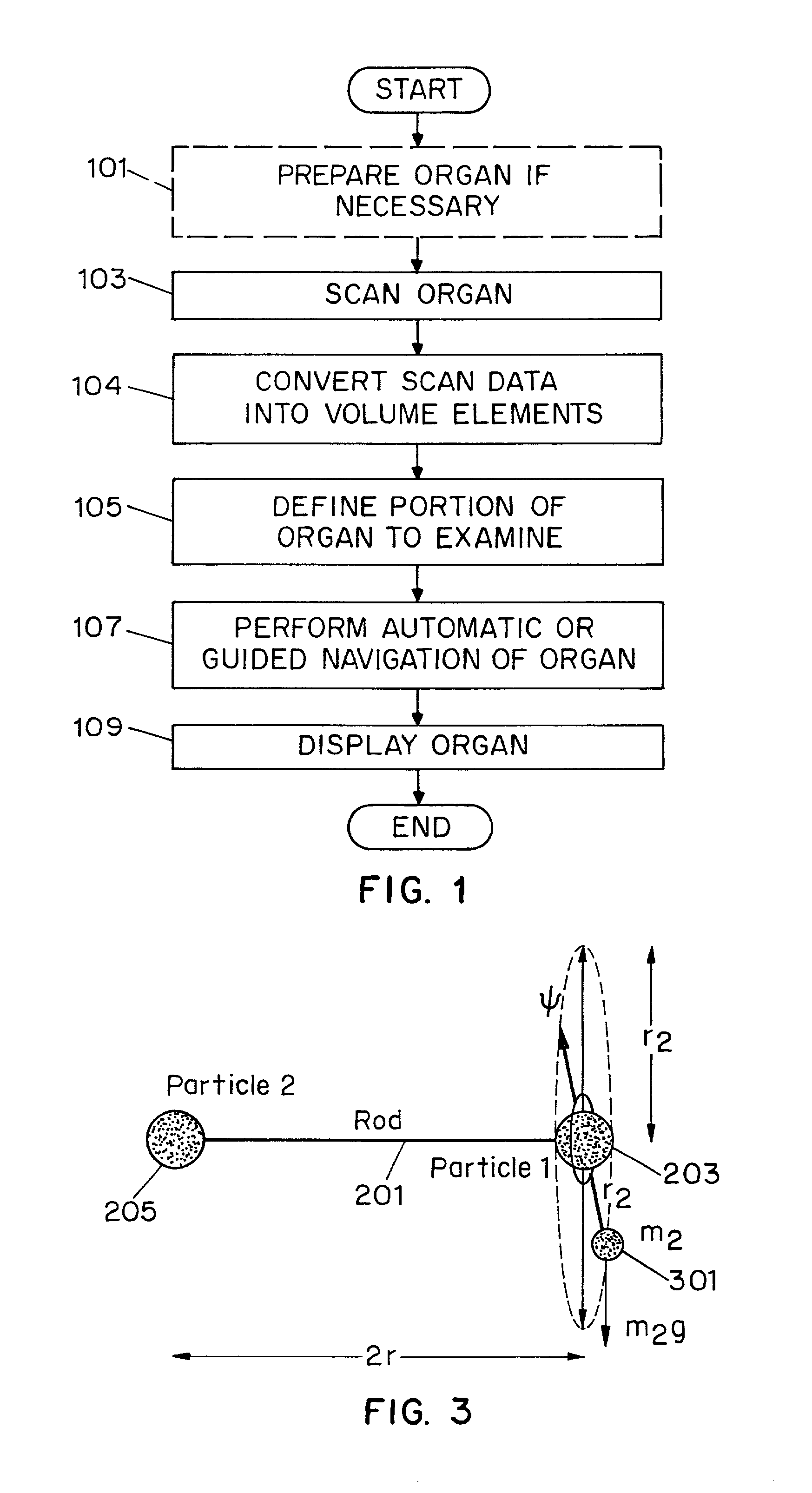
[0054]FIG. 1 illustrates the steps necessary to perform a virtual colonoscopy using volume visualization techniques. Step 101 prepares the colon to be scanned in order to be viewed for examination if required by either the doctor or the particular scanning instrument. This preparation could include cleansing the colon with a “cocktail” or liquid which enters the colon after being orally ingested and passed through the stomach. The cocktail f...


PUM


Abstract

A system and method for generating a three-dimensional visualization image of an object such as an organ using volume visualization techniques and exploring the image using a guided navigation system which allows the operator to travel along a flight path and to adjust the view to a particular portion of the image of interest in order, for example, to identify polyps, cysts or other abnormal features in the visualized organ. An electronic biopsy can also be performed on an identified growth or mass in the visualized object. Virtual colonoscopy can be enhanced by electronically removing residual stool, fluid and non-colonic tissue from the image of the colon, by employing bowel preparation followed by image segmentation operations. Methods are also employed for virtually expanding regions of colon collapse using image segmentation results.

Description

CROSS-REFERENCE to RELATED APPLICATIONS[0001]This application is a divsional application of U.S. patent application, Ser. No. 09 / 343,012, now U.S. Pat No. 6,331,116 filed on Jun. 29, 1999 and entitled “Systems And Method For Performing a Three-Dimensional Virtual Segmentation And Examination,” now issued as U.S. Pat. No. 6,331,116, which is a continuation-in-part of U.S. patent application, Ser. No. 08 / 714,697, now U.S. Pat. No. 5,971,767 filed on Sep. 16, 1996, and entitled “Systems and Method for Performing a Three Dimensional Virtual Examination,” now issued as U.S. Pat. No. 5,971,767, and also claims the benefit of U.S. Provisional Patent Application, Ser. No. 60 / 125,041, filed on Mar. 18, 1999, entitled “Three Dimensional Virtual Examination.”STATEMENT REGARDING FEDERALLY FUNDED RESEARCH[0002]Work related to the present invention was supported by grants from the National Institute of Health, Grant Nos. NIH R21 CA79180, Automatic Colon Segmentation for 3D Virtual Colonoscopy; an...

Claims


Application Information

Patent Timeline
IPC IPC(8): G06T15/00A61B6/03A61B5/055G06T1/00G06T5/00G06T7/00G06T15/04G06T15/08G06T15/40G06T17/00G06V10/147G06V10/28
CPCG06K9/209G06K9/38G06T7/0012G06T7/0081G06T15/04G06T19/20G06T15/08G06K2209/05G06T2219/2021G06T2207/10081G06T2207/20156G06T2207/30032G06T2210/28G06T2210/41G06T7/11G06V10/147G06V10/28G06V2201/03G06T17/00
Inventor KAUFMAN, ARIE E.LIANG, ZHENGRONGWAX, MARK R.WAN, MINGCHEN, DONGQUING
Owner THE RES FOUND OF STATE UNIV OF NEW YORK
Learn More