Method and system for simulating X-ray images

a technology of x-ray images and simulation methods, applied in image data processing, diagnostics, applications, etc., can solve the problems of time-consuming, high cost of sending patients to an x-ray facility for obtaining x-ray images, and large slice images

Active Publication Date: 2006-12-26
MEDICAL INSIGHT
View PDF3 Cites 78 Cited by
  • Summary
  • Abstract
  • Description
  • Claims
  • Application Information

AI Technical Summary

Benefits of technology

[0016]The CT image of a patient is acquired from a repository of patient CT data, for example by using a software application adapted to visualize CT data, i.e. adapted to generate a 3D image from the CT data. The software application is capable of orienting the 3D image with respect to a viewpoint and a virtual image plane, i.e. rotating the CT image. The application may, however, also be capable of other types of manipulation, such as zooming, cutting an area, etc. Positioning the virtual X-ray source relative to a virtual image plane corresponds to the positioning of the actual X-ray source in an X-ray facility. Likewise, the position of the virtual image plane corresponds to the position of the detector plane in an X-ray facility, and the virtual image plane may be viewed upon as a virtual X-ray detector.
[0017]The simulated X-ray image may be generated from a variable and unrestricted viewpoint. That is, the virtual X-ray source may be positioned independently with respect to the CT object. X-ray images may even be generated from a viewpoint, which is not possible in a real X-apparatus. In a real X-ray apparatus a number of viewpoints are not possible, because it is not possible to orient the patient in the required way.
[0018]A 2D X-ray image may be generated in any given orientation of the 3D CT image with respect to the virtual image plane.
[0019]The simulated X-ray images can be generated for any patient who has undergone CT scanning. After the patient has been scanned, the data may be made available by storing the data on a computer system that may be accessed by a user. For example, the user may be a surgeon who makes a diagnosis, plans a surgical operation, or a surgeon who needs further insight during an operation.
[0020]The simulated X-ray image may be simulated using parallel propagating X-rays, so that the X-ray image is simulated free of parallax distortion, i.e. an ideal X-ray image may be simulated. Alternatively the simulated X-ray image may be simulated using divergent X-rays, so that the X-ray image is simulated with parallax distortion as in actual X-ray images.
[0021]The CT data of patient anatomy may preferentially be based on data that conforms to the Digital Imaging and Communications in Medicine standard (DICOM standard) implemented on Picture Archiving and Communications Systems (PACS systems). Use of actual CT patient data in the DICOM format within a PACS system is an advantage since this enables to directly incorporate access to simulated X-ray images in the imaging workflow in a hospital.

Problems solved by technology

Such 2D X-ray projection images are often requested by the surgeons because the slice images from CT data are frequently considered as not providing sufficient information, or the number of slice images is too large for use in an operation situation.
Sending a patient to an X-ray facility for obtaining X-ray images is expensive, it takes time and it exposes the patient to additional X-ray radiation.
Obtaining optimal X-ray images may be difficult due to organs shadowing the area of interest as well as a proper and precise positioning of the X-ray source and the detector plate.

Method used


Image

  • Method and system for simulating X-ray images
  • Method and system for simulating X-ray images
  • Method and system for simulating X-ray images

Examples


Embodiment Construction

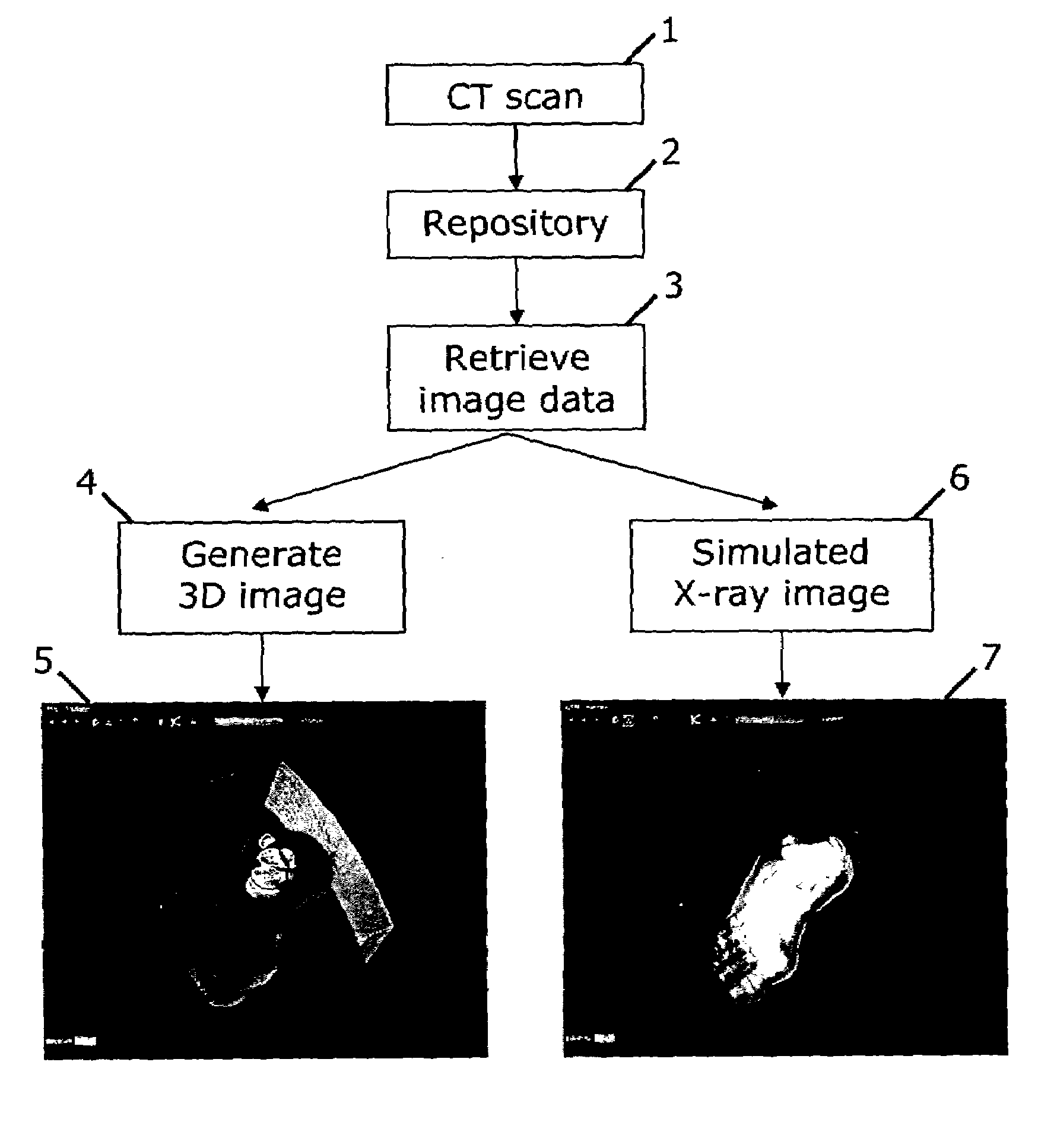
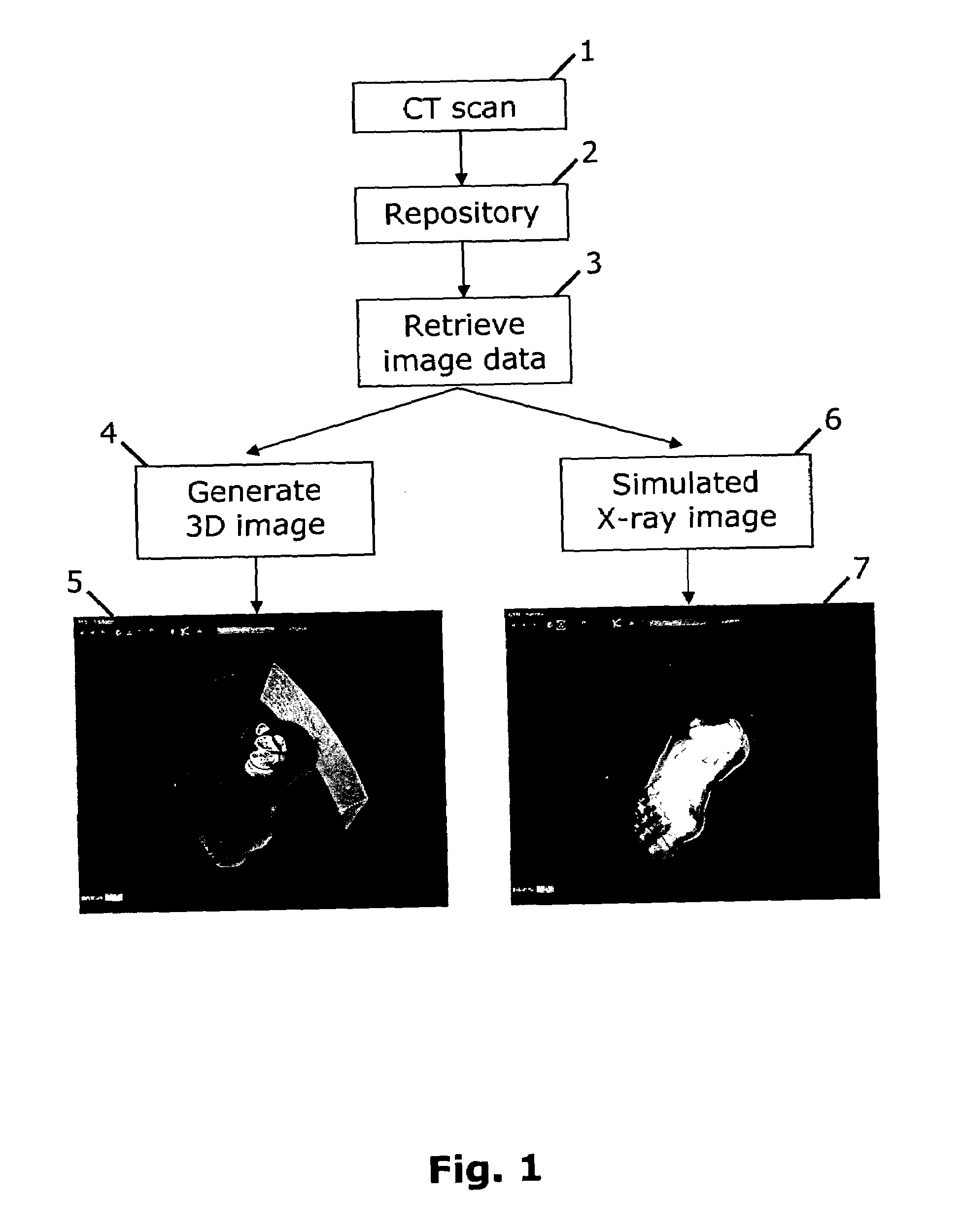
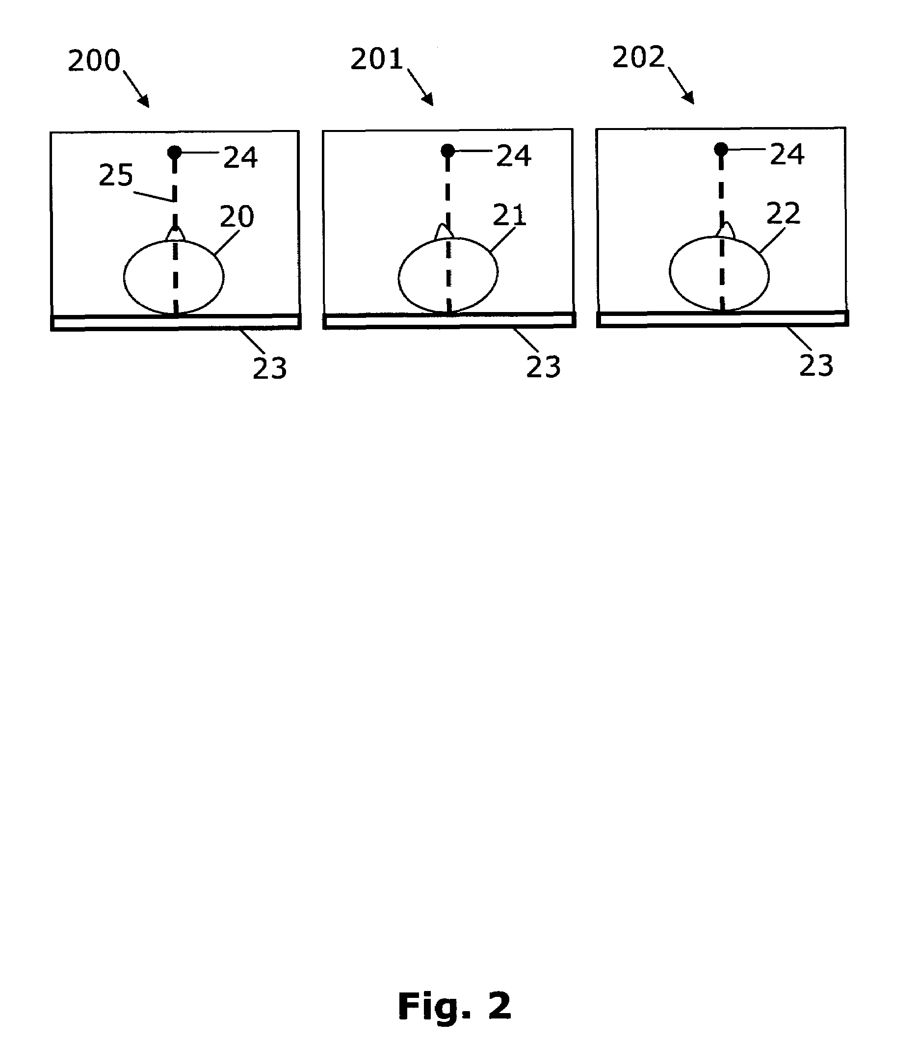
[0007]It is an object of the present invention to facilitate access to 2D X-ray images of a patient under medical treatment who has undergone CT scanning. Accordingly there is provided, in a first aspect, a method of generating a simulated 2D X-ray image from CT data of a patient's anatomy, said method comprising the steps of:[0008]CT scanning the patient to generate corresponding CT data at a first time instance;[0009]storing the CT data in a data repository;[0010]retrieving the CT data at a second time instance after the first time instance;[0011]generating a 3D CT image of the anatomy from the CT data; and[0012]positioning a virtual X-ray source relative to a virtual image plane to generate the corresponding 2D simulated X-ray image, whereby the simulated X-ray image may be generated from an unrestricted viewpoint.

[0013]A patient is CT scanned and the data is stored in a data repository. The repository of patient CT data may be accessible to a user through a computer network. For...


PUM

PropertyMeasurementUnit
CTaaaaaaaaaa
opacityaaaaaaaaaa
Computed Tomographyaaaaaaaaaa

Abstract

The invention relates to simulating 2D X-ray images from 3D CT data. A user can access a repository of CT data of a patient's anatomy 2 by using a computer network system. The CT data has been obtained by a CT scanning of the patient 1. Firstly, the user retrieves the CT image data 3 and a 3D image is generated 4. Secondly, the user may orient the CT image in any orientation desirable with respect to a virtual X-ray source and a virtual image plane. And thirdly, a 2D X-ray image is simulated 6 from the chosen viewpoint and shown to the user.

Description

FIELD OF THE INVENTION[0001]The present invention relates to transforming 3D image data to 2D image data and in particular to simulating 2D X-ray images from CT data.BACKGROUND OF THE INVENTION[0002]Computed Tomography (CT) scanning of patient anatomy is used by radiologists for medical diagnosis. In CT scanning of a patient, a 3D (or volume) image is generated by means of an X-ray source which is rapidly rotated around the patient. A large quantity of images is obtained resulting in slices of the scanned area, which is electronically reassembled to constitute a 3D image of the scanned area. The CT scanning is a measurement of the amount of X-rays absorbed in the specific volume elements constituting the 3D image, and each volume element represents the density of the tissue comprised in the volume element.[0003]The CT image is a 3D counterpart to the traditional 2D X-ray image. In order to obtain a 2D X-ray image, an X-ray source is mounted in a fixed position, and a patient is loca...

Claims


Application Information

Patent Timeline
Patent Type & Authority Patents(United States)
IPC IPC(8): A61B6/03G01N23/04G06T11/00
CPCA61B6/463A61B6/466G01N23/046G06T11/008G01N2223/419G01N2223/612
Inventor DOBBS, ANDREW BRUNOKJ.AE BUTTED.R, NIELS HUSTED
Owner MEDICAL INSIGHT
Why Eureka
Try Eureka