Method for determining density distributions and atomic number distributions during radiographic examination methods

a density distribution and radiographic examination technology, applied in the field of x-ray apparatus, can solve the problems of inability to clearly distinguish between calcification close to the hilus on a thorax overview picture, inability to deduce the material composition and inability to determine the structure of an object to be examined from the attenuation value in an x-ray picture. achieve the effect of satisfying th

Inactive Publication Date: 2007-01-02
SIEMENS HEALTHCARE GMBH
View PDF21 Cites 94 Cited by
  • Summary
  • Abstract
  • Description
  • Claims
  • Application Information

AI Technical Summary

Benefits of technology

[0023]For example, the display of the distribution of the atomic number in tissue etc. permits insight into the biochemical composition of an object to be examined, contrasts based on the chemical structure in organs previously displayed with homogeneous density, a quantitative determination of body constituents such as, for example, iodine or the like, and a demarcation of calcifications based on the atomic number. The isolated density display of an object permits an accurate determination of the centroid and density of objects such as is undertaken, for example, in the case of osteoporosis, inter alia.
[0026]A transformation of the distributions of the X-ray absorption values into a distribution of the density and a distribution of the atomic number is preferably undertaken for each of the assigned X-ray absorption values of the first distribution of the X-ray absorption values and of the further distributions of the X-ray absorption values. This is done on the basis of the ascertainment of a value pair for density and atomic number such that the value pair fulfils the specific functional dependences of the X-ray absorption on density and atomic number for the first X-ray spectrum and at least one further X-ray spectrum. It is thereby possible to calculate density and atomic number for a pixel simply as cut-set of the functional dependences of the mutually assigned X-ray absorption values of the recorded distributions of the X-ray absorption values.
[0030]Furthermore, the recording of at least one subregion of the object to be examined is advantageously performed with an increased X-ray dose so that a satisfactory resolution of the distribution of density and / or atomic number is achieved for this subregion without imposing an increased X-ray dose over the entire zone to be examined and, for example, exposing a patient to a high radiation load.
[0031]In order to record a first distribution of an X-ray absorption of the object to be examined, it is possible in accordance with an advantageous development to introduce into the beam path of the X-radiation a material that is not introduced into the beam path of the X-radiation in order to record a second distribution of an X-ray absorption of the object to be examined. Spectral components of the X-radiation that are specific to the material respectively used can thereby be filtered out when recording the first attenuation value distribution upstream and downstream of the object to be examined, thus achieving a high sensitivity in detecting a substance in the object to be examined that corresponds to this material.

Problems solved by technology

Conversely, on the other hand, it is impossible to deduce the material composition of an object to be examined from the attenuation value in an X-ray picture.
Nevertheless, in some circumstances, structures which occur with increased attenuation values in the imaging process for an X-ray examination cannot be clearly classified.
For example, it is difficult to distinguish between calcification close to the hilus on a thorax overview picture and a vessel which is located orthogonally with respect to the imaging plane.
It is also virtually impossible to distinguish, for example, between diffuse calcification and fresh bleeding.
Nevertheless, it is impossible, for example, for him to distinguish reliably between plastic-bonded explosive mixtures and a non-explosive plastic directly from an X-ray image.

Method used


Image

  • Method for determining density distributions and atomic number distributions during radiographic examination methods
  • Method for determining density distributions and atomic number distributions during radiographic examination methods
  • Method for determining density distributions and atomic number distributions during radiographic examination methods

Examples


Embodiment Construction

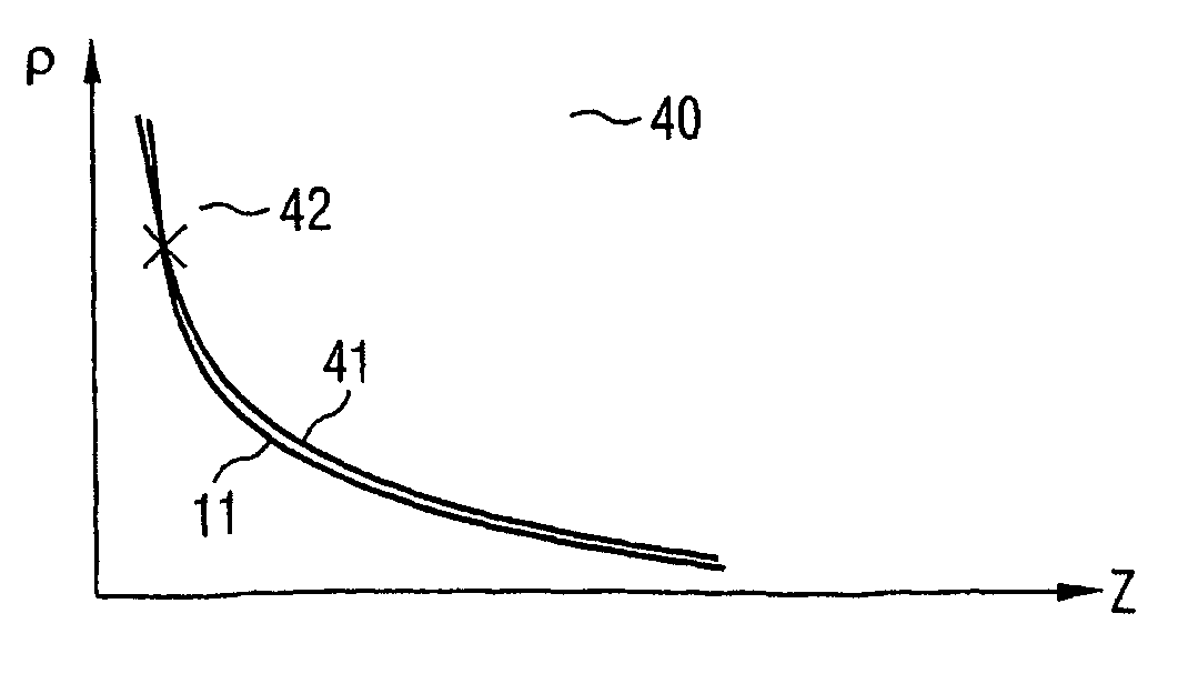
[0040]The isoabsorption line 11 of diagram 10 of FIG. 1 connects all value pairs (ρ, Z) having an attenuation value μ or C identical for a defined X-ray spectrum. The illustration of FIG. 1 makes clear that information on the nature and composition of a tissue or material cannot be derived solely on the basis of the attenuation values of an X-ray image. Usually, in order to identify types of tissue in the X-ray image, a radiologist makes use of his anatomical knowledge and looks for irregularities on this basis. In order to clarify the identity of the irregularities, a medical practitioner is then forced in turn to appeal to empirical values and morphological criteria. Similarly, a person skilled in the art of materials testing and safety testing will base his judgment of the radiographic finding on his store of professional experience.

[0041]X-radiation is attenuated to a greater or lesser extent by different materials and as a function of the energy of the X-radiation. FIG. 2 illus...


PUM

PropertyMeasurementUnit
atomic numberaaaaaaaaaa
energyaaaaaaaaaa
densityaaaaaaaaaa

Abstract

The functional dependency of a first X-ray absorption value of density and atomic number is determined in the instance of a first X-ray spectrum, and at least the functional dependency of a second X-ray absorption value of density and atomic number is determined in the instance of a second X-ray spectrum. Based on a recording of a first distribution of X-ray absorption values of the object to be examined in the instance of a first X-ray spectrum, and on a recording of at least one second distribution of X-ray absorption values of the object to be examined in the instance of a second X-ray spectrum, the values for density and atomic number are determined by comparing the functional dependency of a first X-ray absorption value of the first distribution of X-ray absorption values with the functional dependency(ies) of the X-ray absorption values, which are assigned to the first X-ray absorption value, of the second and / or other distributions of X-ray absorption values.

Description

[0001]This application is the national phase under 35 U.S.C. § 371 of PCT International Application No. PCT / DE02 / 03055 which has an International filing date of Aug. 21, 2002, which designated the United States of America and which claims priority on German Patent Application number DE 101 43 131.7 filed Sep. 3, 2001, the entire contents of which are hereby incorporated herein by reference.FIELD OF THE INVENTION[0002]The present invention generally relates to a method, an X-ray apparatus and a computer software product for ascertaining image data relating to the distribution of physically and chemically relevant data in objects of human or animal type that are to be examined, or from the field of materials testing or safety testing. The invention relates in particular to separating radiographic image data into a distribution of the material density and a distribution of the atomic number in the object to be examined.BACKGROUND OF THE INVENTION[0003]The result of all radiographic met...

Claims


Application Information

Patent Timeline
Patent Type & Authority Patents(United States)
IPC IPC(8): G01N23/087A61B6/00A61B6/03G01N23/04
CPCA61B6/4035A61B6/482A61B6/502G01N23/04A61B6/4241A61B6/50A61B6/583A61B6/505A61B6/508A61B6/501
Inventor HEISMANN, BJORNSTIERSTORFER, KARL
Owner SIEMENS HEALTHCARE GMBH
Why Eureka
Try Eureka