
Prostate boundary segmentation from 2D and 3D ultrasound images

a prostate and ultrasound image technology, applied in the field of medical imaging systems, can solve the problems of increasing the risk of metastasis, difficult segmentation of ultrasound images of the prostate, and insufficient in-segment local image processing operators like edge detectors for finding the boundary

Inactive Publication Date: 2007-01-09
THE JOHN P ROBARTS RES INST
View PDF9 Cites 30 Cited by
  • Summary
  • Abstract
  • Description
  • Claims
  • Application Information

AI Technical Summary

Benefits of technology

[0011]According to the present invention, a fast semi-automatic prostate contouring method is provided using model-based initialization and an efficient Discrete Dynamic Contour (DDC) for boundary refinement. The user initiates the process of the preferred embodiment by identifying four (4) points on the prostate boundary, thereby scaling and shaping a prostate model, and then the final prostate contour is refined with a DDC.

Problems solved by technology

Once the tumour has extended beyond the prostate, the risk of metastasis increases dramatically.
However, ultrasound images of the prostate are very difficult to segment because the contrast is low, and the image suffers from speckle, shadowing and other artifacts.
In performing ultrasound image segmentation, traditional local image processing operators like edge detectors are inadequate in and of themselves for finding the boundary, due to speckle, shadowing and other image artifacts.
Manual contouring of sequential cross-sectional 2D CT or TRUS (transrectal ultrasound) prostate images has reduced this variability, but this approach is time-consuming and arduous, making it impractical during an intra-operative procedure.
Region-based neural net approaches require extensive teaching sets, are slow, and make addition of user specified boundary information difficult.
Contour-based methods, such as “snakes” implementation of active contours are slow, complex, and sensitive to the initial choice of contour.

Method used

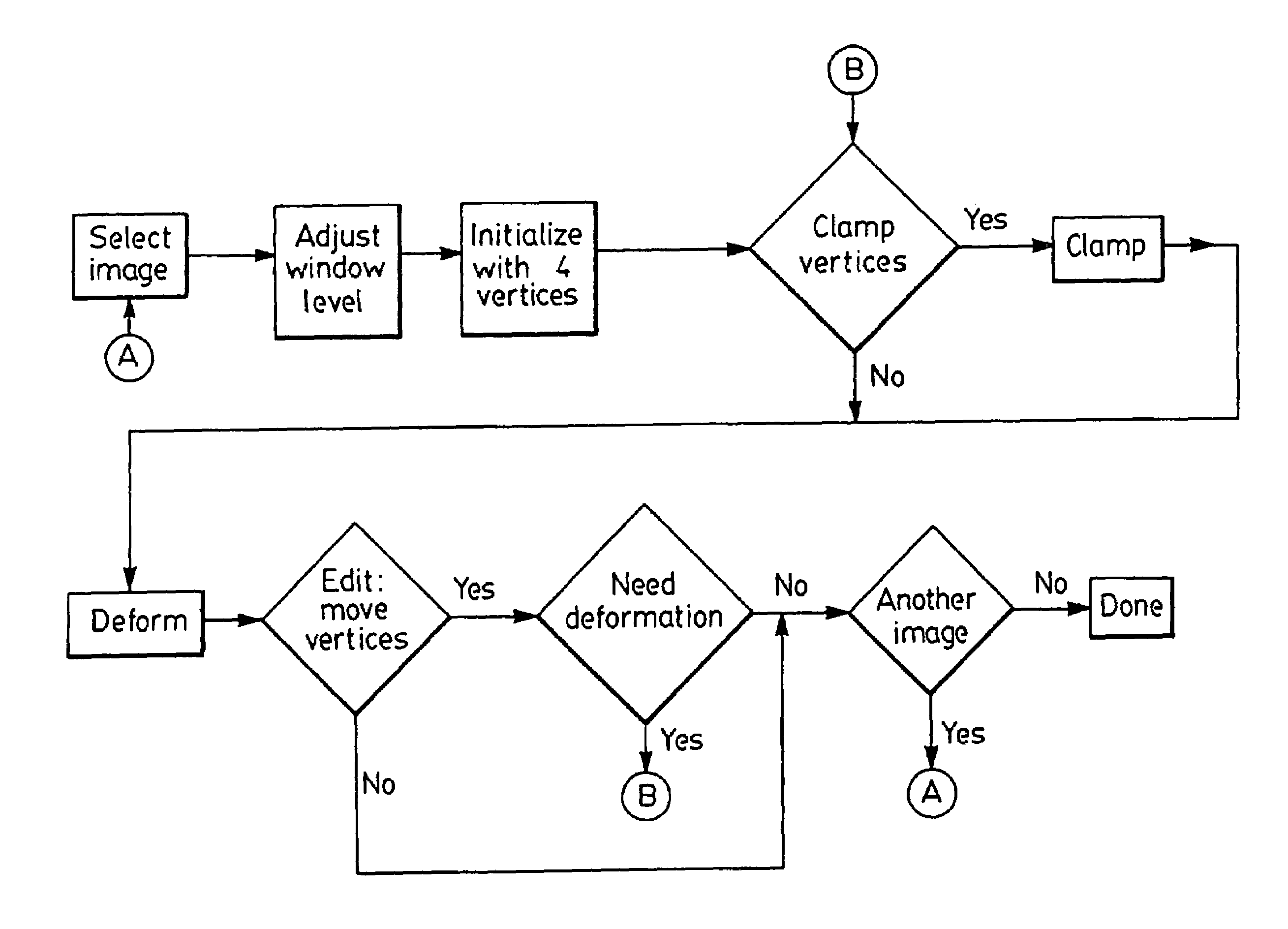

Image

  • Prostate boundary segmentation from 2D and 3D ultrasound images
  • Prostate boundary segmentation from 2D and 3D ultrasound images
  • Prostate boundary segmentation from 2D and 3D ultrasound images

Examples


Embodiment Construction

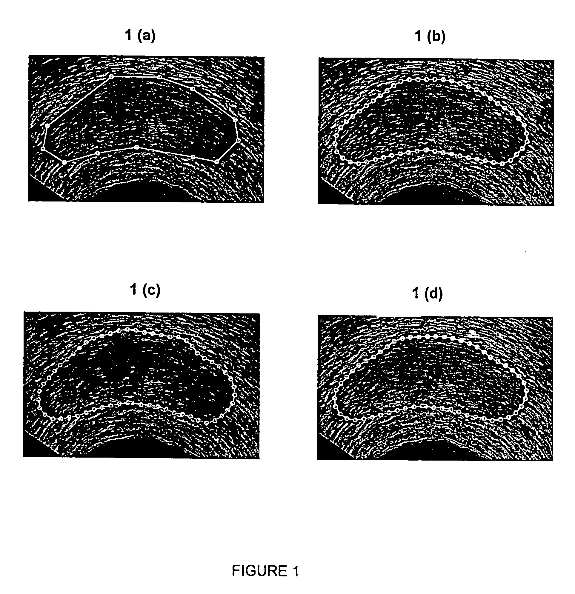
[0029]As discussed above, DDC is a poly-line (i.e., a sequence of points connected by straight-line segments) used to deform a contour to fit features in an image. When using the DDC to segment an object from an image as originally described by Lobregt and Viergever in “A discrete dynamic contour model”, IEEE Trans. Med. Imag. 1995; 14:12–24, the user must first draw an approximate outline of the object. An example outline is shown in FIG. 1(a) for a typical 2D US (ultrasound) image of the prostate. The DDC is initialized to this outline and is automatically deformed to fit features of interest, which in this case consist of edges that define the boundary of the prostate. FIGS. 1(b) and 1(c) show two stages in the deformation of the DDC. Note that points are automatically added to or deleted from the DDC as it deforms to allow it to better conform to the boundary. FIG. 1(d) shows the final deformed DDC. In this example, the final shape conforms well to the prostate boundary except i...


PUM


Abstract

A fast semi-automatic prostate contouring method is provided using model-based initialization and an efficient Discrete Dynamic Contour (DDC) for boundary refinement. The user initiates the process of the preferred embodiment by identifying four (4) points on the prostate boundary, thereby scaling and shaping a prostate model, and then the final prostate contour is refined with a DDC. The method of the present invention has particular application during the pre-implant planning phase of a brachytherapy procedure. However, this method also has uses in any phase of dose planning in the brachytherapy procedure or any other therapy approach.

Description

[0001]This application is a continuation of U.S. application Ser. No. 09 / 373,954 filed on Aug. 13, 1999, now U.S. Pat. No. 6,778,690.FIELD OF THE INVENTION[0002]This invention relates in general to medical imaging systems, and more specifically to a method of prostate boundary segmentation from 2D and 3D ultrasound images.BACKGROUND OF THE INVENTION[0003]Prostate cancer is the most commonly diagnosed malignancy in men over the age of 50, and is found at autopsy in 30% of men at the age of 50, 40% at age 60, and almost 90% at age 90. Worldwide, it is the second leading cause of death due to cancer in men, accounting for between 2.1% and 15.2% of all cancer deaths. In Canada, about 20,000 new prostate cancer cases will be diagnosed and about 4,000 men will die from this disease every year.[0004]Symptoms due to carcinoma of the prostate are generally absent until extensive local growth or metastases develop, accounting for the fact that only 65% of patients are diagnosed with locally c...

Claims


Application Information

Patent Timeline
Patent Type & Authority Patents(United States)
IPC IPC(8): G06K9/00A61B8/00A61N5/10G06T5/00
CPCG06T7/0012G06T7/0083G06T7/0089A61N5/103Y10S128/916G06T2207/10136G06T2207/20101G06T2207/30081G06T7/12G06T7/149A61B8/00
Inventor LADAK, HANIF M.FENSTER, AARONDOWNEY, DONAL B.STEINMAN, DAVID A.
Owner THE JOHN P ROBARTS RES INST
Learn More