Fluid infusion methods for glaucoma treatment

a glaucoma and fluoride technology, applied in the field of animal eye intraocular pressure reduction, can solve the problems of affecting the treatment effect of glaucoma drugs, patients may suffer substantial blindness if untreated, drug therapies for glaucoma are sometimes associated with significant side effects, etc., to improve the treatment of intraocular pressure and reduce the effect of inhibiting or slowing down the effects of glaucoma

Inactive Publication Date: 2007-03-06
GLAUKOS CORP
View PDF125 Cites 270 Cited by
  • Summary
  • Abstract
  • Description
  • Claims
  • Application Information

AI Technical Summary

Problems solved by technology

Glaucoma causes pathological changes in the optic nerve, visible on the optic disk, and it causes corresponding visual field loss, resulting in blindness if untreated.
Patients may suffer substantial, irreversible vision loss prior to diagnosis and treatment.
However, there are secondary open-angle glaucomas that may include edema or swelling of the trabecular spaces (e.g., from corticosteroid use), abnormal pigment dispersion, or diseases such as hyperthyroidism that produce vascular congestion.
However, drug therapies for glaucoma are sometimes associated with significant side effects.
The most frequent and perhaps most serious drawback to drug therapy is that patients, especially the elderly, often fail to correctly self-medicate.
Such patients forget to take their medication at the appropriate times or else administer eye drops improperly, resulting in under- or overdosing.
Because the effects of glaucoma are irreversible, when patients dose improperly, allowing ocular concentrations to drop below appropriate therapeutic levels, further permanent damage to vision occurs.
And, current therapies do not provide for a continuous slow-release of the drug.
However, long-term review of surgical results showed only limited success in adults.
In retrospect, these procedures probably failed due to cellular repair and fibrosis mechanisms and a process of “filling in.” Filling in is a detrimental effect of collapsing and closing in of the created opening in the trabecular meshwork.
Once the created openings close, the pressure builds back up and the surgery fails.
However, the relatively small hole created by this trabeculopuncture technique exhibits a filling-in effect and fails.
This method did not succeed in a clinical trial.
Although ocular morbidity was zero in both trials, success rates did not warrant further human trials.
Failure was again from filling in of surgically created defects in the trabecular meshwork by repair mechanisms.
Neither of these is a viable surgical technique for the treatment of glaucoma.
The risk of placing a glaucoma drainage device also includes hemorrhage, infection, and diplopia (double vision).
All of the above embodiments and variations thereof have numerous disadvantages and moderate success rates.
They involve substantial trauma to the eye and require great surgical skill in creating a hole through the full thickness of the sclera into the subconjunctival space.
However, modifying existing filtering surgery techniques in any profound way to increase their effectiveness appears to have reached a dead end.

Method used


Image

  • Fluid infusion methods for glaucoma treatment
  • Fluid infusion methods for glaucoma treatment
  • Fluid infusion methods for glaucoma treatment

Examples


Embodiment Construction

[0048]The preferred embodiments of the present invention described below relate particularly to surgical and therapeutic treatment of glaucoma through reduction of intraocular pressure. While the description sets forth various embodiment specific details, it will be appreciated that the description is illustrative only and should not be construed in any way as limiting the invention. Furthermore, various applications of the invention, and modifications thereto, which may occur to those who are skilled in the art, are also encompassed by the general concepts described below.

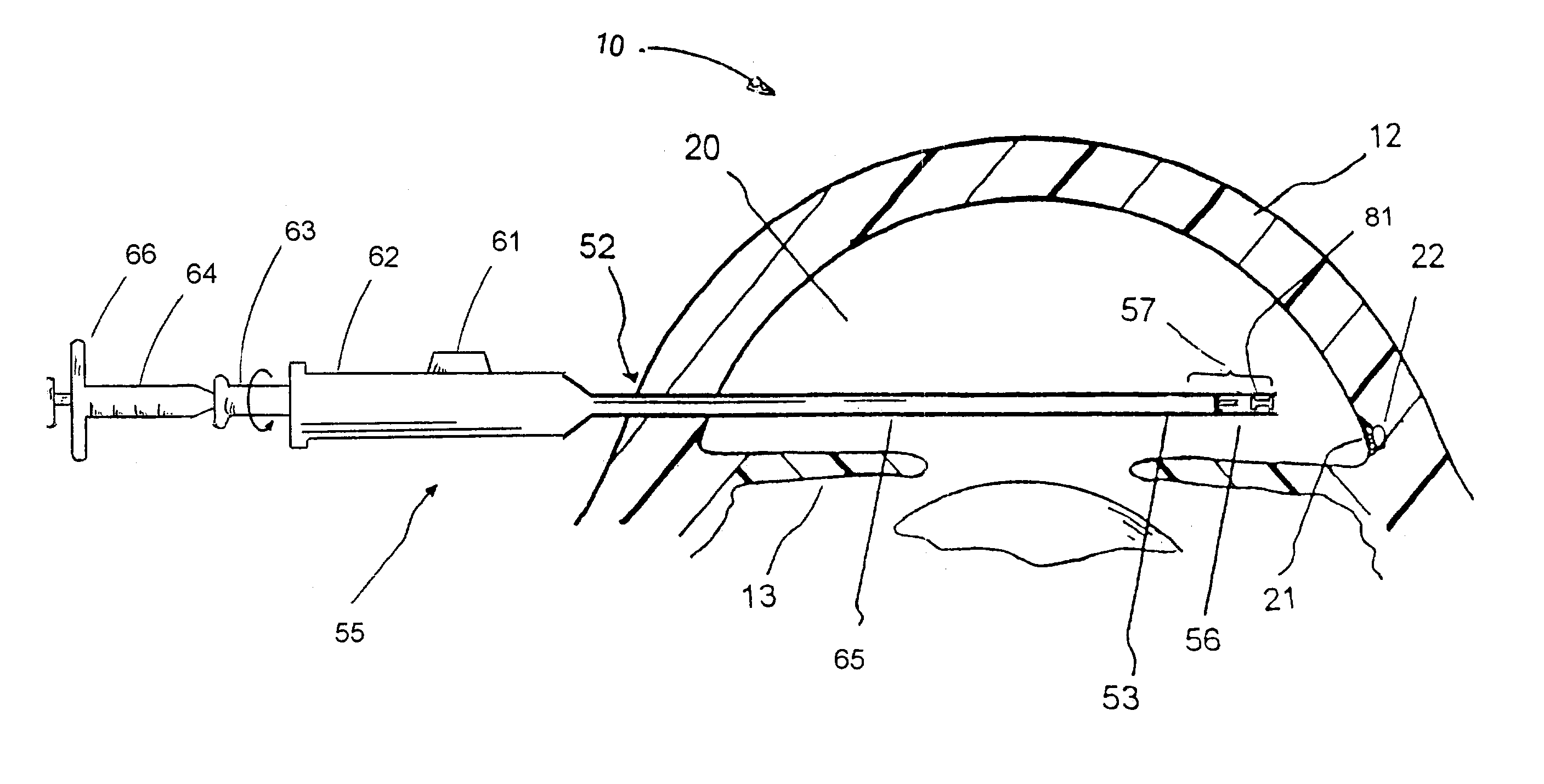
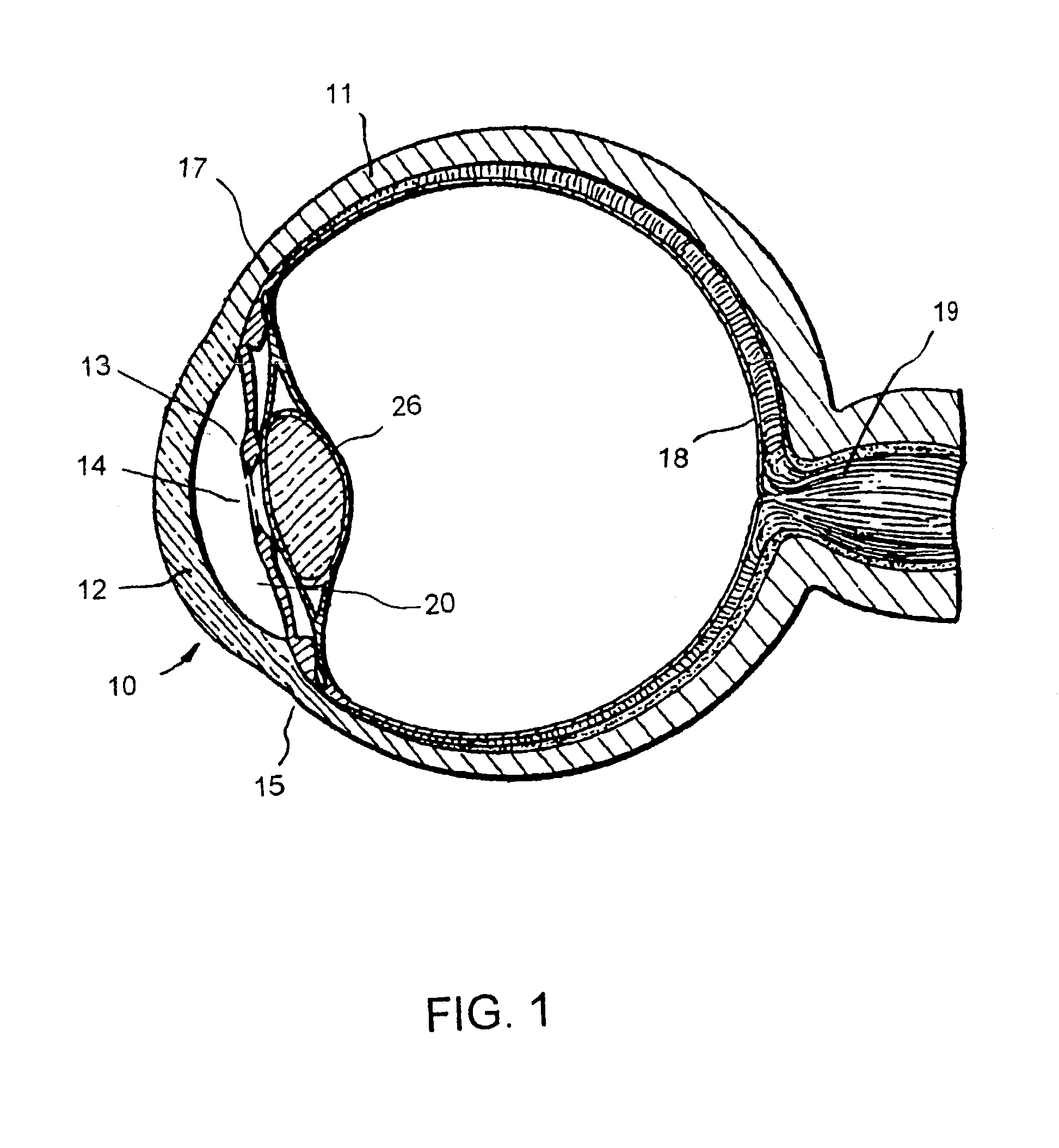
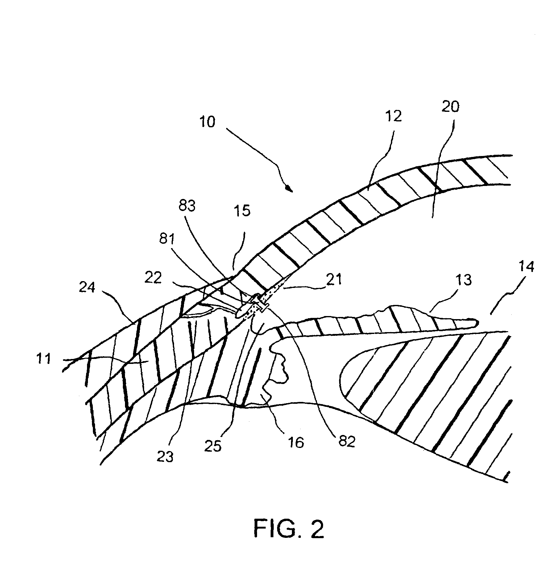
[0049]FIG. 1 is a cross-sectional view of an eye 10, while FIG. 2 is a close-up view showing the relative anatomical locations of a trabecular meshwork 21, an anterior chamber 20, and a Schlemm's canal 22. A sclera 11 is a thick collagenous tissue that covers the entire eye 10 except a portion that is covered by a cornea 12. The cornea 12 is a thin transparent tissue that focuses and transmits light into the eye a...


PUM


Abstract

The invention relates to methods of treating glaucoma, such as a method that includes inserting a stent through an incision in an eye; the stent having an inflow portion that is in fluid communication with an outflow portion of the stent; transporting the stent from the incision through the anterior chamber of the eye to an aqueous cavity of the eye, such that the inflow portion of the stent is positioned in the anterior chamber and the outflow portion of the stent is positioned at the aqueous cavity; and infusing fluid from the inflow portion to the outflow portion of the stent.

Description

CROSS REFERENCE TO RELATED APPLICATION[0001]This application claims the priority benefit of U.S. Provisional Application No. 60 / 362,405, entitled “Apparatus and Combination Therapy for Treating Glaucoma,” filed Mar. 7, 2002, and U.S. Provisional Application No. 60 / 363,980, entitled “Means and Procedures for Implanting a Glaucoma Shunt,” filed Mar. 14, 2002, the entireties of which are hereby incorporated by reference.BACKGROUND OF THE INVENTION[0002]1. Field of the Invention[0003]This invention relates to reducing intraocular pressure within the animal eye. More particularly, this invention relates to a treatment of glaucoma wherein aqueous humor is permitted to flow out of an anterior chamber of the eye through a surgically implanted pathway. Furthermore, this invention relates to directly dilating Schlemm's canal and / or aqueous collector channels by injecting fluid through the implanted pathway of a stent.[0004]2. Description of the Related Art[0005]A human eye is a specialized se...

Claims


Application Information

Patent Timeline
Patent Type & Authority Patents(United States)
IPC IPC(8): A61M5/00A61F11/00A61F2/04A61M31/00A61M35/00
CPCA61F9/00781A61F9/0017A61M5/14A61F9/00745A61F2009/00891A61M27/002A61M2210/0612
Inventor SMEDLEY, GREGORYHAFFNER, DAVIDTU, HOSHENG
Owner GLAUKOS CORP
Why Eureka
Try Eureka