Method for automated window-level settings for magnetic resonance images

Active Publication Date: 2007-05-15
PHILIPS HEALTHCARE INFORMATICS INC
View PDF15 Cites 102 Cited by
  • Summary
  • Abstract
  • Description
  • Claims
  • Application Information

AI Technical Summary

Benefits of technology

[0021]An object of the present invention is to provide a fu

Problems solved by technology

If the window width and level are present, very often this information is not optimal for diagnostic use.
The manual adjustment procedures require the user's time and energy, particularly if a large number of MR slices are acquired for the patient.
However, when W-L values are set to the same value for all images in the same series, the results may not be optimal for all the images and may require additional adjustment for diagnostic use.
As such, the images are often noisy, with maximal and minimal pixel intensity values that vary from image to image, and also within a particular image, e.g. spikes and background noise.
(4) The size and mass distribution of individual patients can affect the values obtained from an MRI acquisition in complex ways.
This method, which is readily calculatable, does not work well for many MR images, in particular, for those acquired with surface coils.
However, in any anatomic regions which are intermediate in brightness betwe

Method used


Image

  • Method for automated window-level settings for magnetic resonance images
  • Method for automated window-level settings for magnetic resonance images
  • Method for automated window-level settings for magnetic resonance images

Examples


Embodiment Construction

[0044]The following is a detailed description of the preferred embodiments of the invention, reference being made to the drawings in which the same reference numerals identify the same elements of structure in each of the several figures.

[0045]The present invention recognizes that the spatial intensity distribution of an image plays an important role in the adjustment of the display window parameters, and different organs may have similar histograms but might be windowed differently. The present invention also recognizes the need to focus on the analysis of radiologist / operator actions while adjusting W-L.

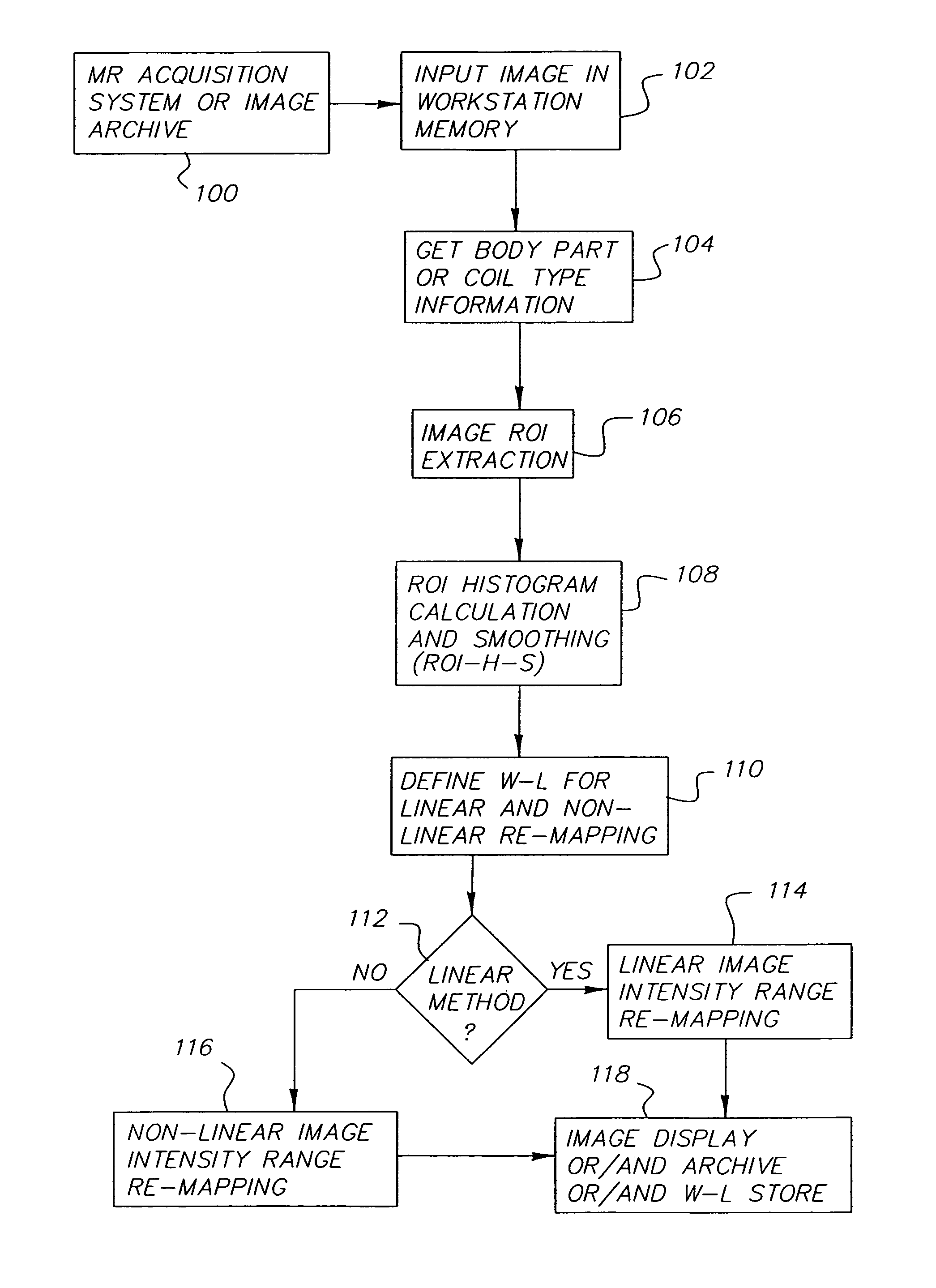
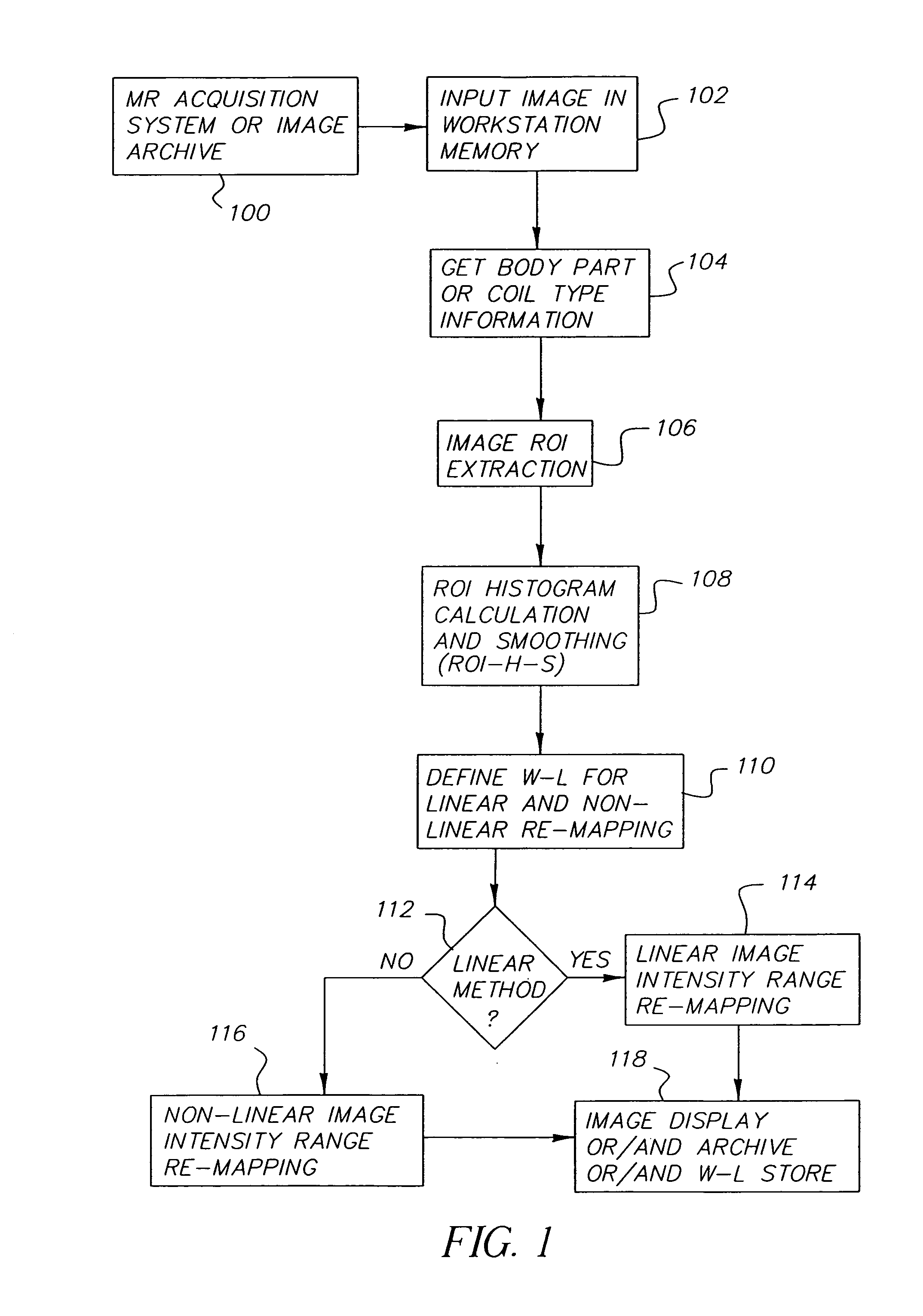
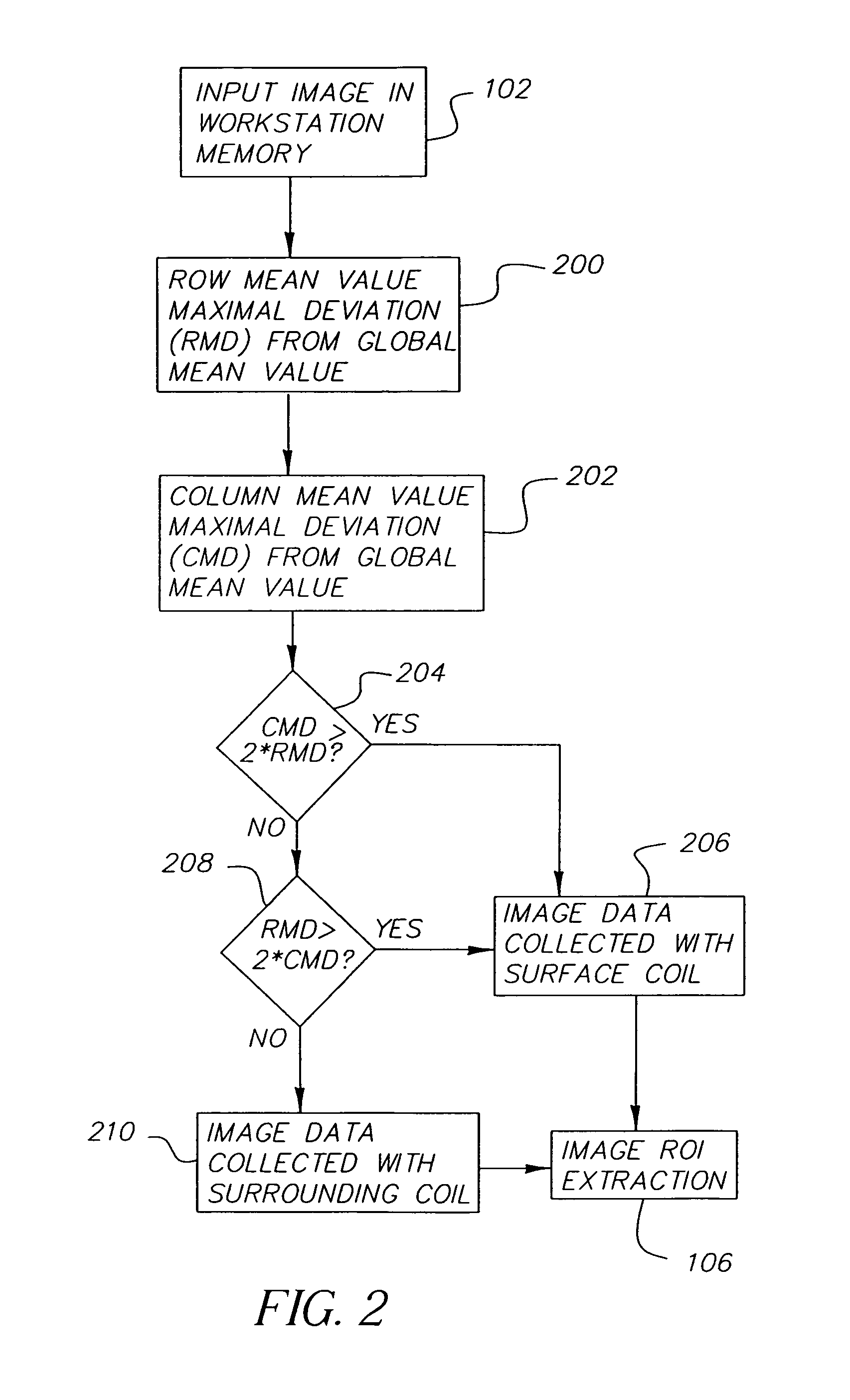
[0046]The present invention provides a method for automatic calculation of the window width and window level values and intensity range re-mapping for MR images for display and / or archive. The algorithm includes five steps: body part or coil type information extraction, ROI extraction, processing of histogram calculated within the ROI, window-level values definition, and linear or ...


PUM


Abstract

A method of automatically determining window-level settings in a digital image of a subject. The method includes the steps of: accessing the digital image; obtaining body part or coil type information; determining a region of interest (ROI) within the digital image; producing and processing a ROI-histogram; defining a W-L value for a linear or non-linear image intensity range re-mapping; and a linear or non-linear re-mapping the image intensity range.

Description

FIELD OF THE INVENTION[0001]The invention relates generally to the field of medical imaging, and in particular to digital image processing and display in Magnetic Resonance Imaging. More specifically, the invention relates to improved image representation for diagnostic purposes.BACKGROUND OF THE INVENTION[0002]Magnetic Resonance (MR) images are acquired during medical examinations on Magnetic Resonance Imaging (MRI) apparatus and stored in digital form in image archives. The digital data of the MR image can be stored and transferred to other image display, analysis, and archiving systems by means of a computer network. The digital data is typically transmitted over a communication network using a DICOM Standard (Digital Imaging and Communications in Medicine (DICOM), Part 3: Information Object Definitions, PS 3.3-2001).[0003]The acquired pixel values in an MR image do not relate directly to the brightness and contrast of display for diagnostic use of the image, and frequently no in...

Claims


Application Information

Patent Timeline
IPC IPC(8): G06K9/00G06T5/40
CPCG06T5/007G06T5/30G06T5/40G06T2207/10088
Inventor BELYKH, IGORCORNELIUS, CRAIG W.
Owner PHILIPS HEALTHCARE INFORMATICS INC
Why Eureka
Try Eureka